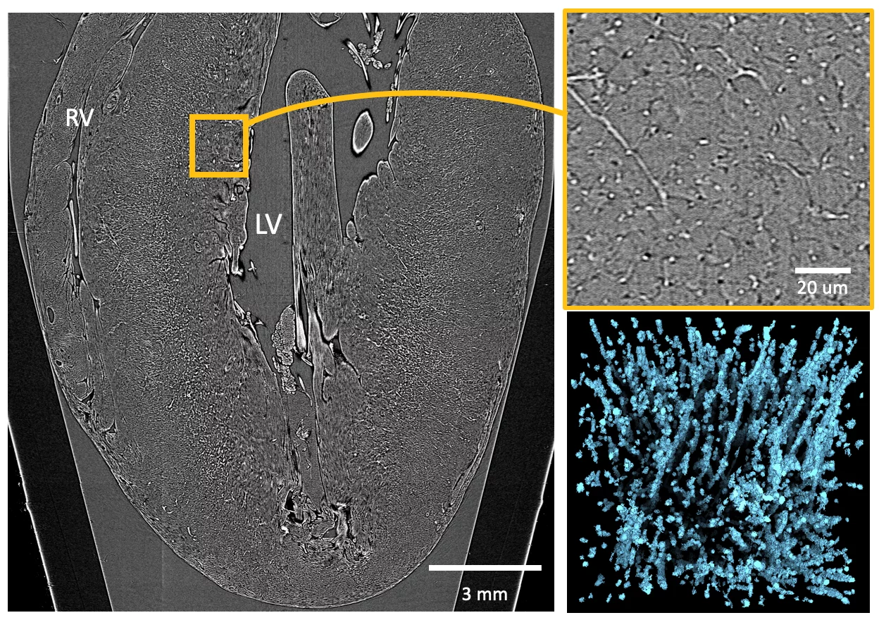
Cardiovascular diseases (CVDs) affect the myocardium and vasculature, inducing remodelling of the heart from cellular to whole organ level. To assess their impact at micro and macroscopic level, multi-resolution imaging techniques that provide high quality images without sample alteration and in 3D are necessary: requirements not fulfilled by most of traditional investigation methods.
A team of researchers from several international institutions, including Paul Scherrer Institute, University College London (United Kingdom), IDIBAPS (Spain) and Universitat Pompeu Fabra (Spain), took advantage of the non-destructive time-efficient 3D multiscale capabilities of synchrotron Propagation-Based X-Ray Phase Contrast Imaging (PB-X-PCI) to study a wide range of cardiac tissue characteristics in healthy and diseased rodent models. With a dedicated image processing pipeline, the presented technique evaluates in detail the overall cardiac morphology, myocyte aggregate orientation, vasculature changes, fibrosis formation and nearly single cell arrangement.
The proposed approach can improve the understanding of the multiscale remodelling processes occurring in CVDs, and the comprehensive and fast assessment of future interventional approaches.
Original Publication
Dejea H, Garcia-Canadilla P, Cook AC, Guasch E, Zamora M, Crispi F, Stampanoni M, Bijnens B & Bonnin A, Comprehensive analysis of animal models of cardiovascular disease using multiscale X-ray phase contrast tomography, Scientific Reports 9(1): 6996 (2019).
Contacts
Dr. Anne Bonnin
Beamline Scientist, Swiss Light Source
Paul Scherrer Institut
Telephone: +41 56 310 4678
E-mail: anne.bonnin@psi.ch

