

Many biological tissue are hierarchical materials with structures spanning from meters down to atomic scales. Small- angle X-ray scattering (SAXS) imaging is in particular valuable for such heterogeneous samples. The SAXS patterns provide statistical information about the nanostructure in each scan point, thus this method is complementary to high-resolution imaging techniques, such as electron microscopy. The information extracted from each scattering pattern can be used to create images with different contrast, such as density of nano-scale features or orientation of nanostructures. With wide-angle X-ray scattering (WAXS) orientation and length scales of crystalline planes can be simultaneously obtained. SAXS/WAXS tensor tomography allows to study the inside of three-dimensional samples. In many biological tissues, adding X-ray fluorescence as additional modality for elemental contrast is highly valuable. The SMAM group is investigating a broad range of biological tissues in various national and international collaborations.
Structure of regenerating bone in and around implants and scaffolds
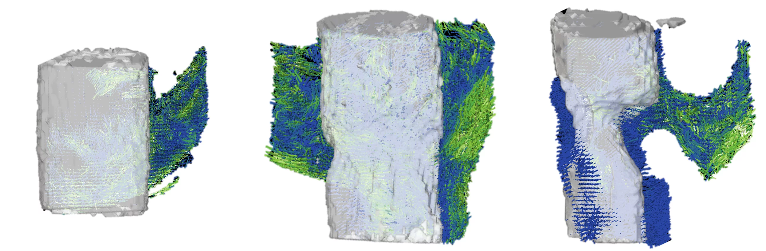
In bone research the mineral structure parameters relevant for the mechanical properties, such as crystallite size and orientation can be obtained from SAXS. The investigation in three-dimensional samples using SAXS tensor tomography allows for an increased understanding of the bone response to the implant materials which we are investigating in various collaboration.
Contribution of Bone Tissue Properties to Strength of the Ageing Human Hip
Osteoporosis is a silent metabolic bone disease reducing bone mass and leading to increased rates of bone fractures. Despite the dominant role of mass in bone mechanical properties, optimal clinical and societal management of osteoporosis requires a better understanding of the role of multiscale biophysical and biomechanical properties of the bone matrix in fracture incidence.
The aim of this project is to apply multi-scale imaging from the whole bone down to the micron level. State-of-the-art biophysical and micro-mechanical experiments will be performed on human tissue to identify the key determinants of fracture resistance. The mechanical response and failure characteristics of bone will be measured with high accuracy in order to retrieve tissue micromechanical properties using an inverse finite-element methodology.
SNF funded project in collaboration with University Bern and Empa.
Narwhal Tusks: A Tale with a Twist
Narwhal tusk is a rare form of external dentitions which show a left-handed spiral. Like other biological materials, the tusk has an impressive hierarchical structure from the atomic scale to the meter which we aim to characterize using a combination of SAXS and WAXS tensor tomography and X-ray phase contrast tomography. In addition, narwhal tusks grow throughout the animal’s life, where signatures of prey items and environmental conditions get stored in the growing tooth which can be characterized by scanning imaging techniques with XRF, SAXS and WAXS contrast. Since the narwhales live for a long time, their tusks proved a history book of changing environmental conditions over the lifetime of the animal.
This Nordforsk funded project is conducted at Chalmers University of Technology, Gothenburg Sweden in collaboration with Aarhus University, Denmark the Greenland Institute of Natural Resources.
Chameleon tongue
The chameleon tongue can be projected up to two-body length from the mouth at incredible speed. In order to withstand the high forces acting upon, the tongue is formed by a unique complex array of cartilage elastic elements and muscles which we try to understand better using SAXS tensor tomography.
This project is performed in close collaboration with Empa, Zurich University and the Zoo Zurich