Making the world go round - a look into the structure of a prominent heterogeneous catalyst
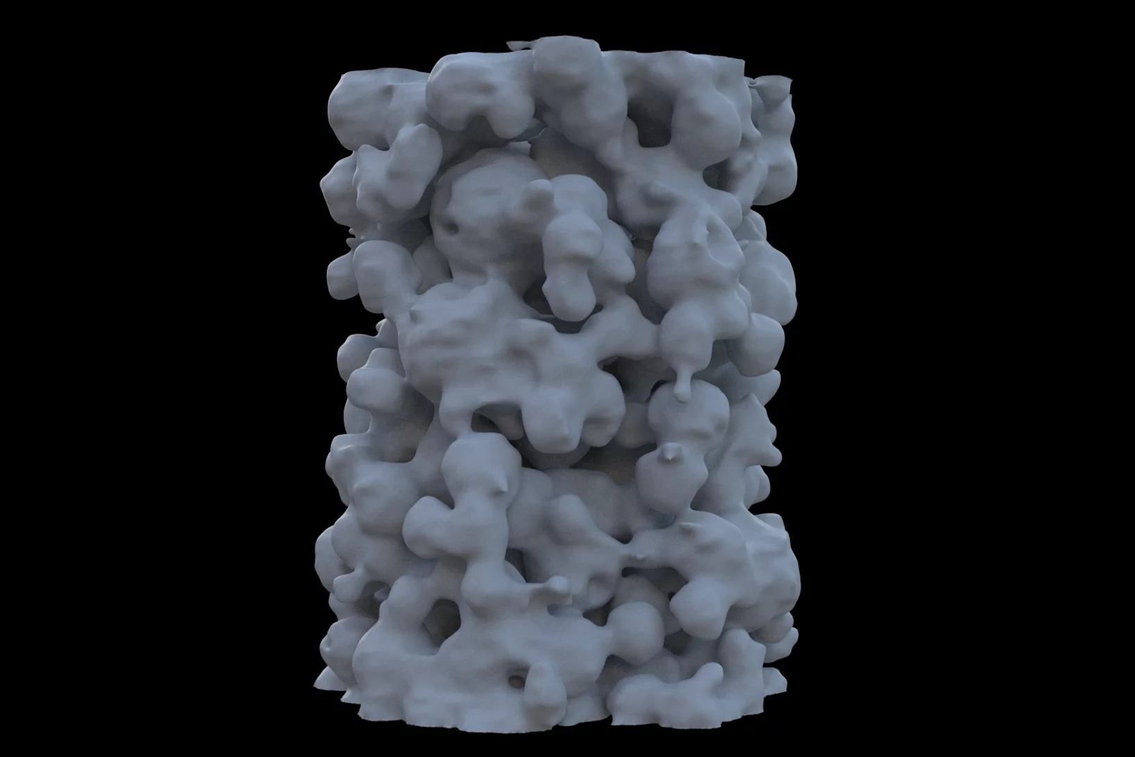
Fluid catalytic cracking catalysts, which are composite particles of hierarchical porosity, were examined using ptychographic X-ray tomography. These particles are essential to the conversion of crude oil into gasoline. Examination of catalysts at decreasing levels of catalytic conversion efficacy allowed the detection of possible deactivation causes.
Swiss Neutron Scattering Prize
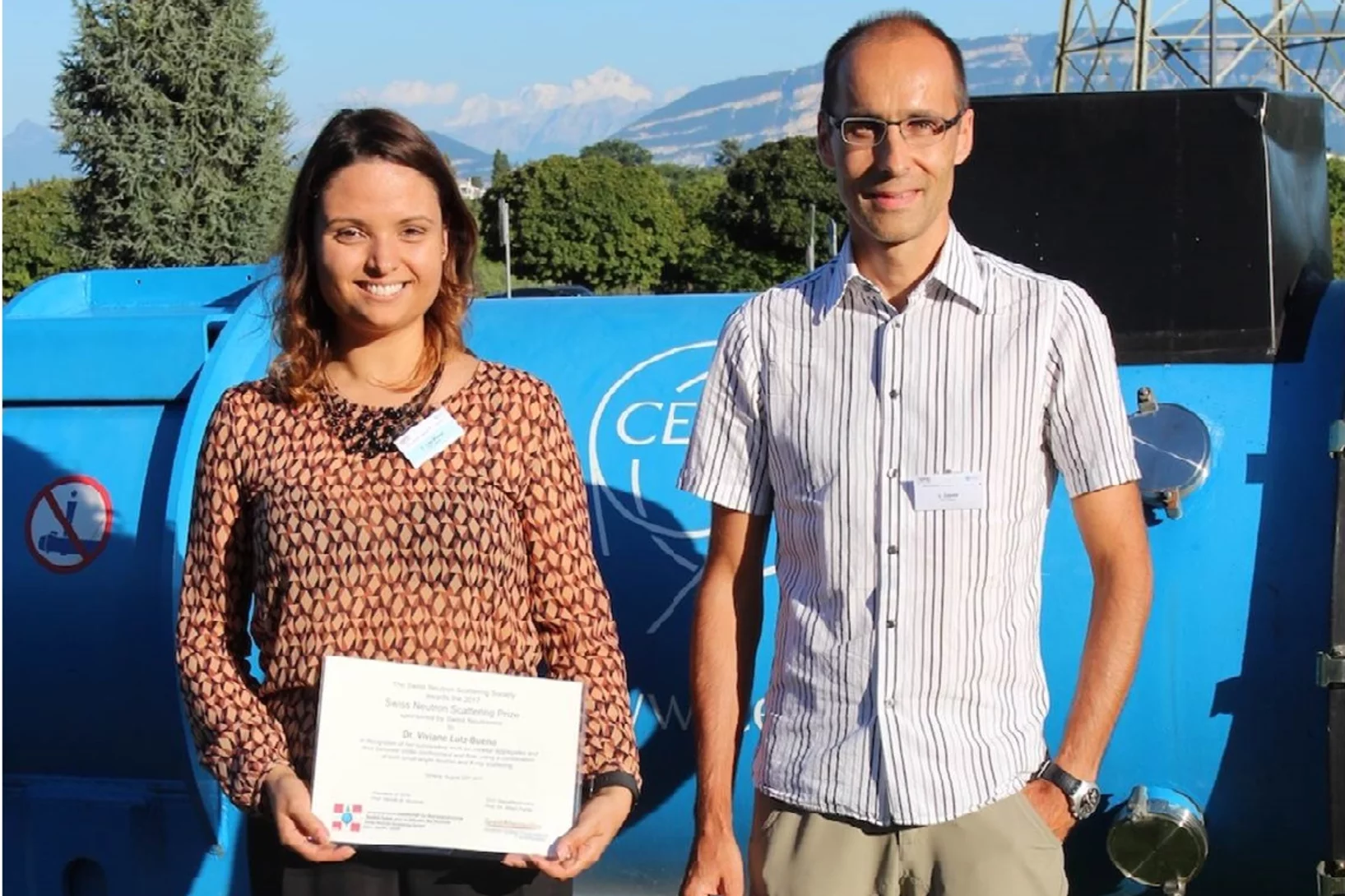
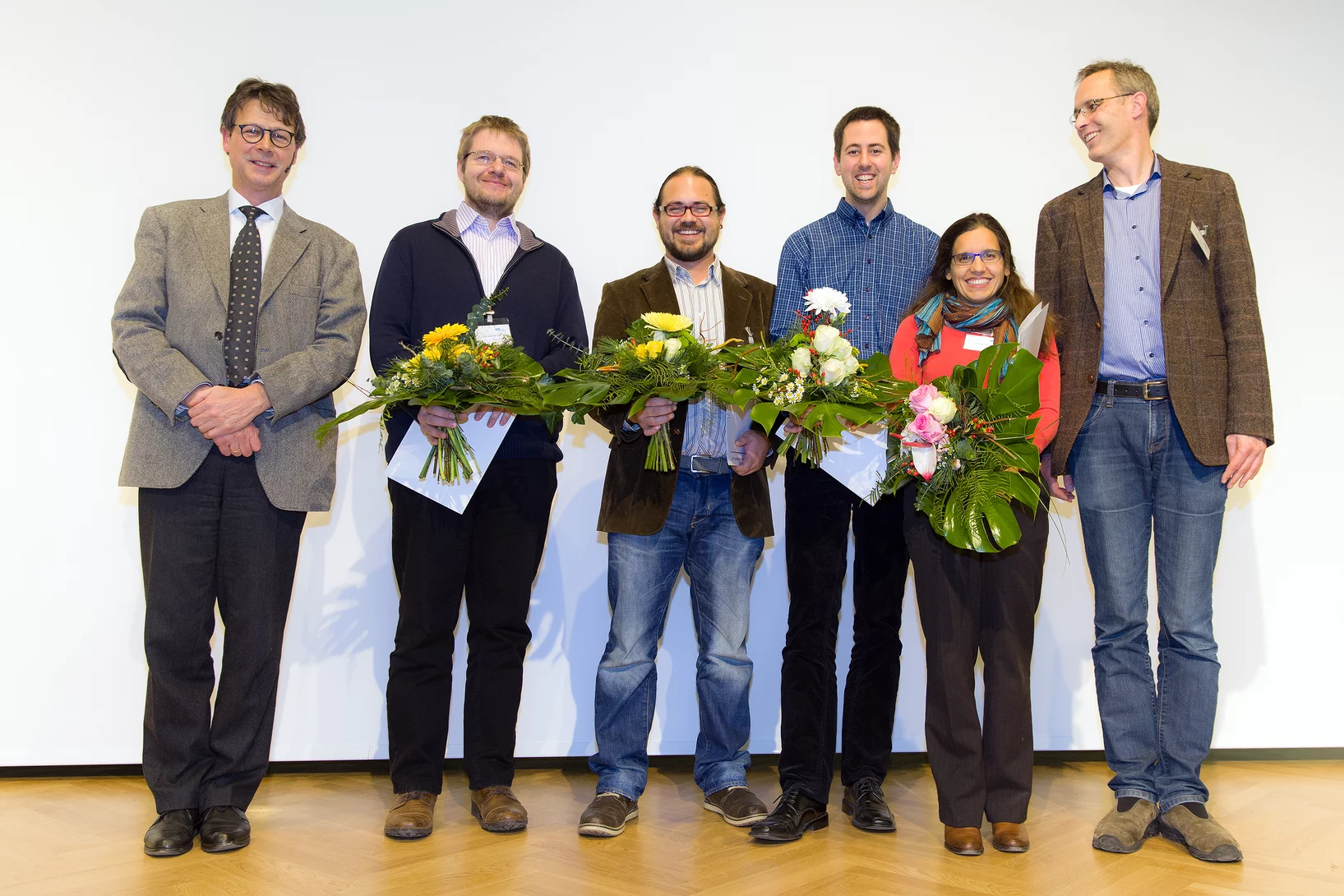
Viviane Lutz-Bueno was awarded the Swiss Neutron Scattering Prize of the Swiss Neutron Scattering Society, at the Joint Annual Meeting of the Swiss and Austrian Physical Society, for her PhD thesis Effects of formulation and flow on the structure of micellar aggregates carried out at ETH Zürich. Viviane is currently a postdoctoral fellow at the CXS group at PSI, developing scanning SAXS analysis methods.
Tauchgang in einen Magneten
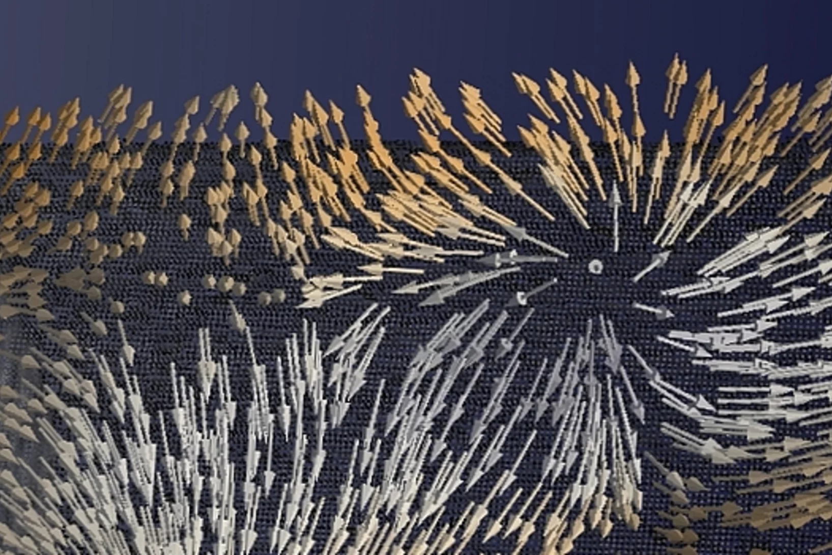
Zum ersten Mal haben Forschende die Richtungen der Magnetisierung in einem dreidimensionalen magnetischen Objekt sichtbar gemacht. Die kleinsten Details in ihrer Visualisierung waren dabei zehntausend Mal kleiner als ein Millimeter. In der sichtbar gemachten magnetischen Struktur stach eine Art von Muster besonders hervor: magnetische Singularitäten namens Bloch-Punkte, die bisher nur in der Theorie bekannt waren.
Photonic structure of white beetle wing scales: optimized by evolution
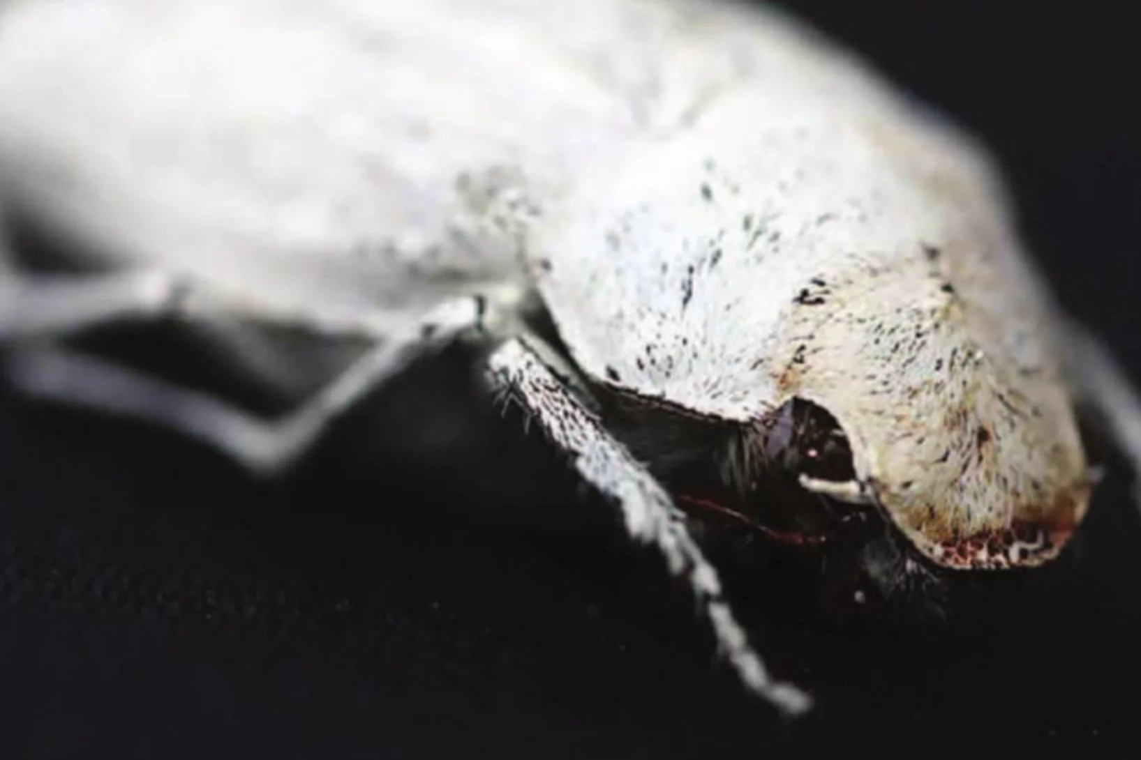
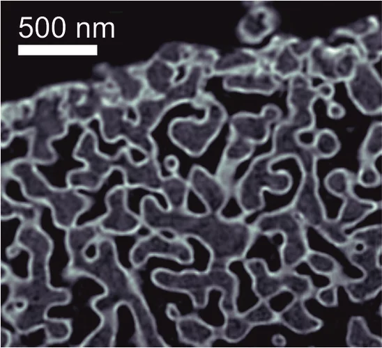
A very thin layer on this beetle’s wings exhibits a complicated structure on the nanoscale that gives them a bright white color. X-ray nanotomography acquired at the Swiss Light Source provides a faithful image of this structure in three dimensions with which scientists can confirm its evolutionary optimization: just enough material for an efficient reflection of white light.
European NESY Winterschool Young Scientist Best Poster Prize
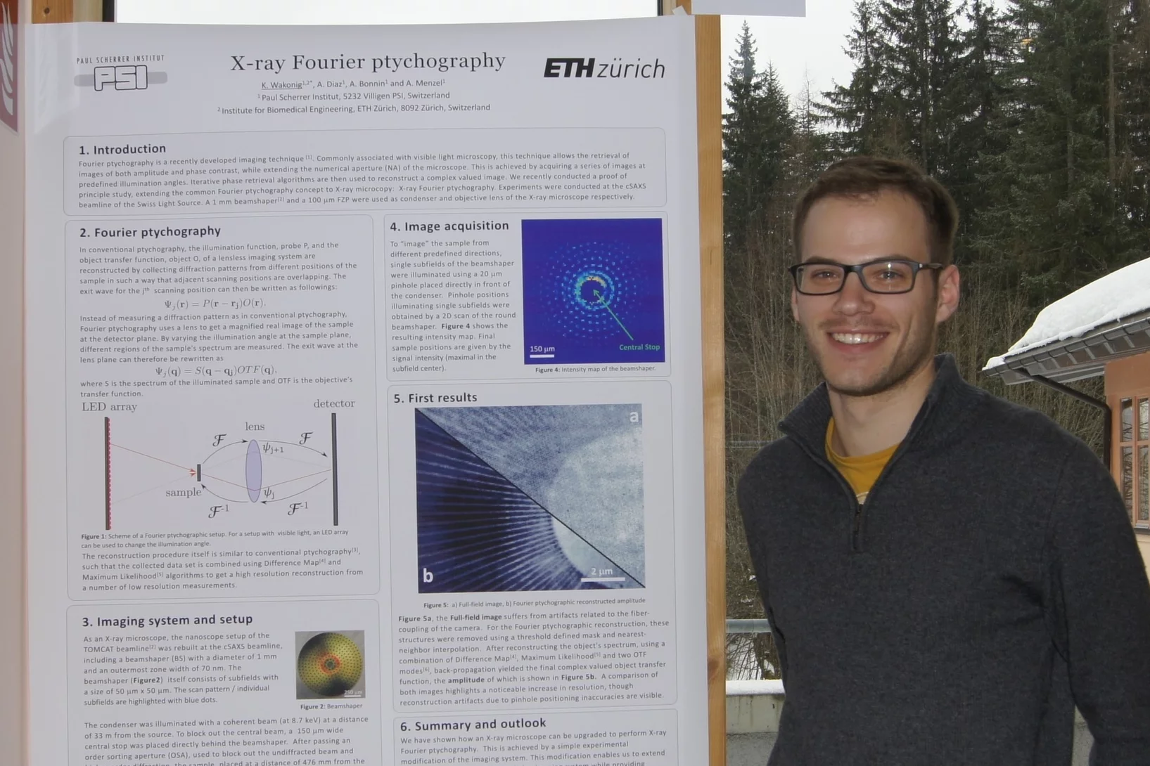
Klaus Wakonig was awarded the "Young Scientist Best Poster Prize" along with a cash prize at the 10th European NESY Winterschool & Symposium on Neutron and Synchrotron Radiation. Klaus is a PhD student at the coherent X-ray scattering group (CXS) in PSI. His poster, entitled "X-ray Fourier ptychography," details his latest results in the implementation of Fourier ptychography at X-ray wavelengths for nanoimaging. Image credit ©NESY/Montanuniversitaet Leoben
3-D-Röntgenbild macht feinste Details eines Computerchips sichtbar
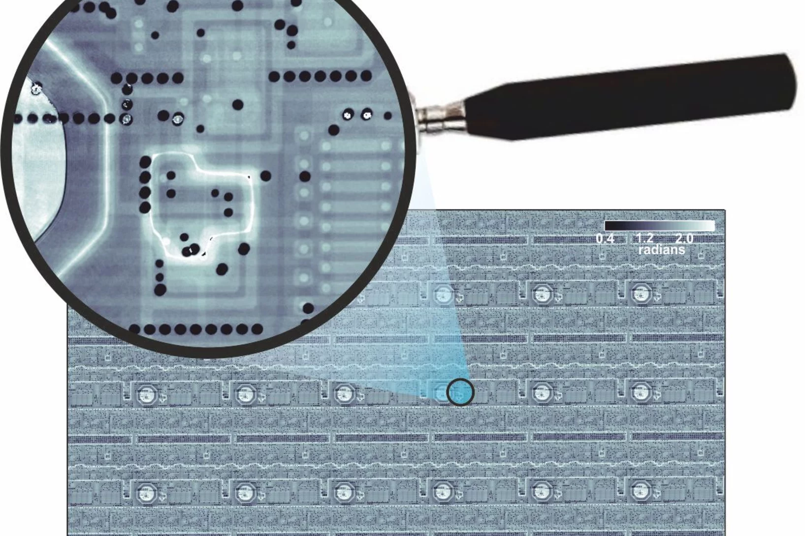
Forschende des PSI haben detaillierte 3-D-Röntgenbilder eines handelsüblichen Computerchips erstellt. In ihrem Experiment haben sie ein kleines Stück aus dem Chip untersucht, das sie zuvor herausgeschnitten hatten. Diese Probe blieb dabei während der Messung unbeschädigt. Für Hersteller ist es eine grosse Herausforderung, zu bestimmen, ob der Aufbau ihrer Chips am Ende den Vorgaben entspricht. Somit stellen diese Ergebnisse eine wichtige Anwendung eines Röntgen-Tomografieverfahrens dar, das die PSI-Forschenden seit einigen Jahren entwickeln.
Interlaced zone plates push the resolution limit in x-ray microscopy
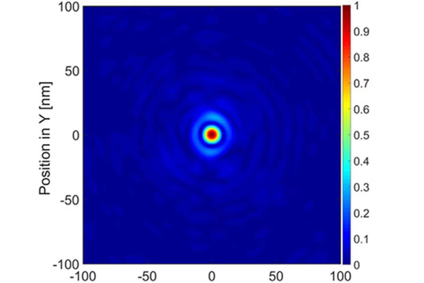
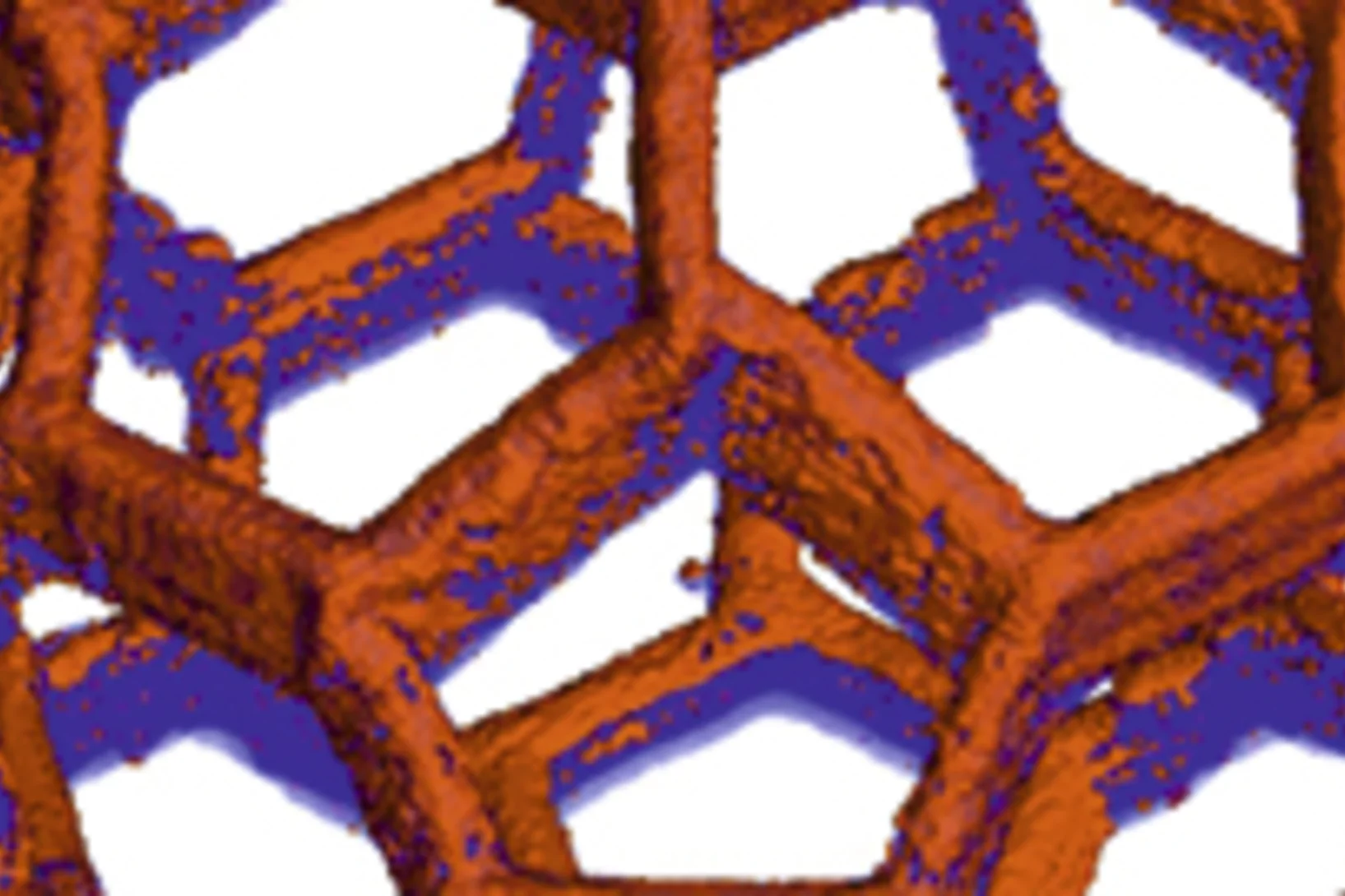
A novel type of diffractive lenses based on interlaced structures enable x-ray imaging at resolutions below 10 nm. The fabrication method and the test results of these novel x-ray lenses have been published in the journal Scientific Reports.
How does food look like on the nanoscale?
The answer to this question could save food industry a lot of money and reduce food waste caused by faulty production. Researchers from the University of Copenhagen and the Paul Scherrer Institut have obtained a 3D image of food on the nanoscale using ptychographic X-ray computed tomography. This work paves the way towards a more detailed knowledge of the structure of complex food systems.
Mass density distribution of intact cell ultrastructure
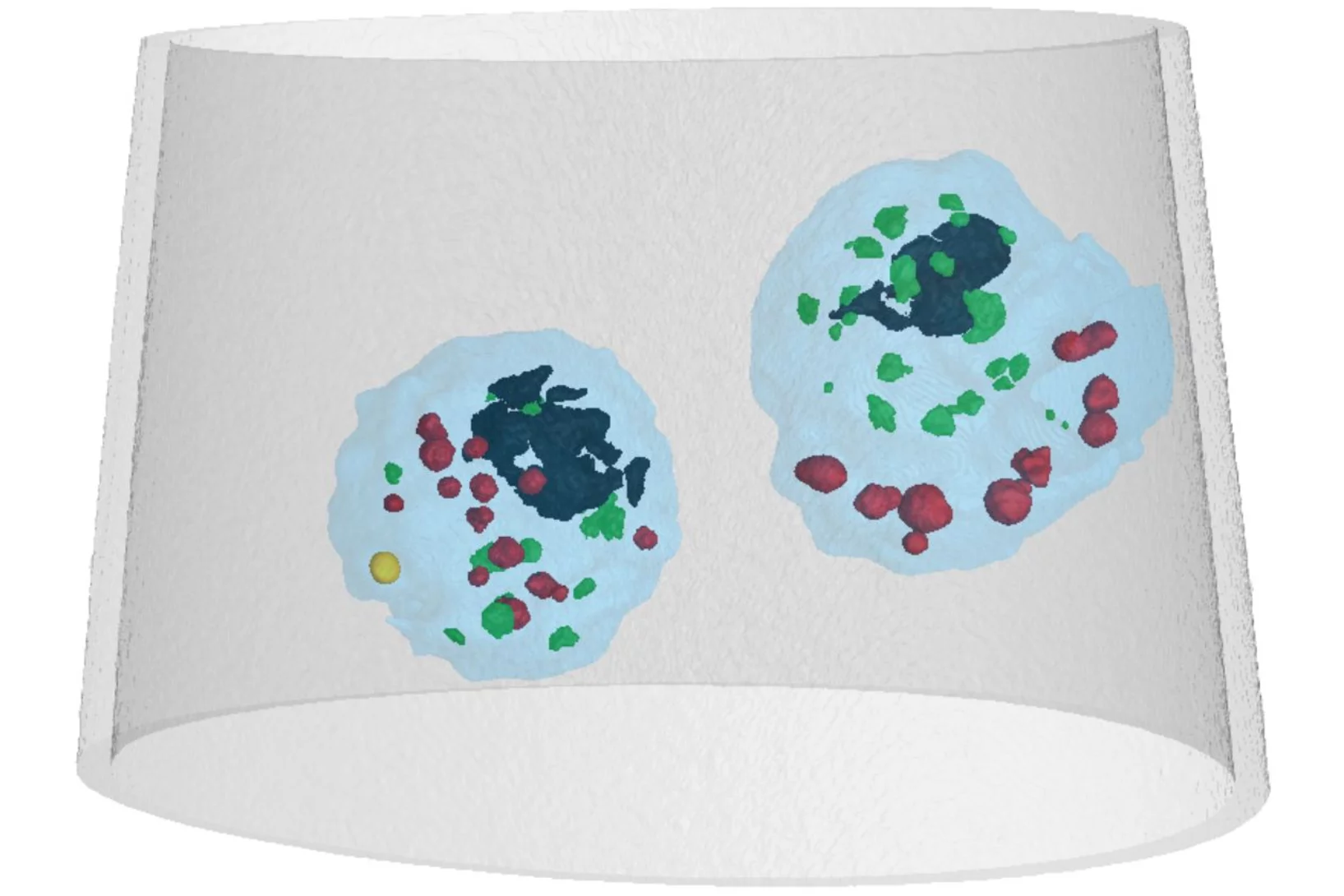
The determination of the mass density of cellular compartments is one of the many analytical tools that biologists need to unravel the extremely complex structure of biological systems. Cryo X-ray nanotomography reveals absolute mass density maps of frozen hydrated cells in three dimensions.
3-D-Nanostruktur eines Knochens sichtbar gemacht
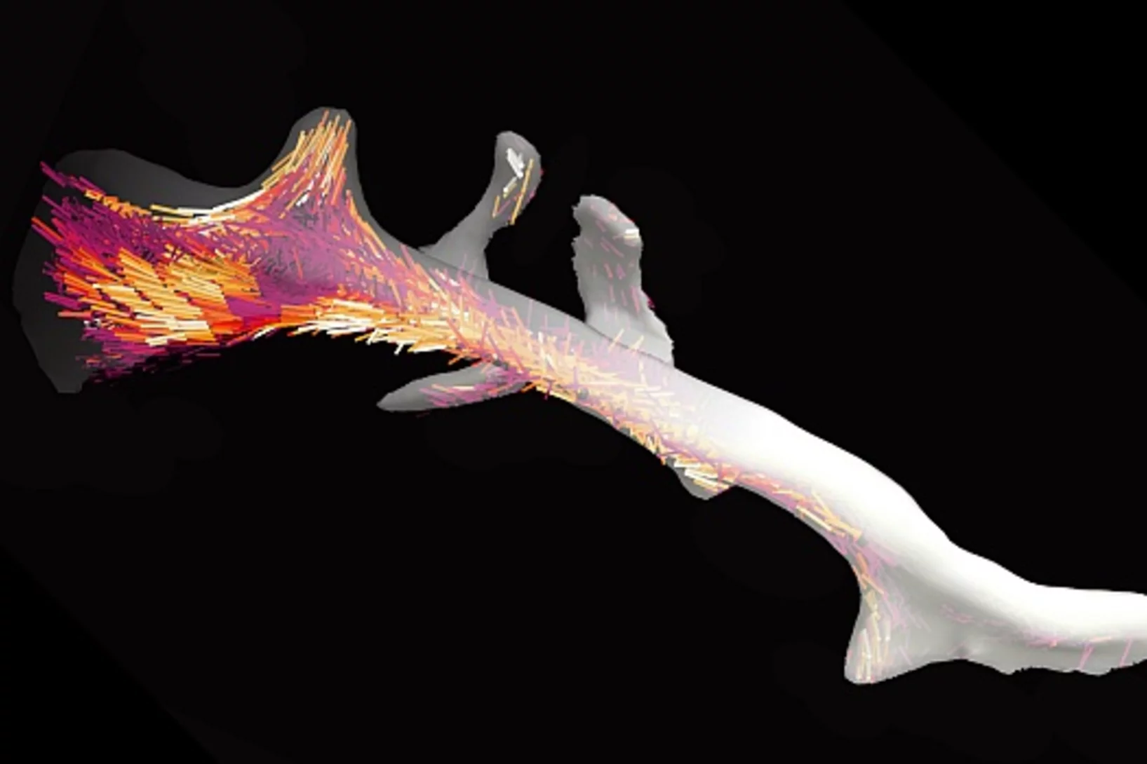
Knochen bestehen aus winzigen Fasern, die etwa tausend Mal feiner sind als ein menschliches Haar. Mit einer neuartigen computerbasierten Auswertungsmethode konnten Forschende des Paul Scherrer Instituts PSI zum ersten Mal die Anordnung dieser Nanostrukturen innerhalb eines gesamten Knochenstücks sichtbar machen.
X-ray nanotomography aids the production of eco-friendly solar cells
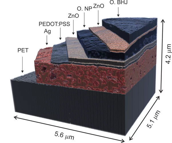
Polymer solar cells are in the spotlight for sustainable energy production of the future. Characterization of these devices by X-ray nanotomography helps to improve their production using environmentally friendly materials.
2015 Otto Kratky award
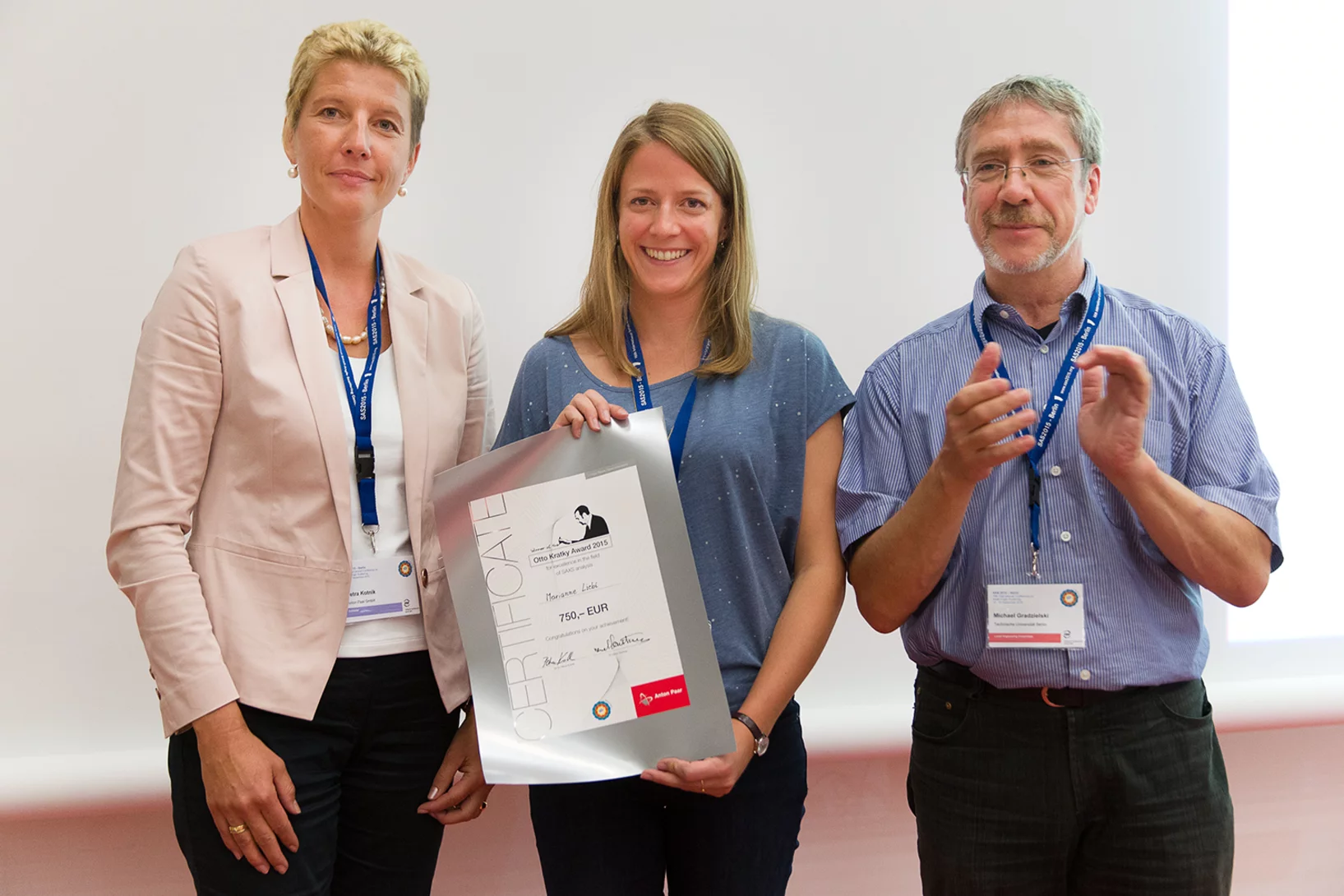
Marianne Liebi was awarded the 2015 Otto Kratky award by the Helmholtz-Centre Berlin for excellence in the field of small-angle X-ray scattering (SAXS) analysis. The award was bestowed in the last SAS2015 conference in Berlin. Marianne is a postdoctoral fellow in the coherent X-ray scattering group (CXS) in PSI, carrying out research in scanning SAXS measurement and analysis in 2D and 3D. Image credit ©HZB/Michael Setzpfandt
Element-Specific X-Ray Phase Tomography of 3D Structures at the Nanoscale
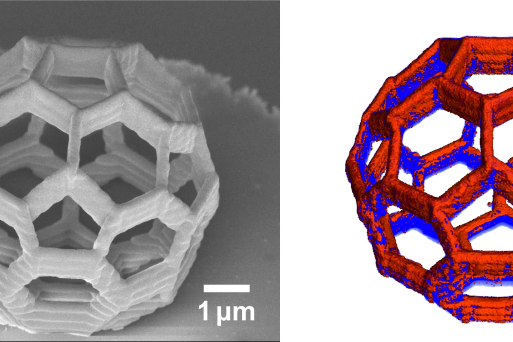
Recent advances in fabrication techniques to create mesoscopic 3D structures have led to significant developments in a variety of fields including biology, photonics, and magnetism. Further progress in these areas benefits from their full quantitative and structural characterization.
Aus dem Innern einer Eierschale
Winzige Bläschen im Innern von Eierschalen liefern die Stoffe, die das Wachstum der Schale stimulieren und steuern. Mit einer neuartigen Tomografie-Technik haben Forschende des Paul Scherrer Instituts PSI, der ETH Zürich und des niederländischen AMOLF-Instituts diese Bläschen erstmals in 3D abbilden können. Sie heben damit eine alte Einschränkung tomografischer Bilder auf und hoffen, dass eines Tages auch die Medizin von ihrer Methode profitiert.
Multiresolution X-ray tomography, getting a clear view of the interior
Researchers at PSI have developed a technique that combines tomography measurements at different resolution levels to allow quantitative interpretation for nanoscale tomography on an interior region of interest of the sample. In collaboration with researchers of the institute AMOLF in the Netherlands and ETH Zurich in Switzerland they showcase their technique by studying the porous structure within a section of an avian eggshell. The detailed measurements of the interior of the sample allowed the researchers to quantify the ordering and distribution of an intricate network of pores within the shell.
Gespaltener Röntgenblitz zeigt schnelle Vorgänge
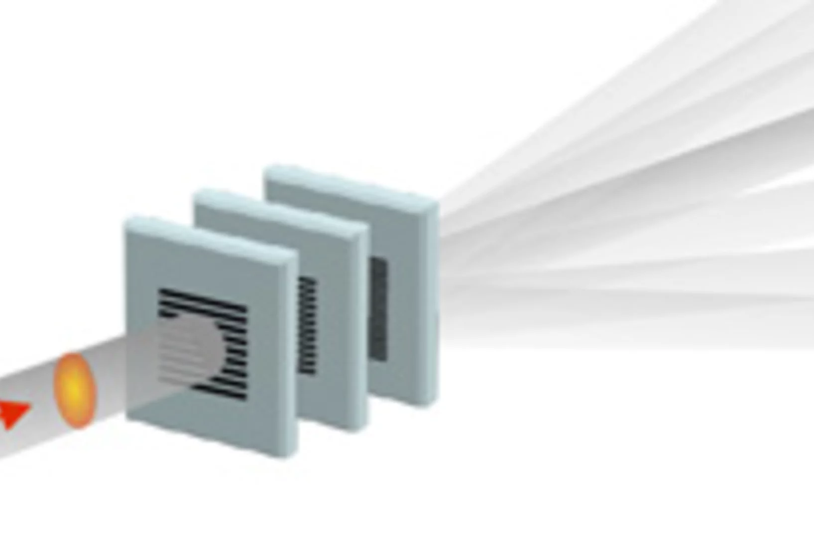
SwissFEL, der Röntgenlaser des PSI, wird die einzelnen Schritte sehr schneller Vorgänge sichtbar machen. Ein neues Verfahren soll besonders genaue Experimente ermöglichen: Dabei werden die einzelnen Röntgenblitze in mehrere Teile aufgespalten, die nacheinander am Untersuchungsobjekt ankommen. Das Prinzip des Verfahrens erinnert an die Ideen der frühesten Hochgeschwindigkeitsfotografie.
Nanometer in 3-D
Forschende haben 3-D-Bilder winziger Objekte erzeugt und konnten dabei sogar 25 Nanometer grosse Details (1 Nanometer = 1 Millionstel eines Millimeters) sichtbar machen. Dabei haben sie nicht nur die Form der Untersuchungsgegenstände bestimmen können, sondern auch gezeigt, wie ein bestimmtes chemisches Element (Kobalt) darin verteilt ist und ob es in einer chemischen Verbindung oder in Reinform vorliegt.
Innovation Award on Synchrotron Radiation 2014 for high-resolution 3D hard X-ray microscopy
The 2014 Innovation Award on Synchrotron Radiation was bestowed to researchers Ana Diaz, Manuel Guizar-Sicairos, Mirko Holler, and Jörg Raabe from the Paul Scherrer Institut, Switzerland, for their contributions to method and instrumentation development, which have set new standards in high-resolution 3D hard X-ray microscopy.
Fast scanning coherent X-ray imaging using Eiger
The smaller pixel size, high frame rate, and high dynamic range of next-generation photon counting pixel detectors expedites measurements based on coherent diffractive imaging (CDI). The latter comprises methods that exploit the coherence of X-ray synchrotron sources to replace imaging optics by reconstruction algorithms. Researchers from the Paul Scherrer Institut have recently demonstrated fast CDI image acquisition above 25,000 resolution elements per second using an in-house developed Eiger detector. This rate is state of the art for diffractive imaging and even on a par with the fastest scanning X-ray transmission instruments. High image throughput is of crucial importance for both materials and biological sciences for studies with representative population sampling.
X-ray tomography reaches 16 nm isotropic 3D resolution
Researchers at PSI reported a demonstration of X-ray tomography with an unmatched isotropic 3D resolution of 16 nm in Scientific Reports. The measurement was performed at the cSAXS beamline at the Swiss Light Source using a prototype instrument of the OMNY (tOMography Nano crYo) project. Whereas this prototype measures at room temperature and atmospheric pressure, the OMNY system, to be commissioned later this year, will provide a cryogenic sample environment in ultra-high vacuum without compromising imaging capabilities. The researchers believe that such a combination of advanced imaging with state-of-the-art instrumentation is a promising path to fill the resolution gap between electron microscopy and X-ray imaging, also in case of radiation-sensitive materials such as polymer structures and biological systems.
Unique insight into carbon fibers on the nanoscale
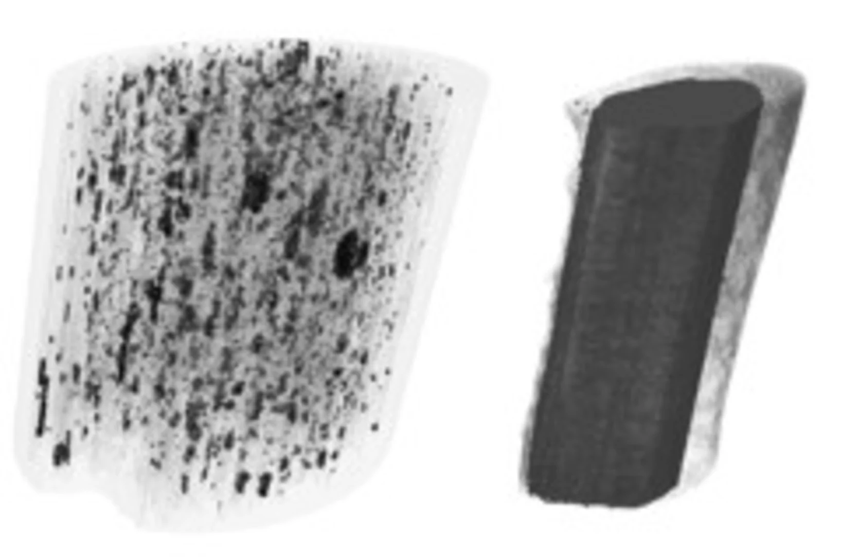
The investigation of the mechanical properties of carbon fibers benefits from highly resolved three-dimensional density maps within representative volumes, but such images are not easily obtained with standard methods. Scientists from the Paul Scherrer Institut in collaboration with Honda R&D in Germany have recently visualized density distributions on the sub-micrometer scale within entire carbon fiber sections, revealing surprising graphite distributions within the fibers. This capability will prove useful for the systematic characterization of fibers, contributing to the development of light and robust materials at lower costs.
Röntgen-Laser: Auf dem Weg zur Strukturbestimmung von Nanoteilchen
An Freie-Elektronen-Röntgen-Lasern wie dem zukünftigen SwissFEL des Paul Scherrer Instituts PSI sollen unter anderem die Strukturen von komplexen Nanoteilchen bis hin zu Biomolekülen untersucht werden. Dabei ist nicht nur die eigentliche Messung eine Herausforderung, sondern auch die Rekonstruktion der Struktur aus den Messdaten. Forscher des PSI haben nun einen optimierten mathematischen Weg aufgezeigt, wie man aus so gewonnen Messdaten eine deutlich bessere Auflösung bei der Bestimmung der Struktur eines einzelnen Teilchens erhält. Das Verfahren wurde an der Synchrotron Lichtquelle Schweiz des PSI erfolgreich getestet.
Fluktuationen mit Röntgenmikroskop sichtbar gemacht
Mit Röntgenstrahlen kann die Nanostruktur von so unterschiedlichen Objekten untersucht werden, wie einzelne Zellen oder magnetische Datenträger. Hochauflösende Bilder sind jedoch nur möglich, wenn sowohl Mikroskop als auch das Untersuchungsobjekt extrem stabil sind. Forscher der TU München des PSI zeigten nun, wie man diese Bedingungen lockern kann, ohne die Bildqualität zu beeinträchtigen. Auch hochdynamische Systeme, wie z.B. magnetische Fluktuationen, die die Lebensdauer von Daten auf Festplatten einschränken, können mit der neuen Methodik untersucht werden.
Nanoforscher untersuchen Karies
Forscher der Universität Basel und des Paul Scherrer Instituts konnten im Nanomassstab zeigen, wie sich Karies auf die menschlichen Zähne auswirkt. Ihre Studie eröffnet neue Perspektiven für die Behandlung von Zahnschäden, bei denen heute nur der Griff zum Bohrer bleibt. Die Forschungsergebnisse wurden in der Fachzeitschrift «Nanomedicine» veröffentlicht.
Röntgen-Methode hilft Hirnerkrankungen besser zu verstehen
Ein internationales Forschungsteam hat eine neue Methode entwickelt, mit der man detaillierte Röntgenbilder von Hirngewebe erstellen kann. Die Methode wurde verwendet, um die Myelinscheide der Nervenfasern sichtbar zu machen. Schäden an der Myelinscheide führen zu verschiedenen Erkrankungen wie etwa Multiple Sklerose. Die Anlage, an der diese Aufnahmen erstellt werden können, wird an der Synchrotron Lichtquelle Schweiz SLS des Schweizer Paul Scherrer Instituts betrieben.
Fortschritt für die Knochen-Forschung
Hochauflösendes Verfahren zur Nano-Computertomographie entwickeltEin neuartiges Nano-Tomographie-Verfahren, das von einem Team der TU München, des Paul Scherrer Instituts und der ETH Zürich entwickelt wurde, erlaubt erstmals computertomographische Untersuchungen feinster Strukturen mit einer Auflösung im Nanometerbereich. Mit Hilfe der neuen Methode können etwa dreidimensionale Innenansichten fragiler Knochenstrukturen erstellt werden.

