Dr. Konstantins Jefimovs wins the 2nd poster prize
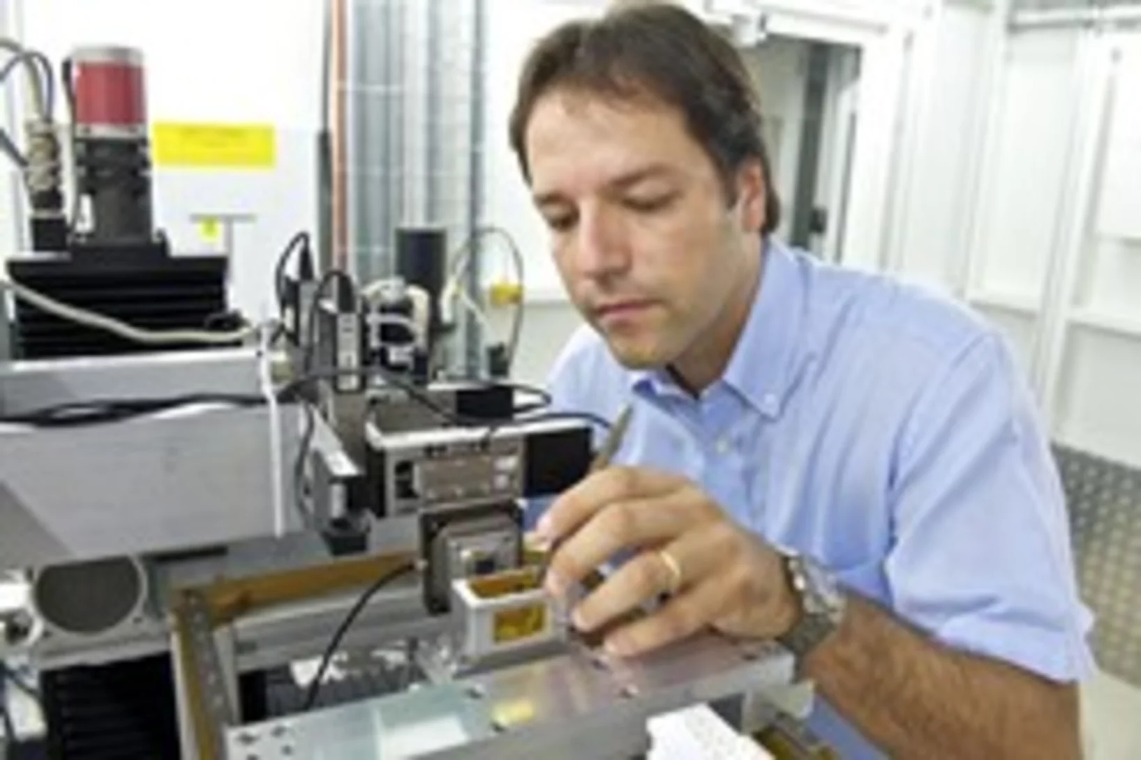
Dr. Konstantins Jefimovs from the TOMCAT team at SLS was awarded with the 2nd poster prize at the 4th XNPIG Conference (X-ray and Neutron Phase Contrast Imaging with Gratings) held in Zürich, September 12th-15th. He presented latest achievements on gratings fabrication under the title: “Large area small pitch gratings for X-ray interferometry by Displacement Talbot Lithography”.
Dr. Matias Kagias receives the William H. F. Talbot Award 2017
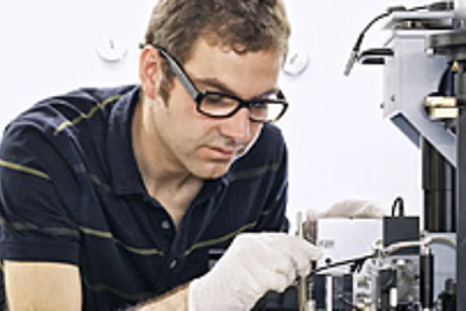
Dr. Matias Kagias from the TOMCAT team at SLS is the recipient of the William H. F. Talbot Award 2017, as announced recently at the 4th XNPIG Conference (X-ray and Neutron Phase Contrast Imaging with Gratings) held in Zürich in September 12th-15th. The award is given to young PhD students for their outstanding contributions in the field of X-ray and Neutron Phase contrast imaging. This award has been introduced this year and Dr. Kagias is the first recipient ever.
Dr. Goran Lovric receives 2nd best abstract prize at the 34th SSAI Congress
Goran Lovric, a Postdoc at TOMCAT and CIBM, received the Best abstract 2nd Prize at the 34th congress of the Scandinavian Society of Anaesthesiology and Intensive Care (SSAI) in Malmö (Sweden), 6-8th of September 2017. He presented the latest results of his work, entitled Spatial distribution of ventilator induced lung injury at the acinar level. An in-vivo synchrotron phase-contrast microscopy study in BALB/c mice.
Hector Dejea presented a talk at the conference Functional Imaging and Modelling of the Heart (FIMH2017) in Toronto
Hector Dejea (PhD student at ETH and TOMCAT) just attended the biennial FIMH conference in Toronto (Canada), which aims to integrate the state-of-the-art scientific advances in the fields cardiovascular imaging, image analysis and heart modelling fields.
Carolina Arboleda presented a talk contribution at the Swiss Congress of Radiology (SCR2017) in Bern
Carolina Arboleda, senior PhD student at TOMCAT, presented a talk entitled “Assessment of breast lesion malignancy using phase contrast imaging” at the Swiss Congress of Radiology, which highlighted the potential of X-ray grating-based phase contrast imaging to distinguish between benign and malignant lesions utilizing the absorption to dark-field signal ratio of associated calcifications.
Lucia Romano gives an invited talk at EIPBN 2017
Lucia Romano (guest professor at ETH and guest scientist at TOMCAT) has just returned from the largest conference devoted to electron, ion and photon beam technology and nanofabrication (EIPBN) in US, where she gave an invited talk about “Fabrication of high aspect ratio metal gratings for X-ray phase contrast interferometry”.
New TOMCAT paper: The GigaFRoST camera and readout system
The PSI in-house developed GigaFRoST high-speed camera and readout system is available for fast imaging experiments at the TOMCAT beamline, opening up exciting new possibilities for the observation of fast dynamic phenomena with X-ray tomography.
Single shot grating interferometry demonstrated using direct conversion detection
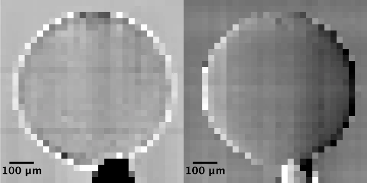
Researchers at the Paul Scherrer Institute's Swiss Light Source in Villigen, Switzerland, have developed an X-ray grating interferometry setup which does not require an analyzer grating, by directly detecting the fringes generated by the phase grating with a high resolution detector. The 25um pitch GOTTHARD microstrip detector utilizes a direct conversion sensor in which the charge generated from a single absorbed photon is collected by more than one channel. Therefore it is possible to interpolate to achieve a position resolution finer than the strip pitch.
Watching lithium move in battery materials
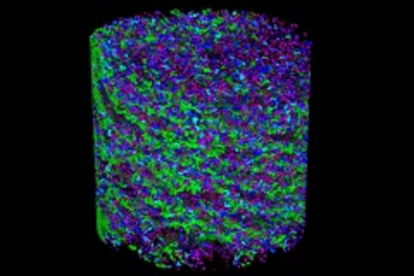
In order to understand limitations in current battery materials and systematically engineer better ones, it is helpful to be able to directly visualize the lithium dynamics in materials during battery charge and discharge. Researchers at ETH Zurich and Paul Scherrer Institute have demonstrated a way to do this.
Preserved Embryos Illustrate Seed Dormancy in Early Angiosperms
The discovery of exceptionally well-preserved, tiny fossil seeds dating back to the Early Cretaceous corroborates that flowering plants were small opportunistic colonizers at that time, according to a new Yale-led study.
Multiresolution X-ray tomography, getting a clear view of the interior
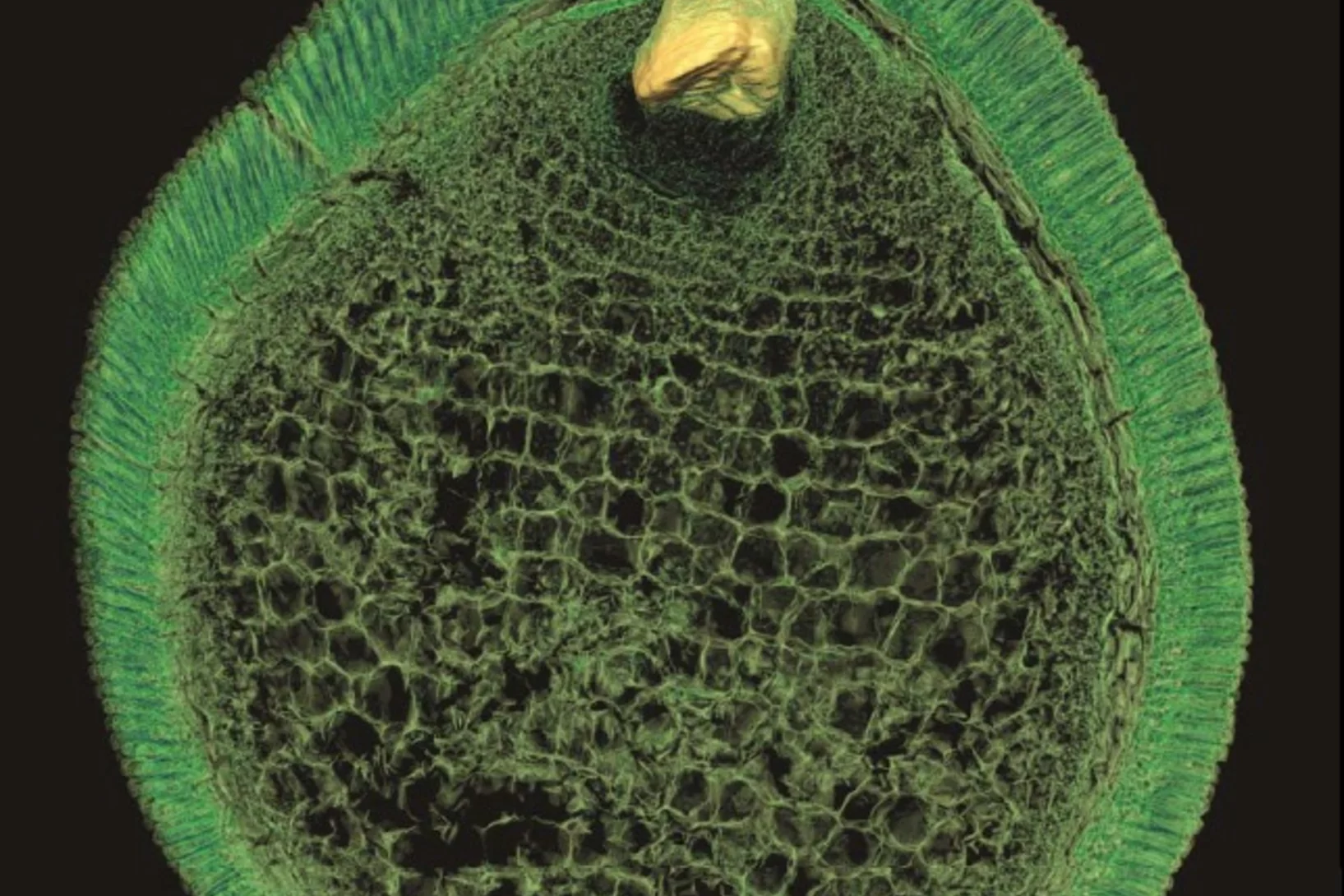
Researchers at PSI have developed a technique that combines tomography measurements at different resolution levels to allow quantitative interpretation for nanoscale tomography on an interior region of interest of the sample. In collaboration with researchers of the institute AMOLF in the Netherlands and ETH Zurich in Switzerland they showcase their technique by studying the porous structure within a section of an avian eggshell. The detailed measurements of the interior of the sample allowed the researchers to quantify the ordering and distribution of an intricate network of pores within the shell.
Schon frühe Säugetiere waren beim Essen wählerisch
Aktuellste Untersuchungen an Fossilien winziger Säugetiere aus dem südlichen Wales liefern neue Erkenntnisse über den Speisezettel unserer frühesten Vorfahren. Dabei stellte sich heraus, dass à anders als bislang vermutet - schon bei den frühesten Säugetieren verschiedene Arten auf verschiedene Nahrung spezialisiert waren. Darüber berichten Forschende Forscher der Universitäten Bristol und Leicester in der neuesten Ausgabe des Fachjournals Nature. Ihre Erkenntnisse beruhen auf Untersuchungen, die mithilfe von Röntgentomografie an der Synchrotron Lichtquelle Schweiz SLS des Paul Scherrer Instituts durchgeführt wurden. Die Ergebnisse helfen zu verstehen, wie Eigenschaften entstanden sind, die heutige Säugetiere auszeichnen.
Phasenkontrast verbessert Mammografie
Mithilfe des Phasenkontrast-Röntgens ist es Forschenden der ETH Zürich, des Paul Scherrer Instituts PSI und des Kantonsspitals Baden gelungen, Mammografien zu erstellen, anhand derer Brustkrebs und dessen Vorstufen präziser beurteilt werden können. Das Verfahren könnte dazu beitragen, Biopsien gezielter einzusetzen und Nachfolgeuntersuchungen zu verbessern.
3-D-Film zeigt das Innere fliegender Insekten
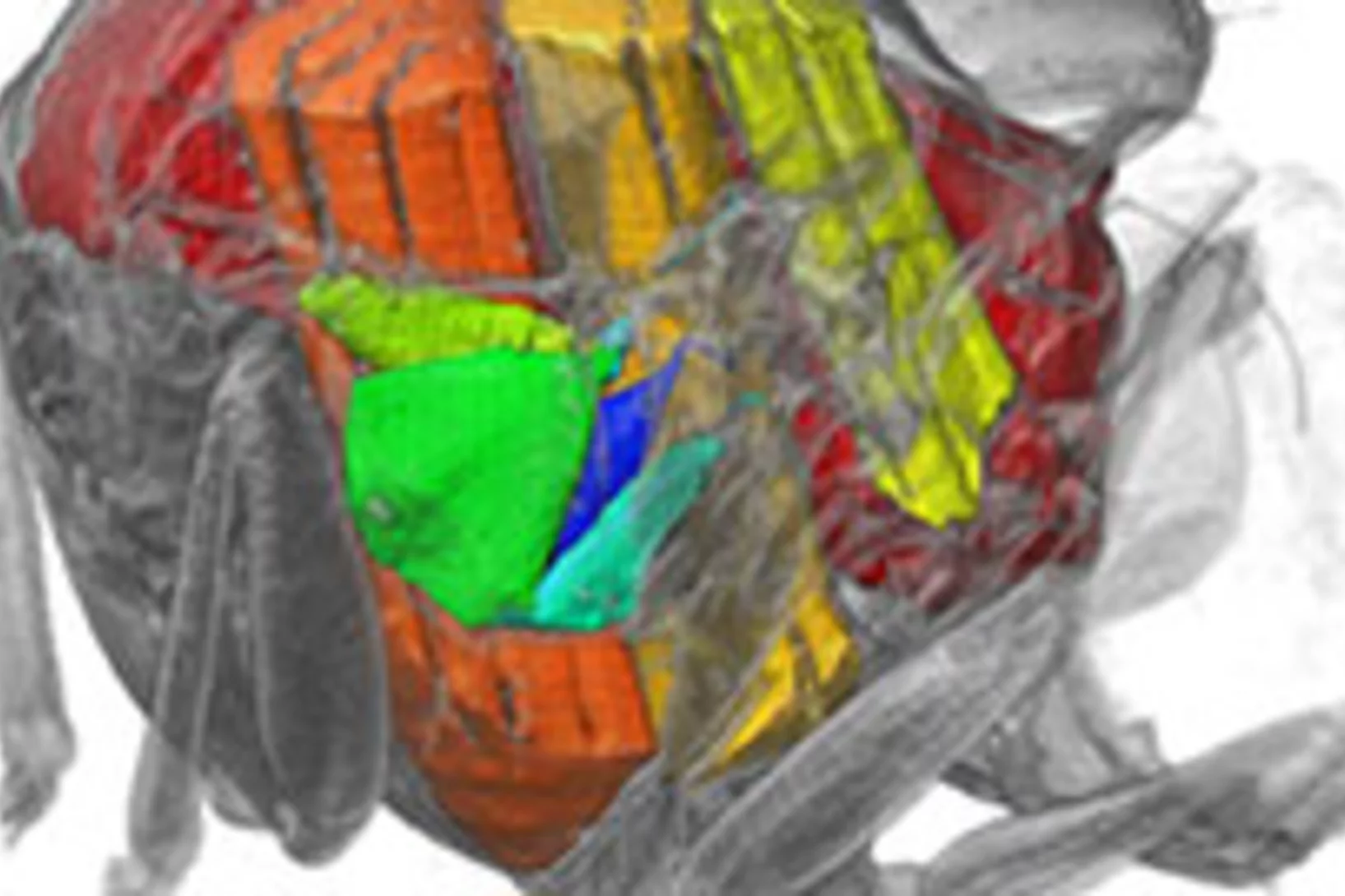
Mithilfe von Röntgenlicht aus der SLS konnten Hochgeschwindigkeitsaufnahmen der Flugmuskeln von Fliegen in 3-D erstellt werden. Ein Team von Wissenschaftlern der Oxford University, des Imperial Colleges (London) und des Paul Scherrer Instituts PSI entwickelte ein bahnbrechendes, neuartiges CT-Aufnahmeverfahren, mit dem sie das Innere von fliegenden Insekten filmen konnten. Die Filme gewähren einen Einblick in die Tiefe eines der komplexesten Mechanismen der Natur und zeigen, dass Strukturverformungen entscheidend dafür sind, wie eine Fliege ihren Flügelschlag steuert.
Neues Diagnoseverfahren bei Brustkrebs vielversprechend
Ein neues Mammografie-Verfahren könnte laut einer soeben veröffentlichten Studie einen deutlichen Mehrwert für die Diagnose von Brustkrebs in der medizinischen Praxis bringen. Die am PSI in Zusammenarbeit mit dem Brustzentrum des Kantonsspitals Baden und dem Industriepartner Philips entwickelte Methode macht bereits kleinste Gewebeveränderungen besser sichtbar. Dies könnte potenziell die Früherkennung von Brustkrebs verbessern. Zukünftig sollen Studien an Frauen mit einer Brustkrebserkrankung den Mehrwert der Methode endgültig belegen.
Mit Röntgenstrahlen der Lebensdauer von Lithium-Ionen-Akkus auf der Spur
Mithilfe von Röntgen-Tomographie haben Forschende die Vorgänge in Materialien von Batterie-Elektroden detailliert untersuchen können. Anhand hochaufgelöster 3D-Filme zeigen sie auf, weshalb die Lebensdauer der Energiespeicher begrenzt ist.
Die Ursprünge der ersten Fische mit Zähnen
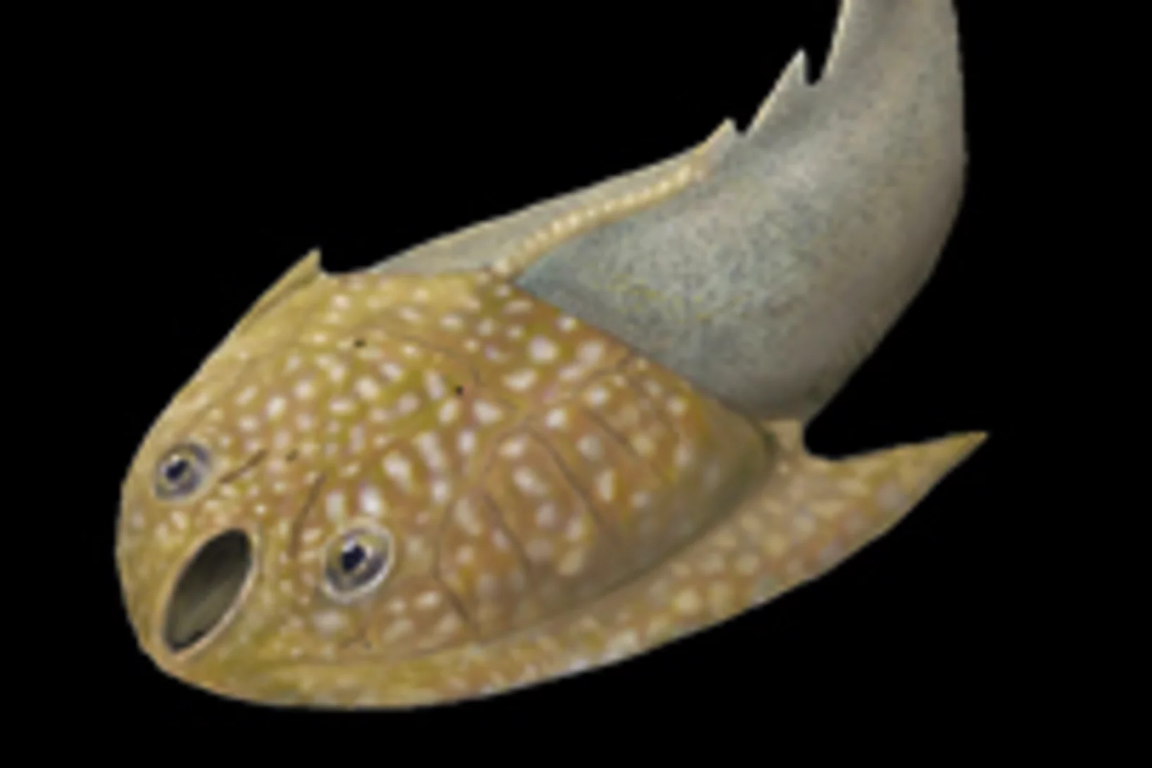
Mit Hilfe von Röntgenlicht aus der Synchrotron Lichtquelle Schweiz des PSI ist es Paläontologen der Universität Bristol gelungen, ein Rätsel um den Ursprung der ersten Wirbeltiere mit harten Körperteilen zu lösen. Sie haben gezeigt, dass die Zähne altertümlicher Fische (der sogenannten Conodonten) unabhängig von den Zähnen und Kiefern heutiger Wirbeltiere entstanden sind. Die Zähne dieser Wirbeltiere haben sich vielmehr aus einem Panzer entwickelt, der dem Schutz vor den Conodonten, den ersten Raubtieren, diente.
Alles im Fluss – Neue Einblicke in das Verhalten von Fluiden in porösem Gestein
Computertomografische Untersuchungen erlauben es erstmals, den Fluss von Öl und Wasser direkt im Gestein zu beobachten à in 3D und mit bisher unerreichter zeitlicher Auflösung. Der neue Ansatz und die daraus gewonnenen Informationen helfen besser zu verstehen, wie sich verschiedene Flüssigkeiten gegenseitig in porösen Materialien verdrängen können.
Der evolutionäre Ursprung unserer Zähne
Bislang war umstritten ob die frühesten Wirbeltiere, die Kiefer hatten, schon Zähne besassen oder nicht. Nun hat ein international zusammengesetztes Forschungsteam gezeigt, dass der urzeitliche Fisch Compagopiscis bereits Zähne hatte. Das deutet darauf hin, dass Zähne in der Evolution gemeinsam mit den Kiefern entstanden sind à oder zumindest kurz danach. Federführend bei dem Projekt waren Forscher der Universität Bristol (England), die entscheidenden Untersuchungen sind an der SLS durchgeführt worden.
Röntgenlicht liefert Einblicke in die Ursachen von Vulkanausbrüchen
Experimente am Paul Scherrer Institut bieten Einblicke in Vorgänge in vulkanischen Materialien, die darüber entscheiden wie heftig ein Vulkan ausbricht. Dabei haben Forschende ein Stück vulkanisches Material so aufgeheizt, dass darin Bedingungen entstanden, wie sie am Beginn eines Vulkanausbruchs herrschen. Sie nutzten dann Röntgenlicht aus der SLS, um in Echtzeit zu verfolgen, was in dem Gestein passiert, während es schmilzt.
ERC Grant for the development of a new imaging method with high potential clinical impact
Marco Stampanoni, Assistant Professor for X-ray microscopy at the ETH Zürich and Head of the 'X-ray Tomography Group' of the SLS has been recently awarded one of the coveted European Research Council (ERC) Starting Grant for the project PhaseX: 'Phase contrast X-ray imaging for medicine'. Marco Stampanoni's project will be supported by the ERC with 1.5 million euros for the next 5 years. The highly competitive ERC Starting Grants are reserved for outstanding young research talents.
Alzheimer-Plaques in 3D
Forschern ist es gelungen, dreidimensionale Aufnahmen der räumlichen Verteilung von Amyloid-Plaques in Gehirnen von an Alzheimer erkrankten Mäusen zu erstellen. In diesen Untersuchungen kam ein neues Verfahren zur Anwendung. Es handelt sich um eine ausserordentlich präzise Methode, die zu einem besseren Verständnis dieser Erkrankung beitragen kann. Die Wissenschaftler hoffen, dass dieses Verfahren künftig die Grundlage einer neuen zuverlässigen Diagnose bilden wird. Die Ergebnisse wurden gemeinsam von zwei Forscherteams à einem am Paul Scherrer Instituts (PSI) und der ETH Zürich und einem an der ETH Lausanne (EPFL) à erzielt.
Fossile Vorläufer der ersten Tiere
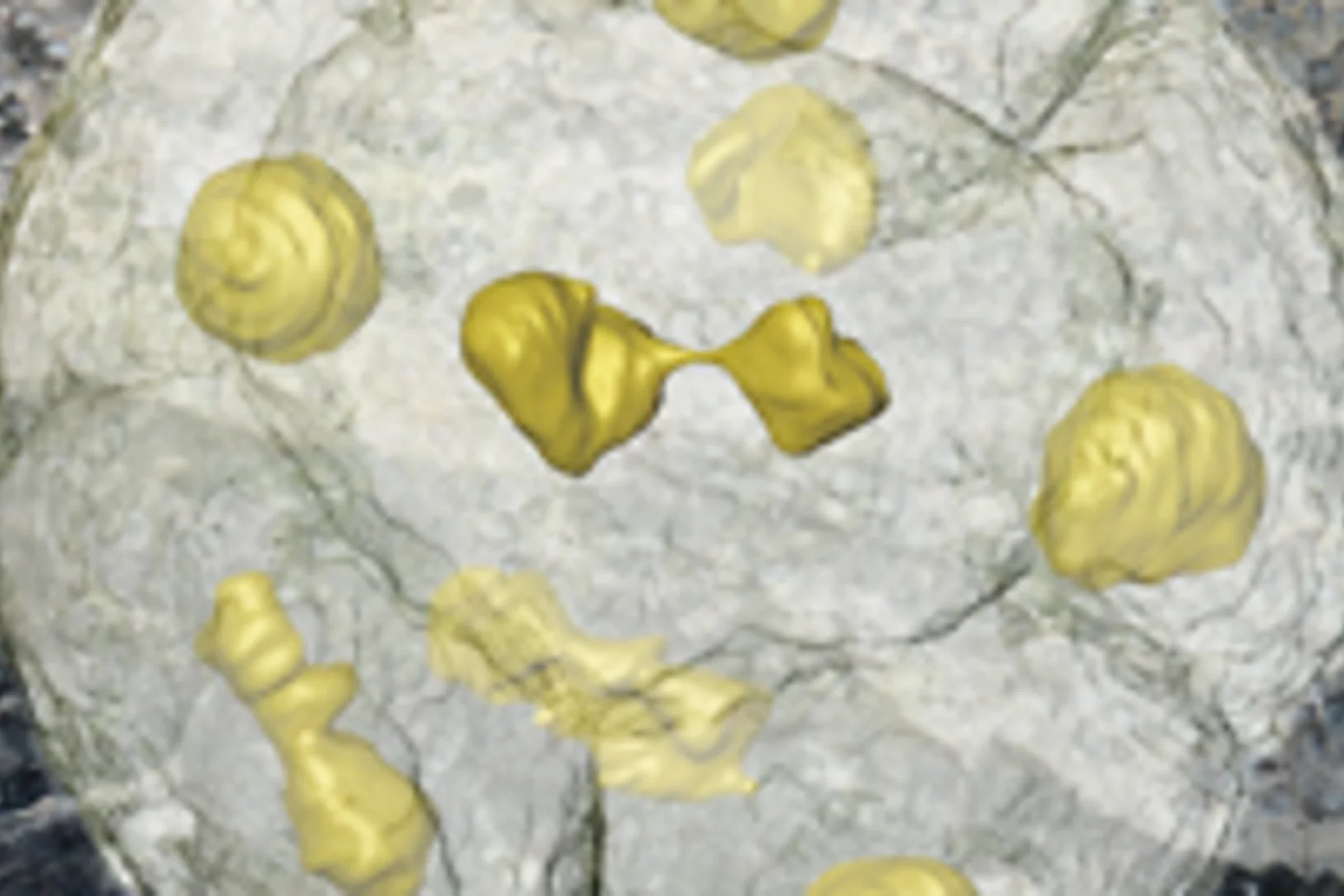
Einzellige Organismen, die vor über einer halben Milliarde Jahre gelebt haben und deren Fossilien in China gefunden wurden, sind wohl die unmittelbaren Vorläufer der frühesten Tiere. Die amöbenartigen Einzeller haben sich in einer Weise in zwei, vier, acht usw. Zellen geteilt, wie es heute tierische (und menschliche) Embryonen tun. Die Forscher glauben, dass diese Organismen einem der ersten Schritte vom Einzeller zum Vielzeller in der Entwicklung richtiger Tiere entsprechen.
Unseren frühen Vorfahren in den Kopf (und in die Nase) geschaut
Der Umbau des Gehirns und der Sinnesorgane dürfte den Erfolg der Wirbeltiere, eines der grossen Rätsel der Evolutionsbiologie, erklären à so die Aussage einer Arbeit, die heute im Wissenschaftsjournal Nature erschienen ist. Die Forschenden konnten das Rätsel durch Untersuchungen des Gehirns eines 400 Millionen Jahre alten versteinerten Fisches à eines evolutionären Bindeglieds zwischen den heute lebenden kiefertragenden Wirbeltieren und den Kieferlosen.
Neue Methode für die Krebserkennung mit Brustgewebe erprobt
Das Paul Scherrer Institut PSI hat eine neue Methode zur Diagnose von Brustkrebs entwickelt und nun zusammen mit dem Kantonsspital Baden AG erstmals an nicht-konserviertem, menschlichem Gewebe erprobt. Dabei wurde erkannt, dass es mit der neuen Methode möglich sein sollte, Strukturen sichtbar zu machen, die mit der herkömmlichen Mammografie nicht abgebildet werden. Wissenschaftler der Forschungsabteilung des Unternehmens Philips untersuchen derzeit auf Grundlage des vorgestellten Verfahrens den Einsatz in der medizinischen Praxis.
Universelles Gesetz für Veränderungen in Werkstoffen gefunden
In vielen wichtigen Werkstoffen findet man mehrere Phasen. Wird ein solcher Werkstoff erwärmt, können Atome von der einen Phase zur anderen wandern, so dass sich die Verteilung der Phasen ändert à und damit oft die Eigenschaften des Werkstoffs. Nun haben Forschende für einen wichtigen Fall einer solchen Veränderung gezeigt, dass es eine universelle Gesetzmässigkeit gibt, die den Vorgang beschreibt. Und zwar für alle Werkstoffklassen.
Neues Röntgenverfahren unterscheidet, was bisher gleich aussah
Auf Bildern, die mit Phasenkontrastverfahren erzeugt werden, kann man Gewebe unterscheiden, das auf gewöhnlichen Röntgenbildern fast gleich aussieht: etwa Muskeln, Knorpel, Sehnen oder Weichteiltumore. Forschende des Paul Scherrer Instituts und der Chinesischen Akademie der Wissenschaften haben das Verfahren so weiterentwickelt, dass es in Zukunft einfacher zu handhaben sein wird. Das könnte helfen, Tumore zu erkennen oder gefährliche Gegenstände im Gepäck sichtbar zu machen.

