Call for expressions of interest: Beamline partners at the SLS for PX II and PX III
We invite companies and institutions to secure access to the beamlines X10SA/PX II and X06DA/PX III through a long term contract.
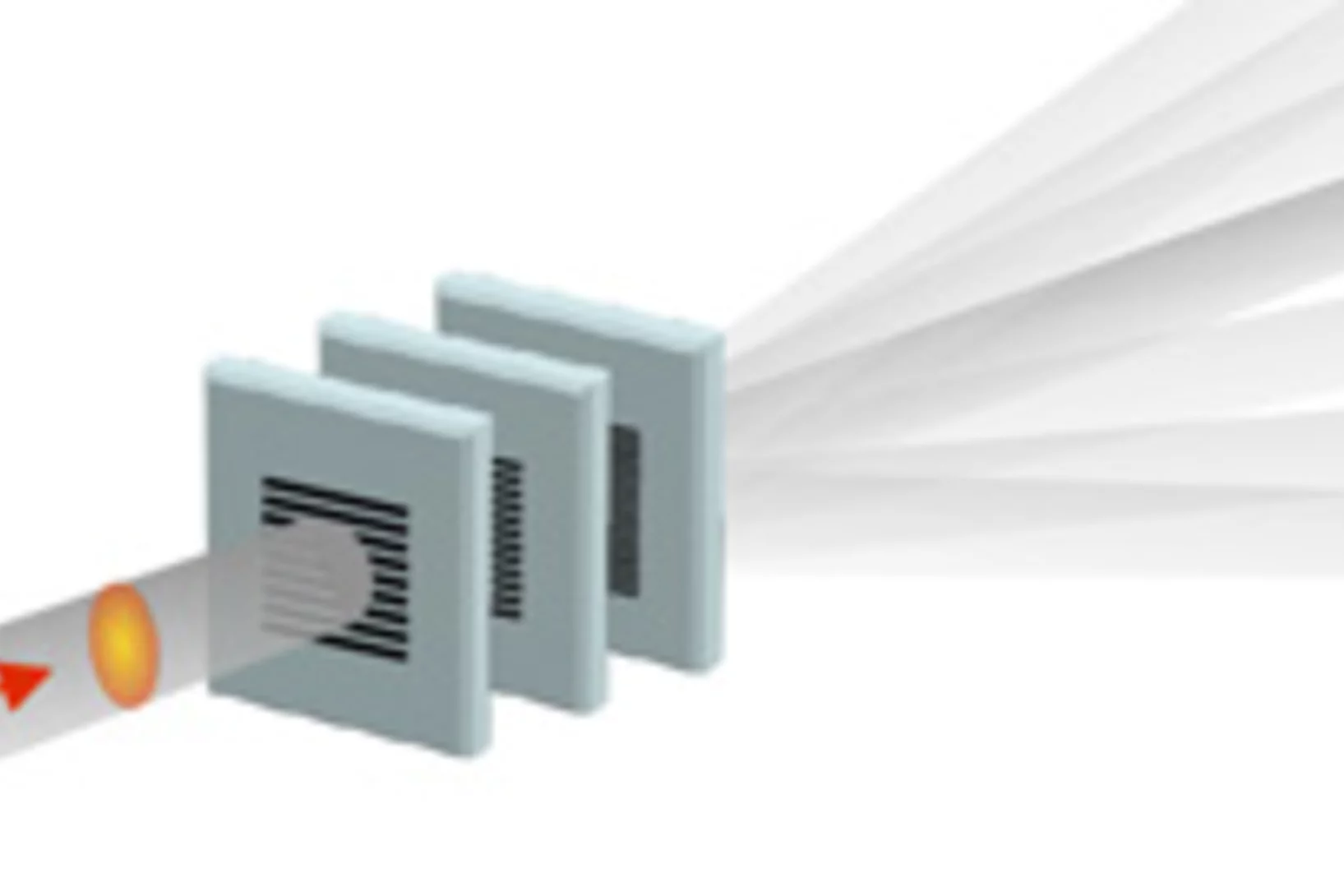
Single shot grating interferometry demonstrated using direct conversion detection
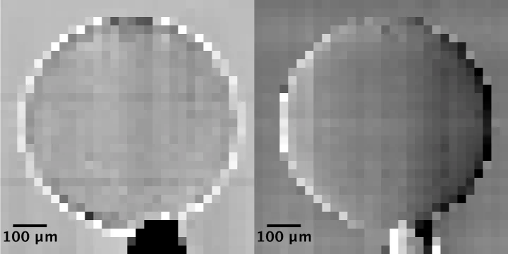
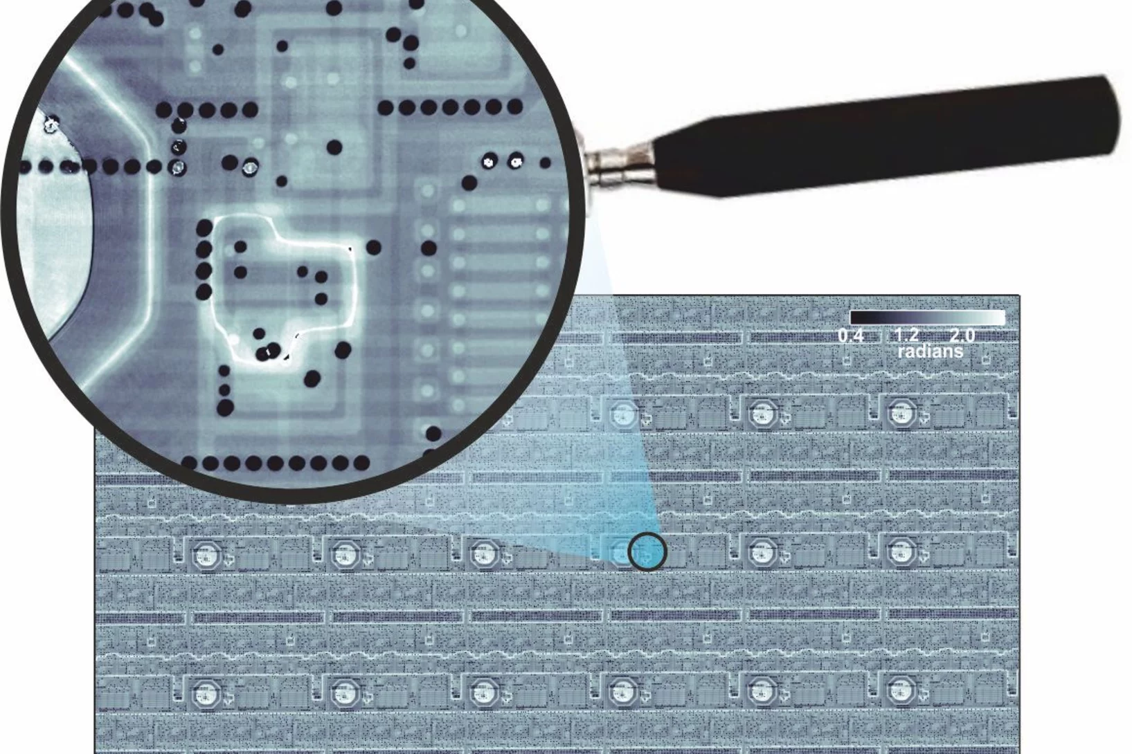
Researchers at the Paul Scherrer Institute's Swiss Light Source in Villigen, Switzerland, have developed an X-ray grating interferometry setup which does not require an analyzer grating, by directly detecting the fringes generated by the phase grating with a high resolution detector. The 25um pitch GOTTHARD microstrip detector utilizes a direct conversion sensor in which the charge generated from a single absorbed photon is collected by more than one channel. Therefore it is possible to interpolate to achieve a position resolution finer than the strip pitch.
Expérience dans une goutte en lévitation
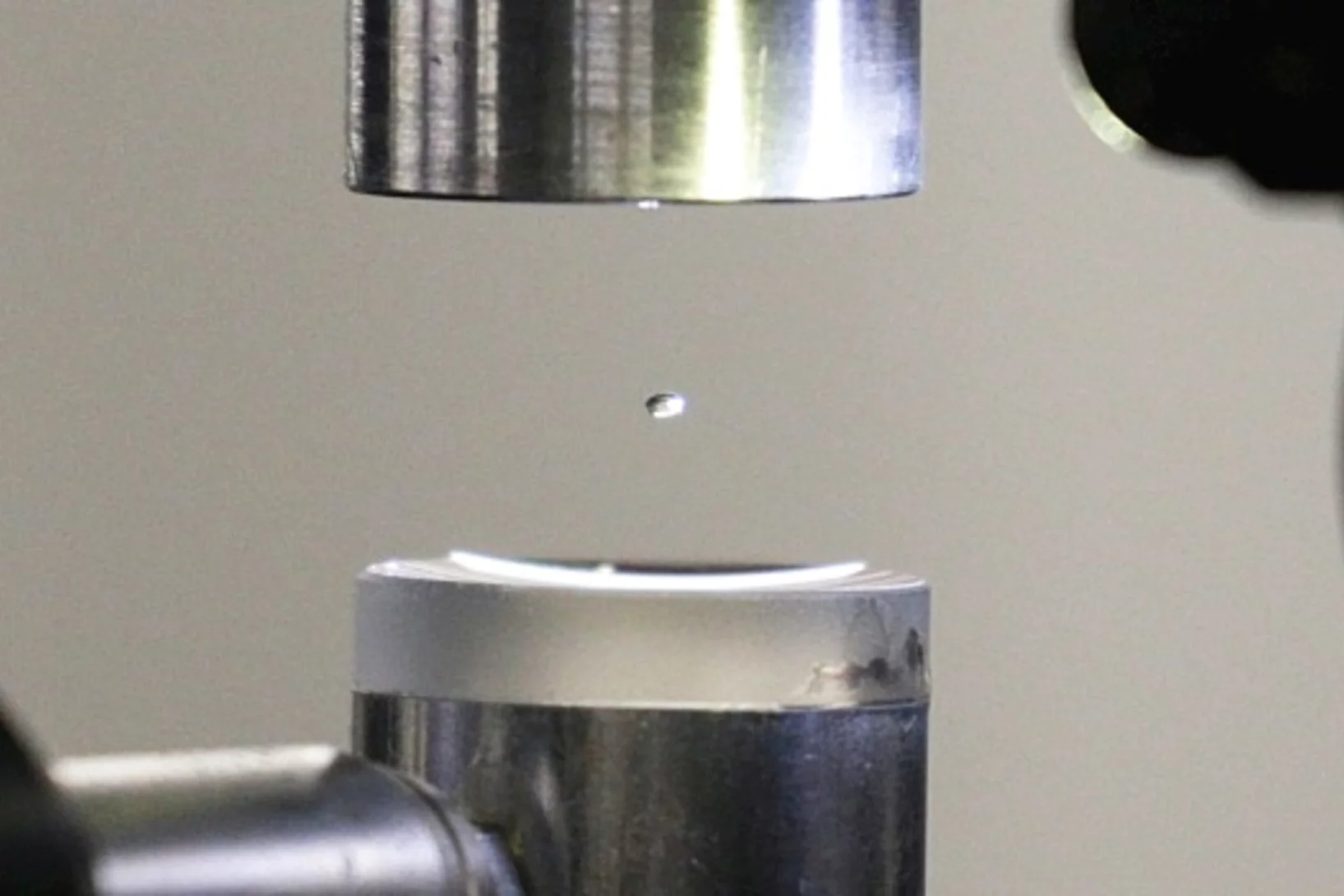
La structure exacte des protéines est normalement déterminée au PSI par la technique de diffraction des rayons X. Deux scientifiques du PSI viennent de l’améliorer de façon astucieuse: au lieu d’immobiliser les protéines, ils les ont étudiées dans une goutte de liquide en lévitation.
How does food look like on the nanoscale?
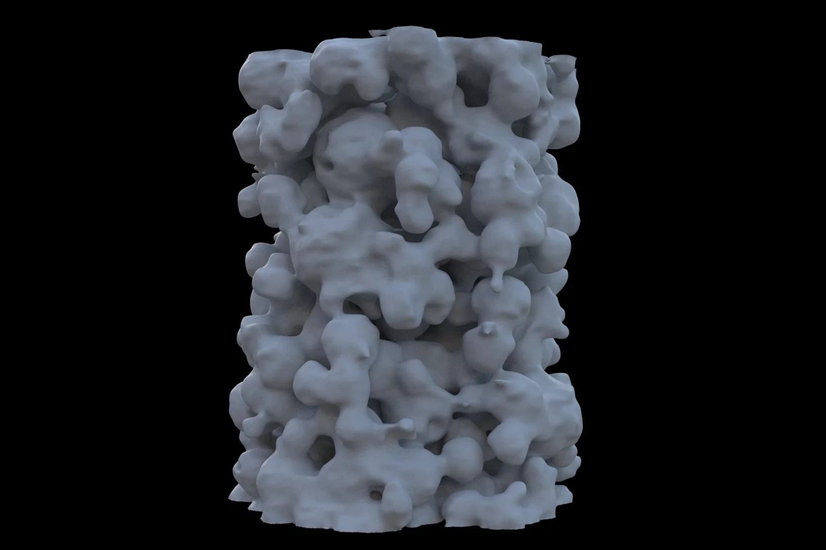
The answer to this question could save food industry a lot of money and reduce food waste caused by faulty production. Researchers from the University of Copenhagen and the Paul Scherrer Institut have obtained a 3D image of food on the nanoscale using ptychographic X-ray computed tomography. This work paves the way towards a more detailed knowledge of the structure of complex food systems.
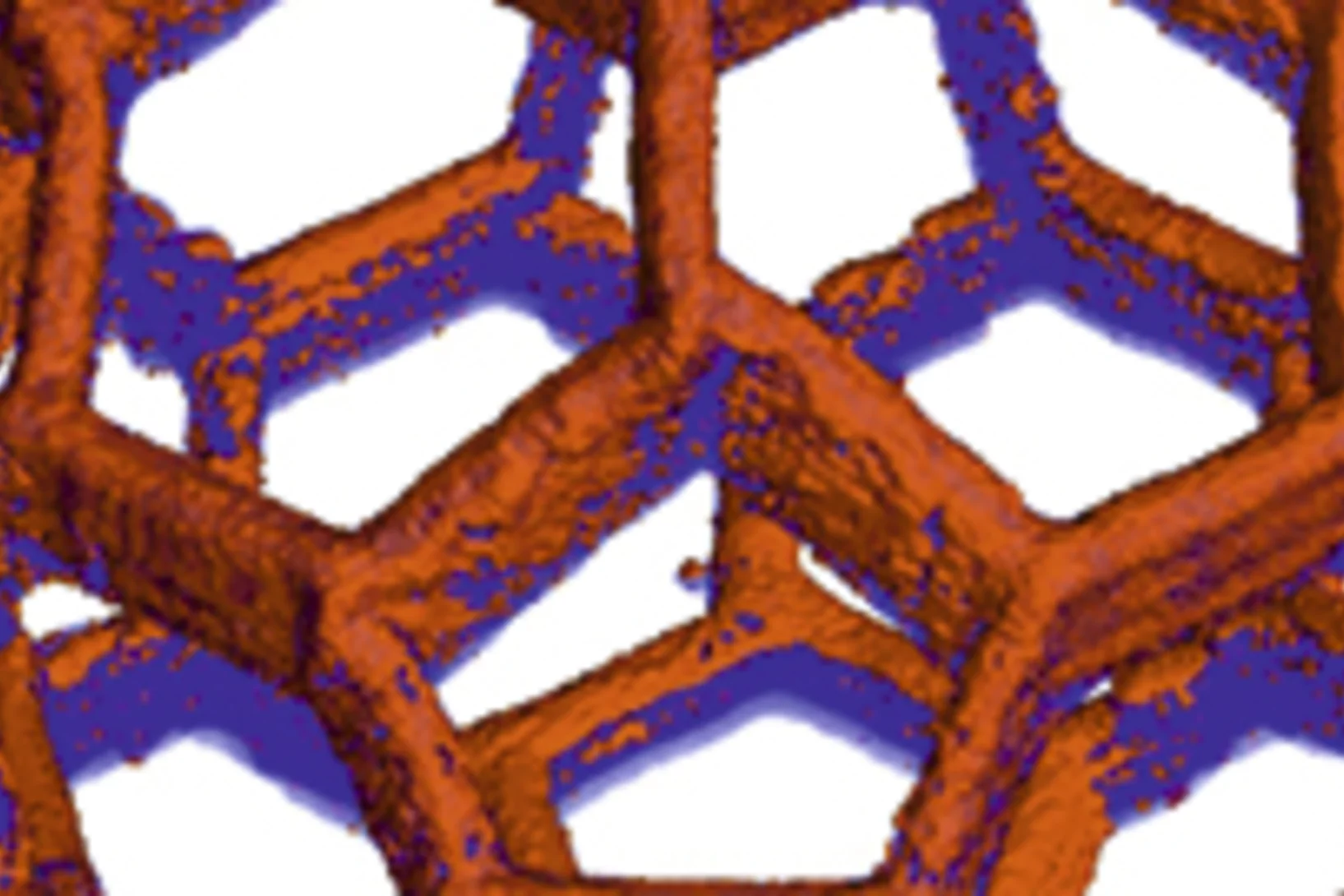
Watching lithium move in battery materials
In order to understand limitations in current battery materials and systematically engineer better ones, it is helpful to be able to directly visualize the lithium dynamics in materials during battery charge and discharge. Researchers at ETH Zurich and Paul Scherrer Institute have demonstrated a way to do this.
Mass density distribution of intact cell ultrastructure
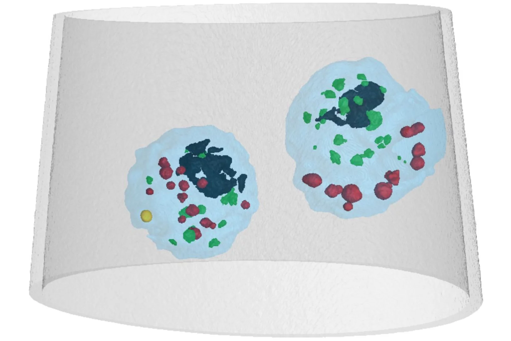
The determination of the mass density of cellular compartments is one of the many analytical tools that biologists need to unravel the extremely complex structure of biological systems. Cryo X-ray nanotomography reveals absolute mass density maps of frozen hydrated cells in three dimensions.
Preserved Embryos Illustrate Seed Dormancy in Early Angiosperms
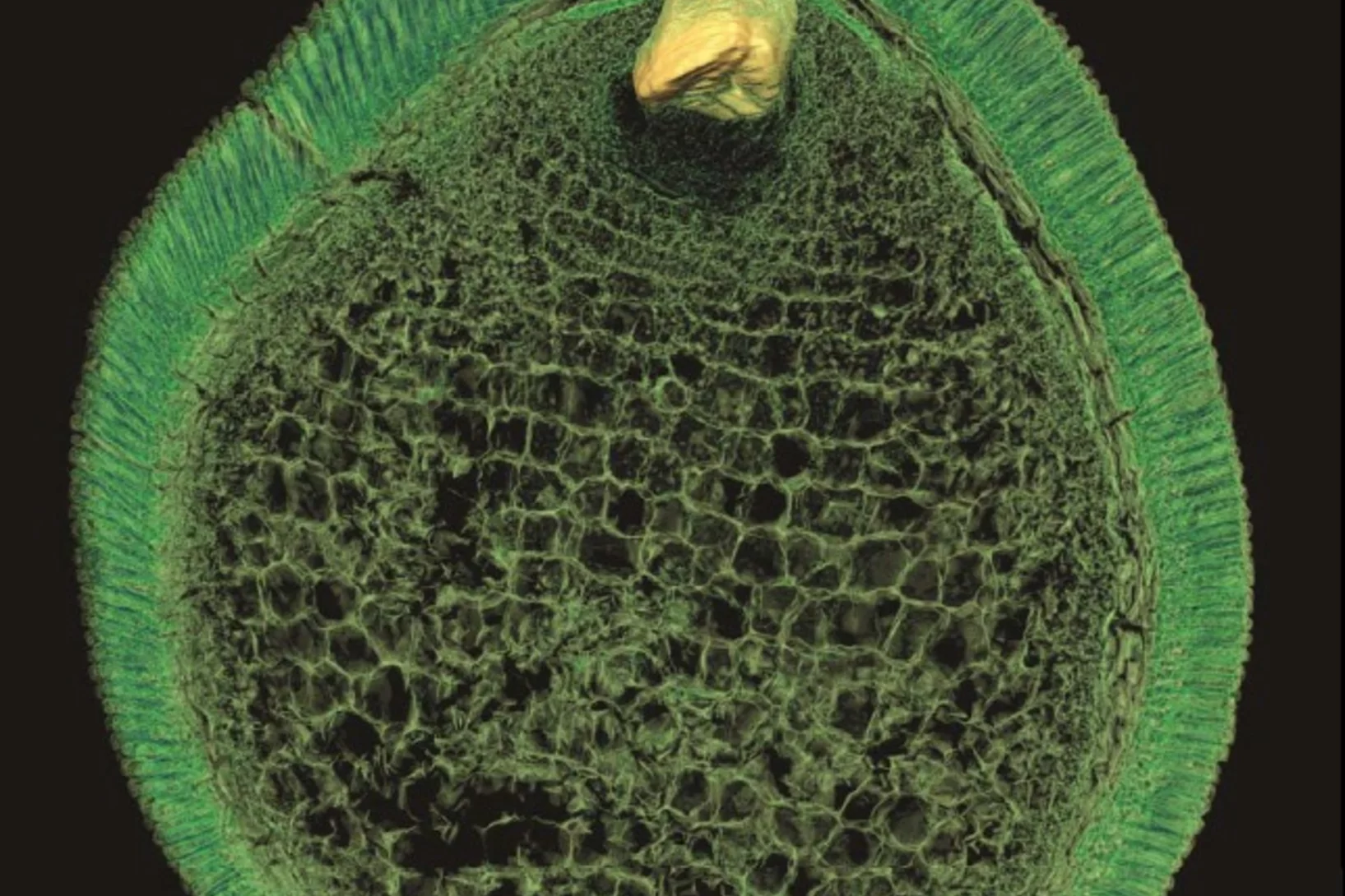
The discovery of exceptionally well-preserved, tiny fossil seeds dating back to the Early Cretaceous corroborates that flowering plants were small opportunistic colonizers at that time, according to a new Yale-led study.
First EIGER X 16M in operation at the Swiss Light Source
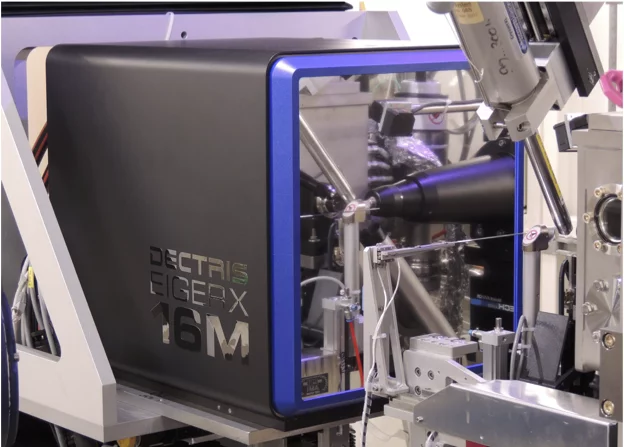
The macromolecular crystallography beamline X06SA at the Swiss Light Source, a synchrotron operated by Paul Scherrer Institute, is the first one in the world to upgrade its detector to an EIGER X 16M.
La nanostructure d’un os dévoilée en 3D
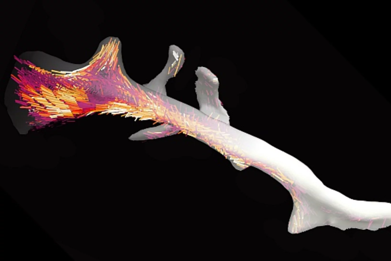
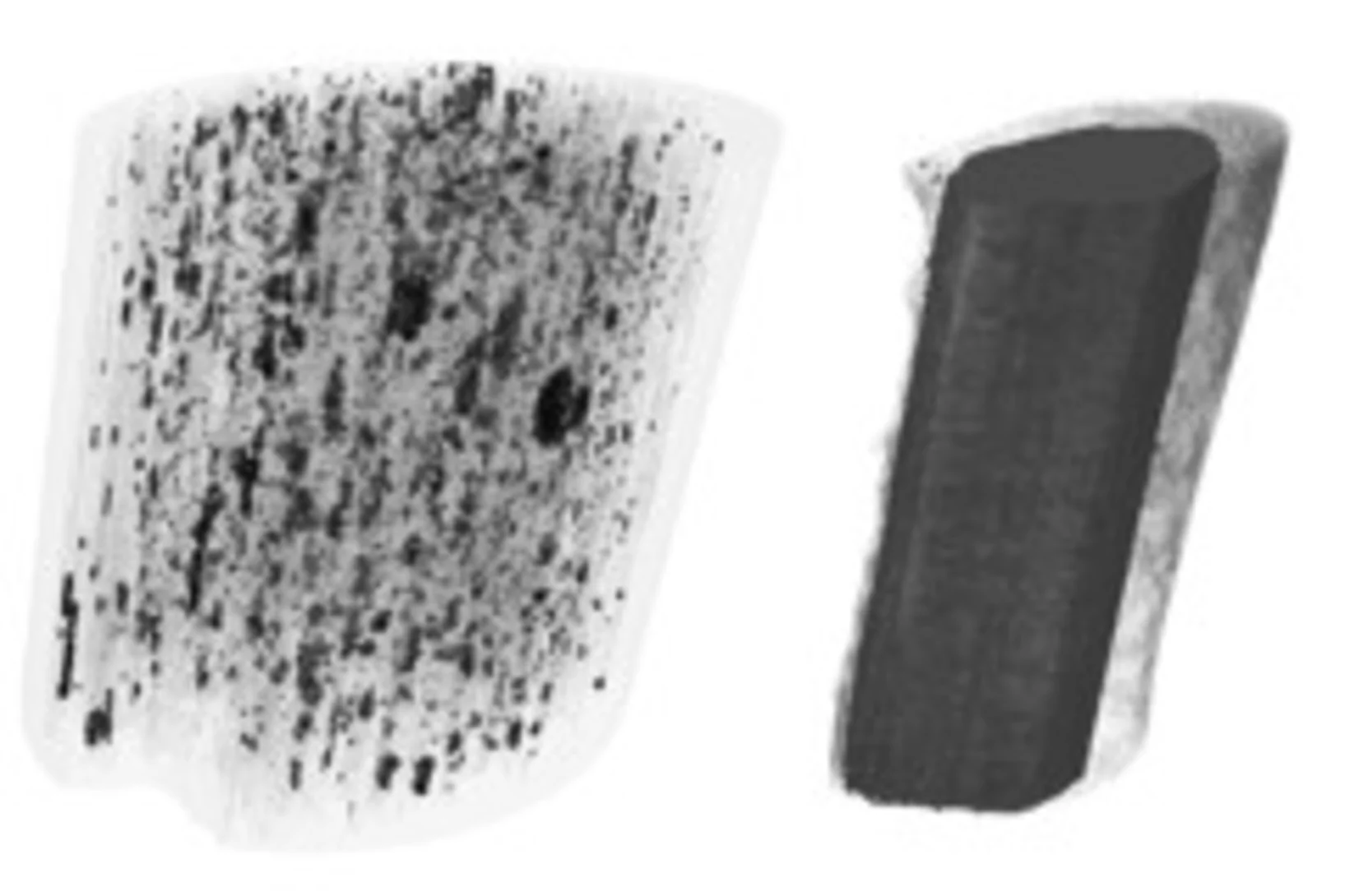
Les os sont composés de minuscules fibres, à peu près mille fois plus fines qu’un cheveu humain. Avec un nouveau type de méthode d’analyse informatique des chercheurs de l'Institut Paul Scherrer PSI étaient en mesure de déterminer pour la première fois l’agencement local et l’orientation de la nanostructure à l’intérieur d’un fragment d’os.
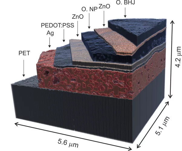
X-ray nanotomography aids the production of eco-friendly solar cells
Polymer solar cells are in the spotlight for sustainable energy production of the future. Characterization of these devices by X-ray nanotomography helps to improve their production using environmentally friendly materials.
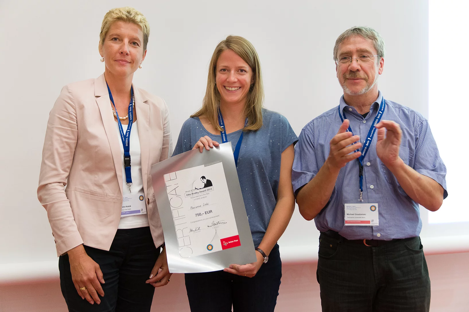
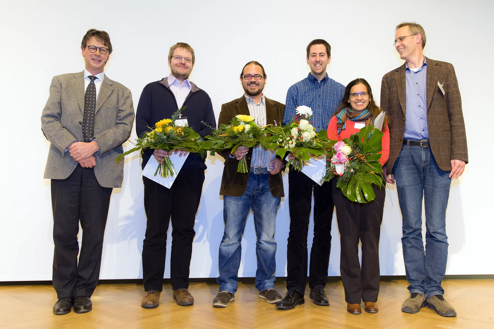
2015 Otto Kratky award
Marianne Liebi was awarded the 2015 Otto Kratky award by the Helmholtz-Centre Berlin for excellence in the field of small-angle X-ray scattering (SAXS) analysis. The award was bestowed in the last SAS2015 conference in Berlin. Marianne is a postdoctoral fellow in the coherent X-ray scattering group (CXS) in PSI, carrying out research in scanning SAXS measurement and analysis in 2D and 3D. Image credit ©HZB/Michael Setzpfandt
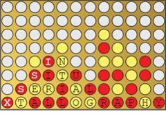
In Situ Serial Crystallography Workshop at the SLS
The Macromolecular Crystallography group at SLS is organizing a three days workshop on in situ serial crystallography (http://indico.psi.ch/event/issx) between November 17 and 19, 2015. It will be dedicated in the presentation of a novel method facilitating the structure determination of membrane proteins, which are highly important pharmaceutical targets but are difficult to handle using 'classical' crystallographic tools. Designed for 20 Ph.D. students, postdocs and young scientists from both academia and industry, the workshop will consist of introductory lectures, followed by hands-on practicals on in meso or lipidic cubic phase (LCP) crystallization, on in situ serial crystallography data collection using a micro-sized beam and on data processing.
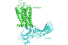
New insight into receptor signalling
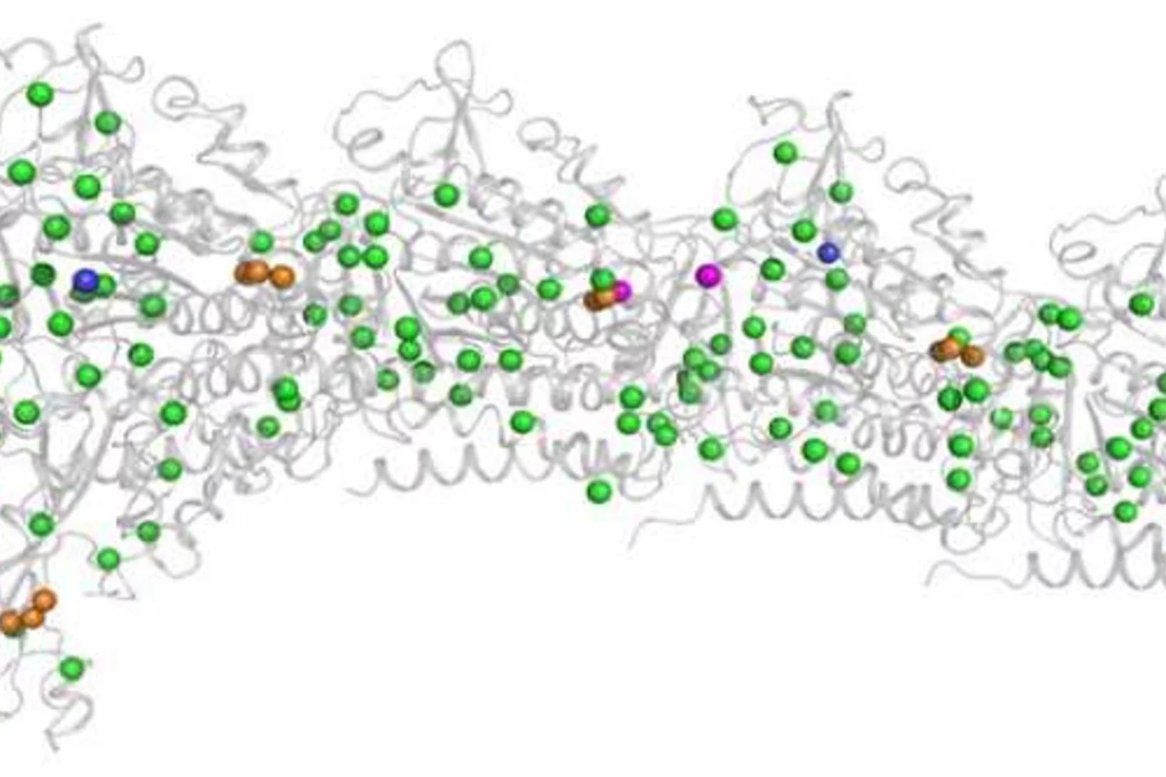
A team of 72 investigators across 25 institutions including researchers from the Paul Scherrer Institut obtained the X-ray structure of a rhodopsinàarrestin complex, which represents a major milestone in the area of G-protein-coupled-receptor (GPCR), a protein family recognized in the award of the 2012 Nobel Prize in Chemistry.
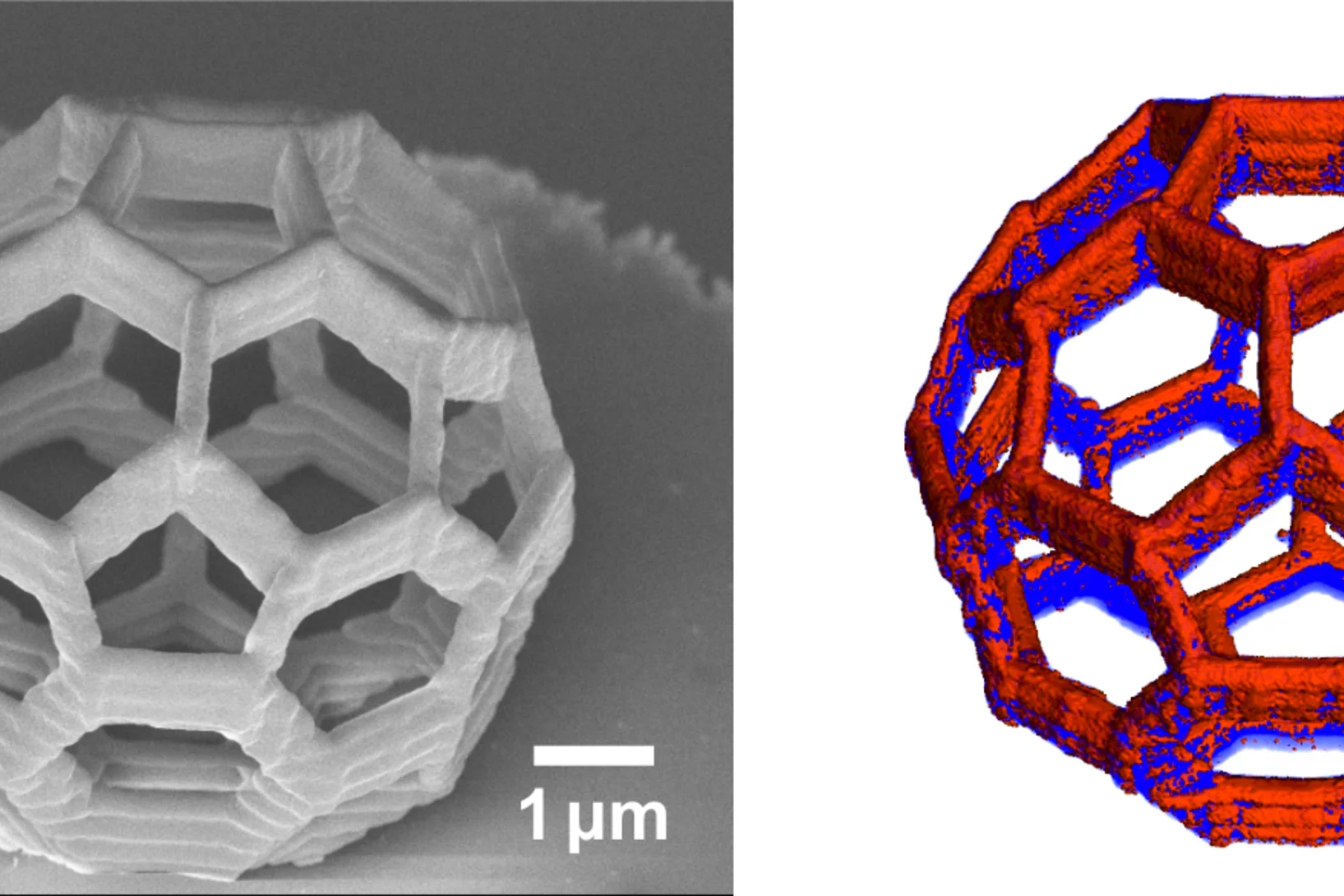
Element-Specific X-Ray Phase Tomography of 3D Structures at the Nanoscale
Recent advances in fabrication techniques to create mesoscopic 3D structures have led to significant developments in a variety of fields including biology, photonics, and magnetism. Further progress in these areas benefits from their full quantitative and structural characterization.
L’union fait la force
Décrypter les molécules au SwissFEL et à la SLSLes protéines sont un objet de recherche convoité, mais récalcitrant. Leur étude est aujourd’hui facilitée par une nouvelle méthode développée à l’aide d’un laser à rayons X à électrons libres comme le futur SwissFEL du PSI. Elle consiste à exposer à intervalles rapprochés de petits échantillons identiques de protéines à de la lumière de type rayons X. On contourne ainsi un problème majeur auquel la recherche sur les protéines s’est heurtée jusqu’ici: produire des échantillons de taille suffisante.
De l’intérieur d’une coquille d’œuf
La coquille d’un uf abrite de minuscules vésicules. Elles fournissent les substances qui stimulent et contrôlent la croissance de cette enveloppe solide. Grâce à une technique de tomographie novatrice, des chercheurs de l’Institut Paul Scherrer (PSI), de l’EPF Zurich et de l’Institut AMOLF aux Pays-Bas, ont réussi pour la première fois à obtenir une image en 3D de ces vésicules. Ils surmontent ainsi une limite à l’imagerie tomographique, et espèrent qu’un jour leur méthode profitera aussi à la médecine.
Multiresolution X-ray tomography, getting a clear view of the interior
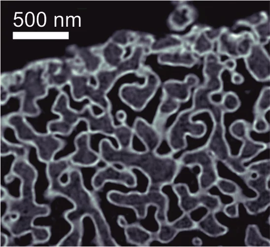
Researchers at PSI have developed a technique that combines tomography measurements at different resolution levels to allow quantitative interpretation for nanoscale tomography on an interior region of interest of the sample. In collaboration with researchers of the institute AMOLF in the Netherlands and ETH Zurich in Switzerland they showcase their technique by studying the porous structure within a section of an avian eggshell. The detailed measurements of the interior of the sample allowed the researchers to quantify the ordering and distribution of an intricate network of pores within the shell.
Fractionner une impulsion de rayons X pour visualiser des processus ultra rapides
Le laser à rayons X SwissFEL du PSI permettra de visualiser les différentes étapes de processus très rapides. Un nouveau procédé devrait rendre possibles des expériences encore plus précises : il consiste à fractionner chaque impulsion de rayons X, et à faire en sorte que chaque fraction de l’impulsion atteigne l’une après l’autre l’objet étudié. Le principe de ce processus rappelle celui de l’ancienne chronophotographie.
La 3D, au nanomètre près
Des chercheurs de l'Institut Paul Scherrer et de l'ETH Zurich ont créé des images en 3D de minuscules objets, et ont même réussi à visualiser au niveau de ces derniers des détails de 25 nanomètres (1 nanomètre = 1 million de millimètre). En plus de déterminer la forme de leurs objets d'étude, ils ont pu également mettre en évidence la façon dont un élément chimique donné (le cobalt) était réparti au sein de ces derniers, tout en étant capables d'établir si ce même élément était présent sous forme de liaison chimique ou sous forme pure.
Obtenir plus rapidement le portrait d’une protéine
Tous les êtres vivants, de la bactérie à l’être humain, ont besoin de protéines pour l’accomplissement de leurs fonctions vitales. La manière dont les protéines remplissent leurs tâches dépend de leur structure. Des chercheurs de l’Institut Paul Scherrer (PSI) ont développé une méthode novatrice, qui permet de déterminer plus rapidement la structure cristalline des protéines, grâce à de la lumière de type rayons X. Elle pourrait ainsi accélérer le développement de nouveaux médicaments. Leur étude sera publiée le 15 décembre dans la revue spécialisée « Nature Methods ».
Innovation Award on Synchrotron Radiation 2014 for high-resolution 3D hard X-ray microscopy
The 2014 Innovation Award on Synchrotron Radiation was bestowed to researchers Ana Diaz, Manuel Guizar-Sicairos, Mirko Holler, and Jörg Raabe from the Paul Scherrer Institut, Switzerland, for their contributions to method and instrumentation development, which have set new standards in high-resolution 3D hard X-ray microscopy.
Côté menu, les premiers mammifères avaient déjà leurs préférences
De toutes nouvelles analyses de petits mammifères fossiles livrent de nouveaux éléments sur la connaissance du menu de nos plus lointains ancêtres. Les résultats permettent de comprendre comment sont apparues certaines caractéristiques typiques des mammifères actuels. Ces conclusions reposent sur des analyses, qui ont été réalisées à la Source de Lumière Suisse (SLS) de l’Institut Paul Scherrer, au moyen de la tomographie par rayons X.
Fast scanning coherent X-ray imaging using Eiger
The smaller pixel size, high frame rate, and high dynamic range of next-generation photon counting pixel detectors expedites measurements based on coherent diffractive imaging (CDI). The latter comprises methods that exploit the coherence of X-ray synchrotron sources to replace imaging optics by reconstruction algorithms. Researchers from the Paul Scherrer Institut have recently demonstrated fast CDI image acquisition above 25,000 resolution elements per second using an in-house developed Eiger detector. This rate is state of the art for diffractive imaging and even on a par with the fastest scanning X-ray transmission instruments. High image throughput is of crucial importance for both materials and biological sciences for studies with representative population sampling.
Le contraste de phase améliore la mammographie
Grâce à l'imagerie par contraste de phase, des chercheurs de l’EPF Zurich, de l’Institut Paul Scherrer (PSI) et de l’hôpital cantonal de Baden ont réussi à obtenir des mammographies, grâce auxquelles il est possible de procéder à une évaluation plus précise des cancers et des précancers du sein. Ce procédé pourrait contribuer à une utilisation plus ciblée des biopsies, et à l’amélioration des examens de suivi.
Un film en 3D montre ce qui se passe à l’intérieur d’insectes en plein vol
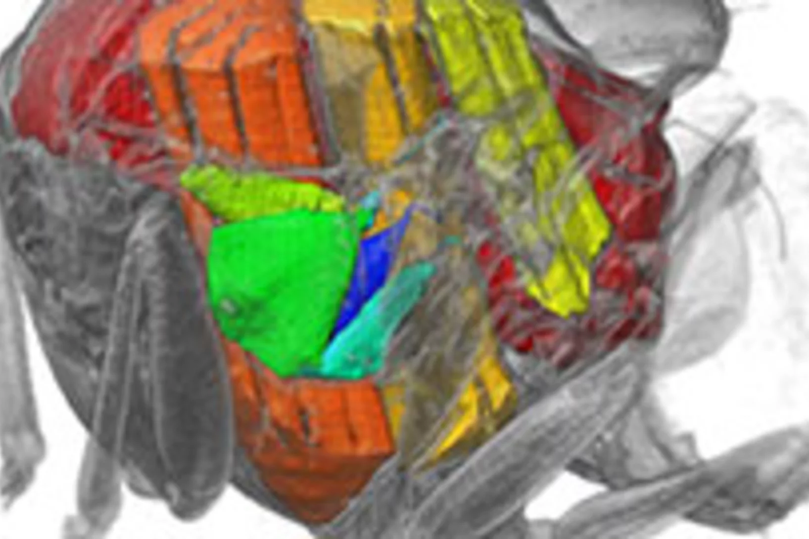
Grâce aux rayons X produits par la Source de Lumière Suisse SLS, des prises de vue inédites des muscles que les mouches utilisent pour voler (muscles alaires) ont pu être réalisées, à haute vitesse et en 3D. Une équipe de scientifiques de l’Université d’Oxford, de l’Imperial College de Londres et de l’Institut Paul Scherrer (PSI) a développé un procédé de prise de vue tomographique révolutionnaire. Grâce à lui, ils ont pu filmer ce qui se passe à l’intérieur d’insectes en plein vol. Ces films permettent de découvrir en profondeur l’un des mécanismes naturels les plus complexes, et montrent que les déformations structurelles sont la clé pour comprendre la manière dont la mouche contrôle son battement d’aile.
X-ray tomography reaches 16 nm isotropic 3D resolution
Researchers at PSI reported a demonstration of X-ray tomography with an unmatched isotropic 3D resolution of 16 nm in Scientific Reports. The measurement was performed at the cSAXS beamline at the Swiss Light Source using a prototype instrument of the OMNY (tOMography Nano crYo) project. Whereas this prototype measures at room temperature and atmospheric pressure, the OMNY system, to be commissioned later this year, will provide a cryogenic sample environment in ultra-high vacuum without compromising imaging capabilities. The researchers believe that such a combination of advanced imaging with state-of-the-art instrumentation is a promising path to fill the resolution gap between electron microscopy and X-ray imaging, also in case of radiation-sensitive materials such as polymer structures and biological systems.
Unique insight into carbon fibers on the nanoscale
The investigation of the mechanical properties of carbon fibers benefits from highly resolved three-dimensional density maps within representative volumes, but such images are not easily obtained with standard methods. Scientists from the Paul Scherrer Institut in collaboration with Honda R&D in Germany have recently visualized density distributions on the sub-micrometer scale within entire carbon fiber sections, revealing surprising graphite distributions within the fibers. This capability will prove useful for the systematic characterization of fibers, contributing to the development of light and robust materials at lower costs.
Neues Diagnoseverfahren bei Brustkrebs vielversprechend
Ein neues Mammografie-Verfahren könnte laut einer soeben veröffentlichten Studie einen deutlichen Mehrwert für die Diagnose von Brustkrebs in der medizinischen Praxis bringen. Die am PSI in Zusammenarbeit mit dem Brustzentrum des Kantonsspitals Baden und dem Industriepartner Philips entwickelte Methode macht bereits kleinste Gewebeveränderungen besser sichtbar. Dies könnte potenziell die Früherkennung von Brustkrebs verbessern. Zukünftig sollen Studien an Frauen mit einer Brustkrebserkrankung den Mehrwert der Methode endgültig belegen.Cette actualité n'existe qu'en anglais et allemand.
Mit Röntgenstrahlen der Lebensdauer von Lithium-Ionen-Akkus auf der Spur
Mithilfe von Röntgen-Tomographie haben Forschende die Vorgänge in Materialien von Batterie-Elektroden detailliert untersuchen können. Anhand hochaufgelöster 3D-Filme zeigen sie auf, weshalb die Lebensdauer der Energiespeicher begrenzt ist.Cette actualité n'existe qu'en anglais et allemand.
Die Ursprünge der ersten Fische mit Zähnen
Mit Hilfe von Röntgenlicht aus der Synchrotron Lichtquelle Schweiz des PSI ist es Paläontologen der Universität Bristol gelungen, ein Rätsel um den Ursprung der ersten Wirbeltiere mit harten Körperteilen zu lösen. Sie haben gezeigt, dass die Zähne altertümlicher Fische (der sogenannten Conodonten) unabhängig von den Zähnen und Kiefern heutiger Wirbeltiere entstanden sind. Die Zähne dieser Wirbeltiere haben sich vielmehr aus einem Panzer entwickelt, der dem Schutz vor den Conodonten, den ersten Raubtieren, diente.

