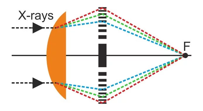
A team of scientists from the Paul Scherrer Institut, the University of Basel and DESY have demonstrated the first-ever realization of apochromatic X-ray focusing using a tailored combination of a refractive lens and a Fresnel zone plate. This innovative approach enables the correction of the chromatic aberration suffered by both refractive and diffractive lenses over a wide range of X-ray energies. This groundbreaking development in X-ray optics have been just published in the scientific journal Light: Science & Applications.
Diffractive and refractive lenses are widely used in X-ray analysis and high-resolution X-ray microscopy with many scientific applications in materials science, energy sciences and biology. Nevertheless, both types of X-ray optics suffer from a strong chromatic aberration, meaning that they focus different X-ray energies/wavelengths at different distances along the optical axis. As a result, many high-resolution X-ray analysis techniques can only be implemented with monochromatic X-ray beams, meaning that in many cases a large portion of the X-ray intensity is sacrificed.
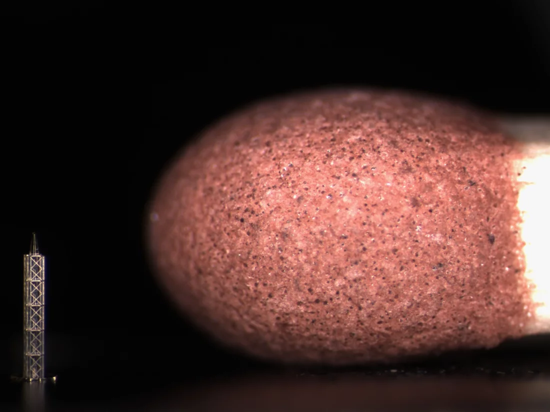
The apochromatic X-ray optic consisted of two independent optical elements separated by a distance: a divergent refractive X-ray lens produced by state-of-the-art 3D printing with submicrometer accuracy and a Fresnel zone plate fabricated by electron beam lithography and gold electroplating. These elements were produced in-house using the cleanrooms and nanofabrication facilities at Paul Scherrer Institut. The apochromatic X-ray optics was characterized by scanning transmission X-ray microscopy and ptychographic X-ray imaging. Sub-micrometer apochromatic focusing was achieved for an X-ray energy range from 7 to 12 keV. The X-ray measurements was carried out at beamline P06 at PETRA III, DESY (Hamburg, Germany).
In the future, apochromatic X-ray lenses could emerge a cost-effective and compact alternative to mirror-based systems, with the further advantage of being on-axis imaging optics. They are likely to play increasingly important role in the field of X-ray imaging and microscopy and for their scientific applications in both accelerator-based and laboratory X-ray sources.
This investigation received funding from the Europen Union’s Horizon 2020 research and innovation program under the Marie Sklodowska-Curie grant agreement No 884104 (PSI-FELLOW-III-3i) and from the Swiss Nanoscience Institute Argovia project 16.01 ACHROMATIX.
Contact
Dr. Joan Vila-Comamala
Laboratory for X-ray Nanoscience and Technologies
Paul Scherrer Institute
Forschungsstrasse 111, 5232 Villigen PSI, Switzerland
Telephone: +41 56 310 5133
e-mail: joan.vila-comamala@psi.ch
Original Publication
Apochromatic X-ray focusing
Umut T. Sanli, Griffin Rodgers, Marie-Christine Zdora, Qi Peng, Jan Garrevoet, Ken Vidar Falch, Bert Müller, Christian David and Joan Vila-Comamala
Light: Science & Applications (2023)
DOI: 10.1038/s41377-023-01157-8


