Welcome to the news archive for the TOMCAT beamline!
The complete news archive for the X-ray Tomography Group can be found here.
News
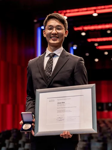
Jisoo Kim receives PSI Thesis Medal 2023
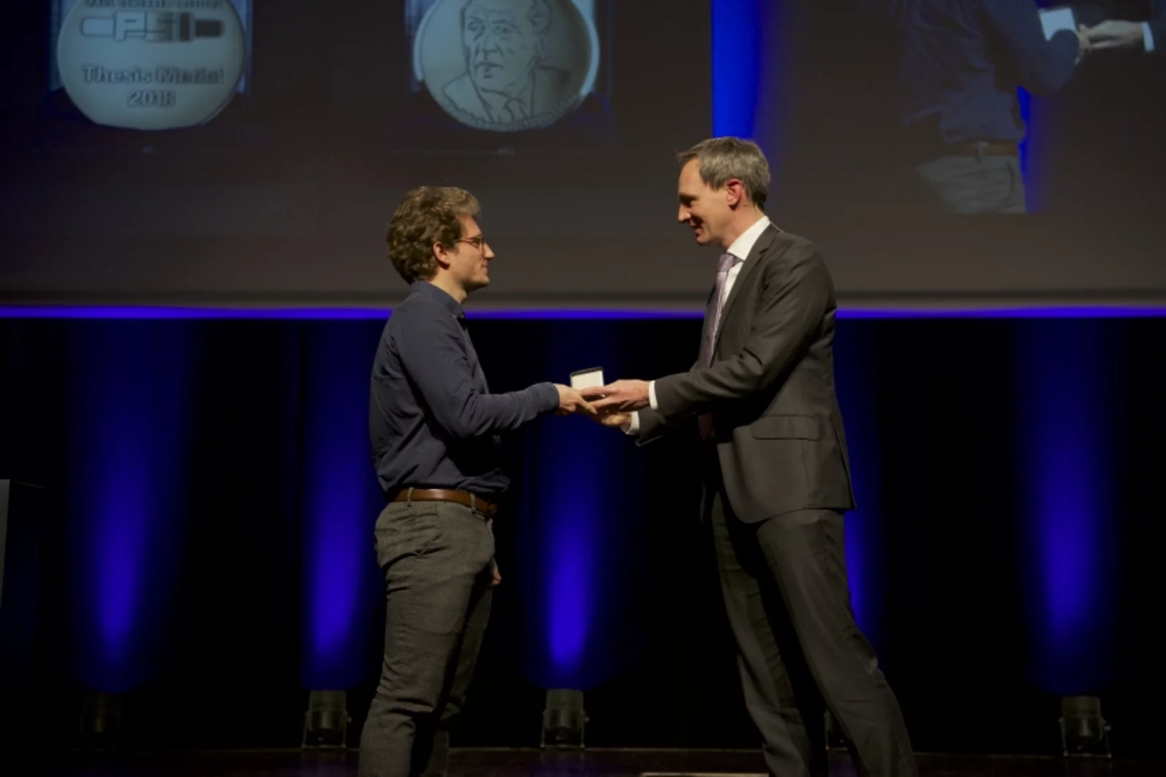
Jisoo Kim receives the PSI Thesis Medal 2023. With this award, PSI recognises outstanding PhD theses, achieving a high degree of innovation and potentially leading to scientific breakthroughs. Jisoo holds a Master of Science from the Korean Advanced Institute of Science &Technology and defended his thesis entitled “Towards time-resolved X-ray scattering tensor tomography” at ETH Zürich.
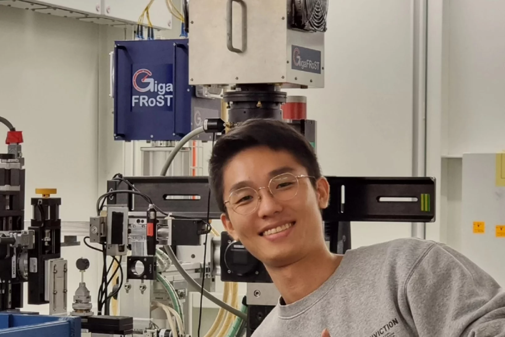
Jisoo Kim bags the 2022 Werner Meyer-Ilse Award
Jisoo Kim was awarded the 2022 Werner Meyer-Ilse Memorial Award. The WMI Award is given to young scientists for exceptional contributions to the advancement of X-ray microscopy through either outstanding technical developments or applications, as evidenced by their presentation at the International Conference on X-ray Microscopy and supporting publications. Jisoo was awarded for his development of the method "Time-resolved x-ray scattering tomography for rheological studies", and is co-recipient of the award with Yanqi Luo from the Advanced Photons Source for her work on applications. The award was presented during the 15th International Conference on X-ray Microscopy XRM2022 hosted by the National Synchrotron Radiation Research Center (NSRRC) in Hsinchu, Taiwan on 19 - 24 June, 2022.
TOMCAT welcomes on board two scientists
The X-ray Tomography group welcomes on board Mariana Verezhak and Goran Lovric as members of the TOMCAT beamline crew. They will both contribute to the further development and realization of TOMCAT 2.0 (S- and I-TOMCAT branches on SLS2.0).
SLS 2.0 approved - TOMCAT 2.0 cleared for takeoff!
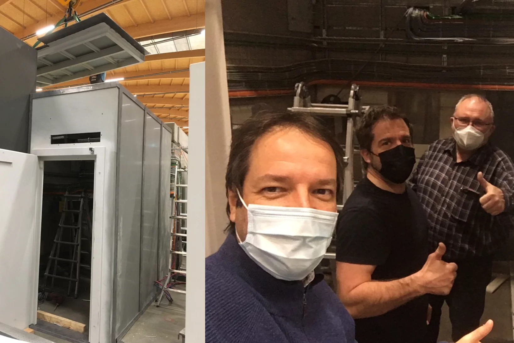
In December 2020 the Swiss parliament approved the Swiss Dispatch on Promotion of Education, Research and Innovation (ERI) for 2021 to 2024 which includes funding for the planned SLS 2.0 upgrade. The new machine will lead to significantly increased brightness, thus providing a firm basis for keeping the SLS and its beamlines state-of-the-art for the decades to come. The TOMCAT crew is very excited that the TOMCAT 2.0 plans (deployment of the S- and I-TOMCAT branches, see SLS 2.0 CDR, p. 353ff) have been included in the Phase-I beamline upgrade portfolio. These beamlines will receive first light right after the commissioning of the SLS 2.0 machine around mid 2025. A first milestone towards this goal has just been achieved, with the successful installation of the S-TOMCAT optics hutch during W1 of 2021. The TOMCAT scientific and technical staff would like to thank Mr. Nolte and his Innospec crew for delivering perfectly on schedule.
BEATS beamline scientist from SESAME synchrotron trains at TOMCAT
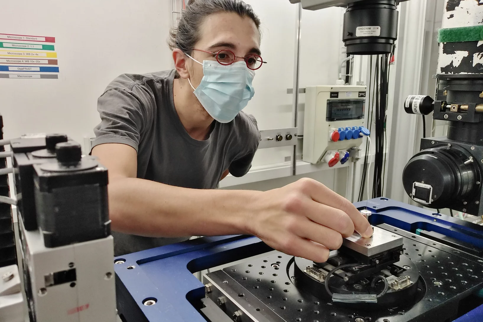
TOMCAT welcomes Gianluca Iori, beamline scientist from BEATS - the new beamline for tomography at the SESAME synchrotron in Jordan, to a 3-month training on beamline operations. Gianluca’s visit is part of the Staff Training (BEATS Work Package 2) organized for BEATS scientific staff and SESAME control engineers. BEATS is a European project, funded under the EU’s Horizon 2020 research and innovation programme and coordinated by the ESRF.
3 new Post Docs and 1 PhD student join TOMCAT
The X-ray Tomography group welcomes Stefan Gstöhl (Post-Doc), Maxim Polikarpov (Post-Doc), Margaux Schmeltz (Post-Doc) and Aleksandra Ivanovic (PhD Student) as new members. The group also thank everybody who helped making it possible for our Post-Docs and PhD student to join PSI amidst the challenges brought by the COVID-19 pandemic.
Automatic extraction of dynamic features from sub-second tomographic microscopy data
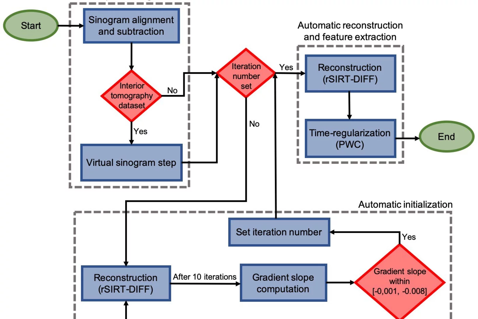
A fully automatized iterative reconstruction pipeline designed to reconstruct and segment dynamic processes within a static matrix has been developed at TOMCAT. The algorithm performance is demonstrated on dynamic fuel cell data where it enabled automatic extraction of liquid water dynamics from sub-second tomographic microscopy data. The work is published in Scientific Reports on 2 October 2020.
4 times compression factor for tomographic data feasible
In a recent study, TOMCAT has shown that lossy compression by a factor of at least 3 to 4 of raw acquisitions generally does not affect the reconstruction quality and that higher factors (six to eight times) can be achieved for tomographic volumes with a high signal-to-noise ratio as it is the case for phase-retrieved datasets. This finding is relevant to current challenges on large tomography data management and storage especially at synchrotron facilities. The results of this study was published in Journal of Synchrotron Radiation.
Rapid 3D directional small-angle scattering imaging achieved at TOMCAT
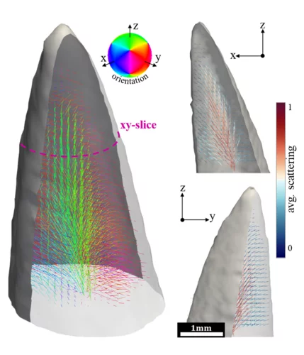
Researchers from the TOMCAT beamline have developed a small-angle scattering tensor tomography method to visualize microscopic features within a macroscopic field of view with unprecedented data acquisition speed. The results of the study were published in Applied Physics Letters on April 1, 2020.
Towards dynamic feedback control during time-resolved CT at TOMCAT
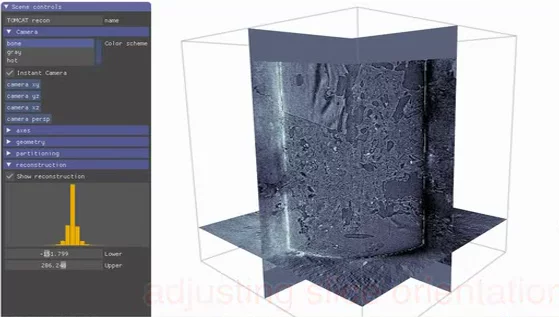
Researchers from the CWI in Amsterdam and the TOMCAT beamline have developed and implemented a real-time CT reconstruction, visualisation, and on-the-fly analysis approach to monitor dynamic processes as they occur. With processes of multiple sets of CT slices per second, this represents the next crucial step towards adaptive feedback control of time-resolved in situ tomographic experiments. The results of this study were published in Scientific Reports on December 5, 2019.
High-numerical-aperture optics is key to ultra-fast tomographic microscopy
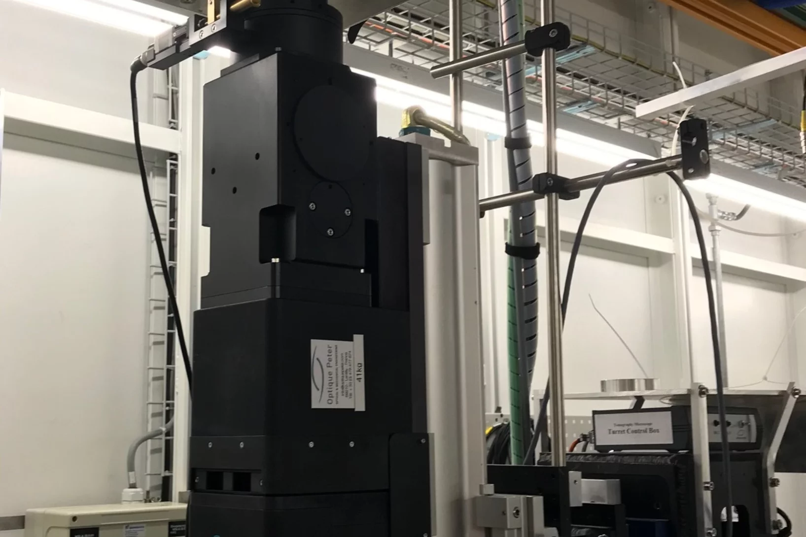
A novel high-numerical-aperture macroscope optics dedicated to high-temporal and high-spatial resolution X-ray tomographic microscopy is available at TOMCAT. Coupled with the in-house developed GigaFRoST camera, this highly efficient imaging setup enables tomographic microscopy studies at 20 Hz and beyond, opening up new possibilities in tomographic investigations of dynamic processes. A detailed characterization of the macroscope performance was published in Journal of Synchrotron Radiation on May 21, 2019.
From whole organ imaging down to single cell analysis
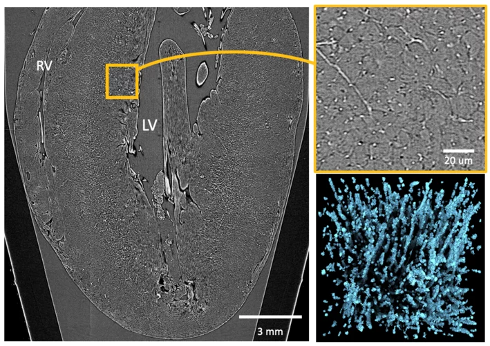
Researchers from the TOMCAT beamline, University College London (UCL), IDIBAPS and Universitat Pompeu Fabra (UPF) have developed a methodology that allows the multiscale analysis of the structural changes resulting from remodelling cardiovascular diseases, from whole organ down to single-cell level. This methodology has been published as an article in the journal Scientific Reports on May 6th 2019.
PSI Thesis Medal goes to Dr. Matias Kagias
The PSI Thesis Medal is awarded every second year to the best PhD thesis performed at the Paul Scherrer Institut. Matias received the prize for his excellent thesis entitled "Direct Self-Imaging Methods for X-ray Differential Phase and Scattering Imaging". Congratulations!
TOMCAT paper on hard X-ray multi-projection imaging published
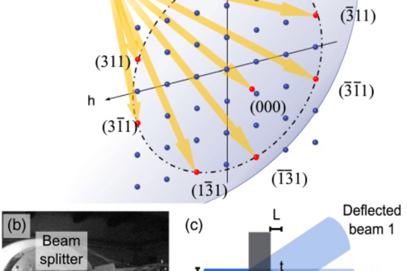
The TOMCAT team in collaboration with scientists from CFEL, MaxIV and ESRF developed a method for hard X-ray multi-projection imaging, using a single crystal to split the beam into multiple beams with different directions.
New TOMCAT paper: The GigaFRoST camera and readout system
The PSI in-house developed GigaFRoST high-speed camera and readout system is available for fast imaging experiments at the TOMCAT beamline, opening up exciting new possibilities for the observation of fast dynamic phenomena with X-ray tomography.

