Mapping the Nanoscale Architecture of Functional Materials
A new X-ray technique reveals the 3D orientation of ordered material structures at the nanoscale, allowing new insights into material functionality.
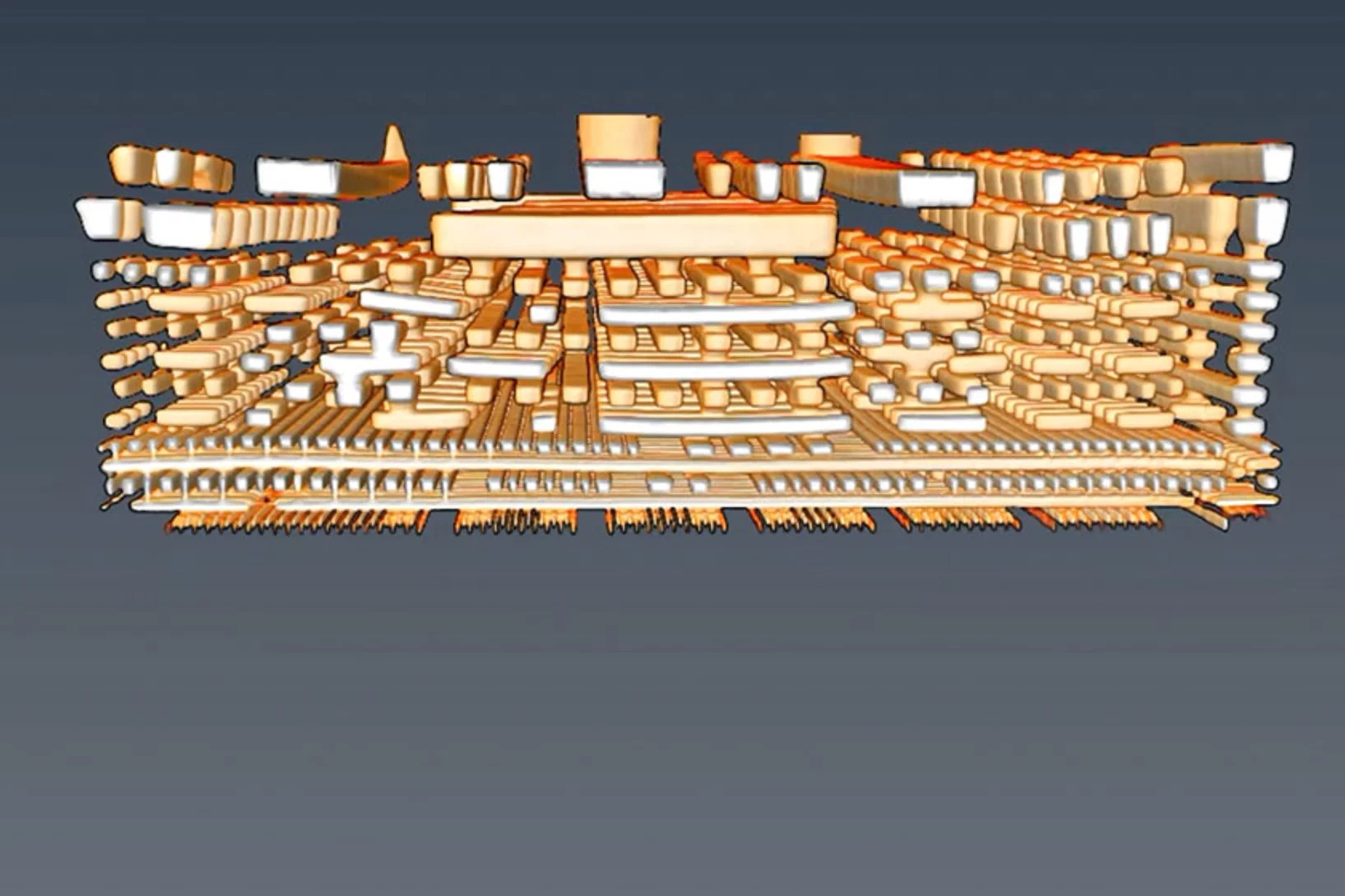
Neuer Röntgenweltrekord: Blick in einen Computerchip auf 4 Nanometer genau
Mit einer Rekordauflösung von 4 Nanometern gelang es Forschenden am PSI, die räumliche Struktur eines Computerchips mithilfe von Röntgenlicht abzubilden.
Manuel Guizar-Sicairos appointed as Associate Professor at EPF Lausanne and head of the Computational X-ray Imaging group at PSI
Dr. Manuel Guizar-Sicairos, currently Senior Scientist at PSI, was appointed as Associate Professor of Physics in EPF Lausanne and head of the Computational X-ray Imaging group in PSI.
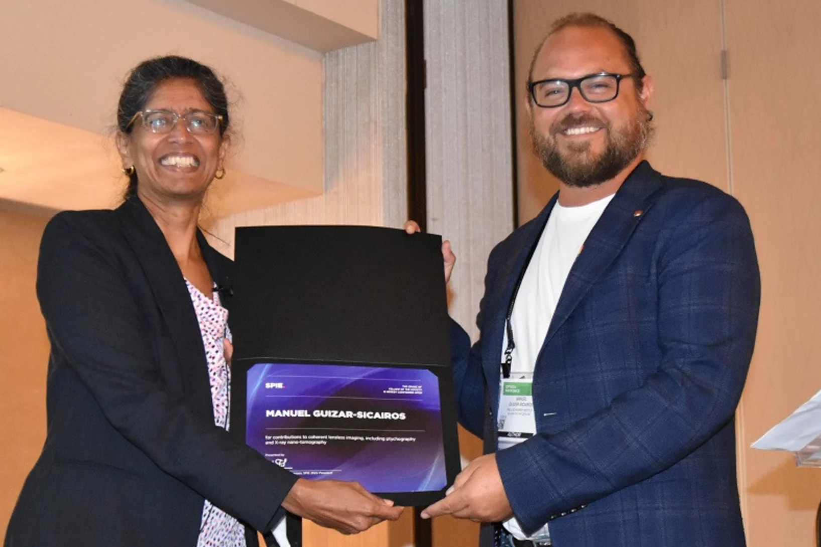
Dr. Manuel Guizar-Sicairos elected as SPIE Fellow member
Dr. Manuel Guizar-Sicairos was elected as a 2022 SPIE Fellow Member for his contributions to coherent lensless imaging, including ptychography and X-ray nano-tomography. The distinction was awarded in the SPIE’s Optics & Photonics conference in San Diego, California.
Nanomaterial aus dem Mittelalter
Das Geheimnis des Zwischgolds wurde am PSI gelüftet.
Ground-breaking technology development recognised
PSI researchers win the international Innovation Award on Synchrotron Radiation for 3D mapping of nanoscopic details in macroscopic specimens, such as bone.
Neurodegenerative disease studied by cryogenic X-ray nanotomography
Hard X-ray cryo-tomography scanning of retina from healthy and inherited blindness specimen paves the way for correlative analysis after imaging at the cSAXS beamline.
Dr. Manuel Guizar-Sicairos is awarded ICO prize
Dr. Manuel Guizar-Sicairos, beamline scientist at the cSAXS beamline, is the 2019 recipient of the International Commission for Optics (ICO) Prize. The distinction was awarded in the EOSAM conference in Rome.
Wie Katalysatoren altern
Katalysatoren, die in der Industrie eingesetzt werden, verändern über die Jahre ihre Materialstruktur. Mit einer neuen Methode haben PSI-Forschende dies nun auf der Nano-Skala untersucht.
Quantifying oriented myelin in mouse and human brain
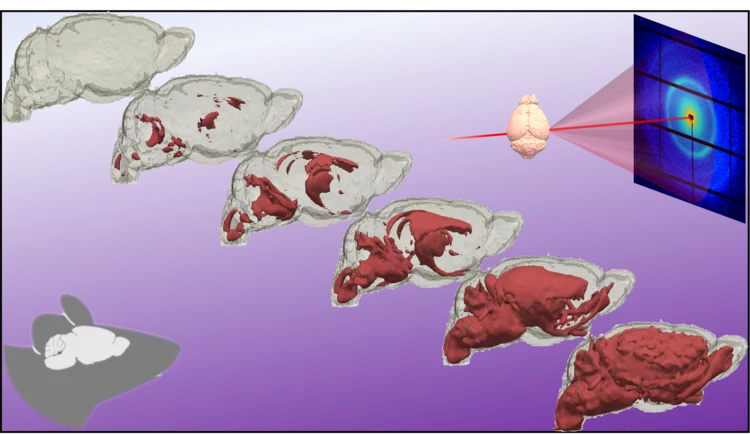
Myelin 'insulates' our neurons enabling fast signal transduction in our brain. Myelin levels, integrity, and neuron orientations are important determinants of brain development and disease. Small-angle X-ray scattering tensor tomography (SAXS-TT) is a promising technique for non-destructive, stain-free imaging of brain samples, enabling quantitative studies of myelination and neuron orientations, i.e. of nano-scale properties imaged over centimeter-sized samples.
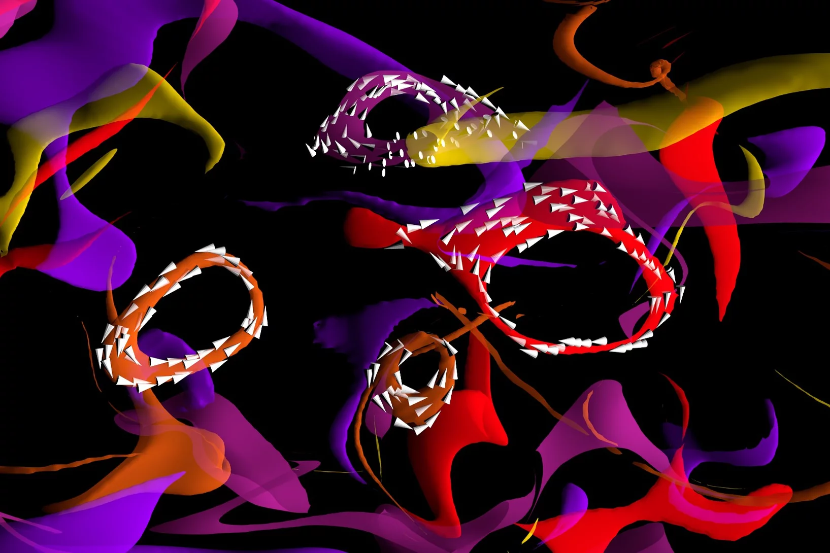
Magnetic vortices come full circle
The first experimental observation of three-dimensional magnetic ‘vortex rings’ provides fundamental insight into intricate nanoscale structures inside bulk magnets, and offers fresh perspectives for magnetic devices.
Dr. Manuel Guizar-Sicairos elected as Fellow member of The Optical Society (OSA)
Dr. Manuel Guizar-Sicairos, beamline scientist at the cSAXS beamline, was elected as a Fellow Member of The Optical Society (OSA) for seminal contributions to methods and applications of coherent lensless imaging, ptychography, x-ray nanotomography, and new modalities of x-ray microscopy.
Nanowelten in 3-D
Tomogramme aus dem Inneren von Fossilien, Hirnzellen oder Computerchips liefern neue Erkenntnisse über feinste Strukturen. Die 3-D-Bilder gelingen mithilfe der Röntgenstrahlen der Synchrotron Lichtquelle Schweiz SLS dank eigens entwickelter Detektoren und raffinierter Computeralgorithmen.
Kurzfilm eines magnetischen Nanowirbels
Mit einer neu entwickelten Untersuchungsmethode konnten Forschende die magnetische Struktur im Inneren eines Materials mit Nanometer-Auflösung abbilden. Ihnen gelang ein kurzer «Film» aus sieben Bildern, der erstmalig in 3-D zeigt, wie sich winzige Wirbel der Magnetisierung tief im Inneren eines Materials verändern.
3D imaging for planar samples with zooming
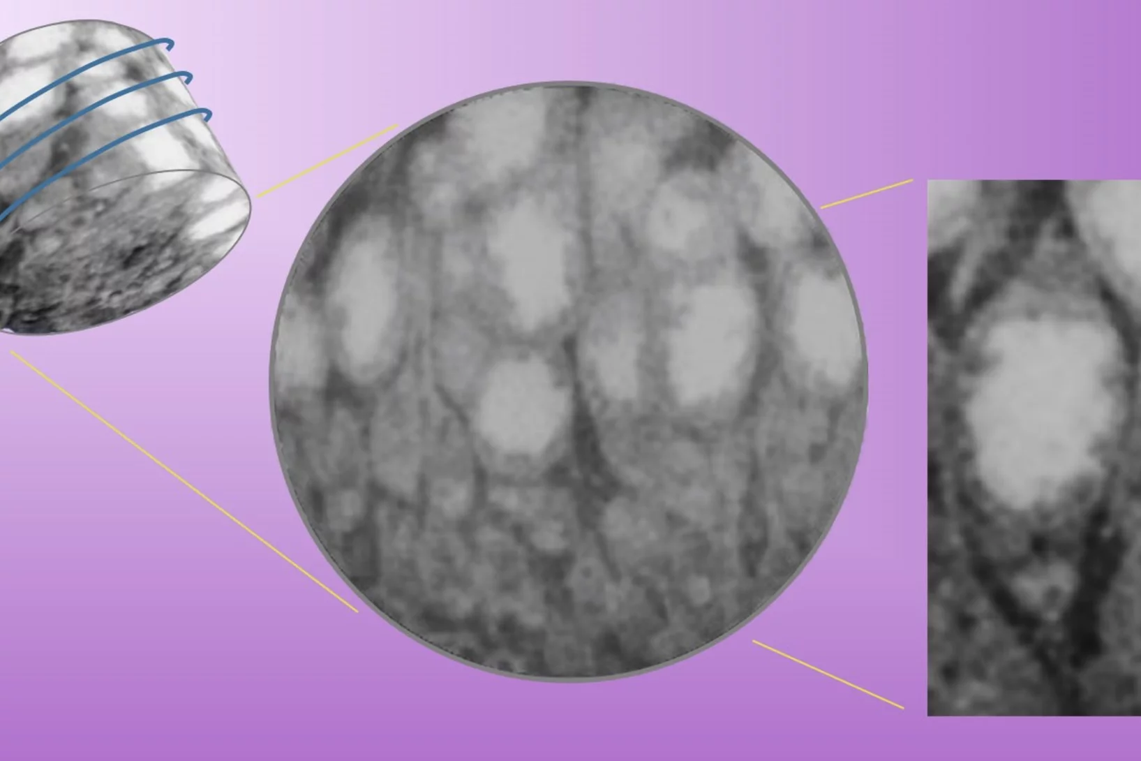
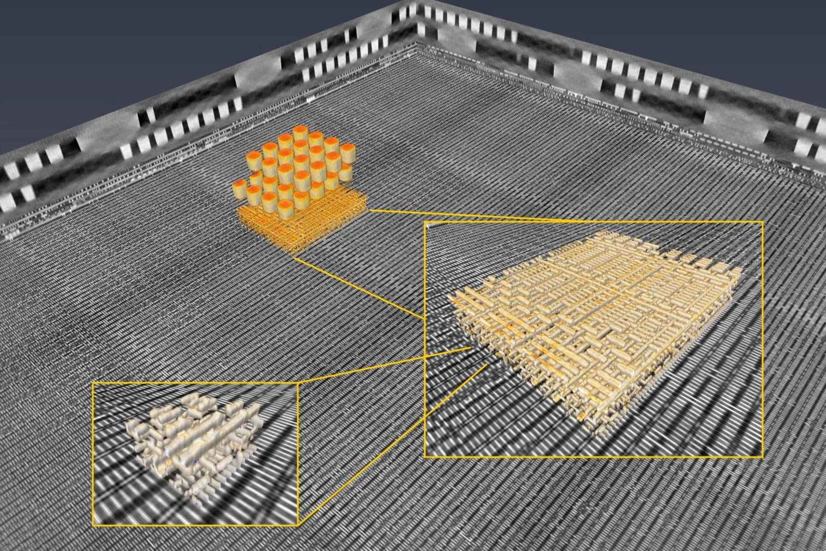
Researchers of the Paul Scherrer Institut have previously generated 3-D images of a commercially available computer chip. This was achieved using a high-resolution tomography method. Now they extended their imaging approach to a so-called laminography geometry to remove the requirement of preparing isolated samples, also enabling imaging at various magnification. For ptychographic X-ray laminography (PyXL) a new instrument was developed and built, and new data reconstruction algorithms were implemented to align the projections and reconstruct a 3D dataset. The new capabilities were demonstrated by imaging a 16 nm FinFET integrated circuit at 18.9 nm 3D resolution at the Swiss Light Source. The results are reported in the latest edition of the journal Nature Electronics. The imaging technique is not limited to integrated circuits, but can be used for high-resolution 3D imaging of flat extended samples. Thus the researchers start now to exploit other areas of science ranging from biology to magnetism.
Inside Batteries
Lithium ion batteries (LIB) are essential in modern everyday life, with increasing interest in enhancing their performance and lifetime. Secondary particles of Li-rich cathode material were examined with correlated ptychographic X-ray tomography and diffraction microscopy at different stages of cycling to probe the aging mechanism.
Virtuelle Linse verbessert Röntgenmikroskopie
Eine von PSI-Forschenden neu entwickelte Methode macht Röntgenbilder von Materialien noch besser. Die Forschenden bewegten dafür eine optische Linse und nahmen dabei Einzelbilder auf. Mit Hilfe von Computeralgorithmen errechneten sie daraus ein Gesamtbild.

