
From inside an eggshell
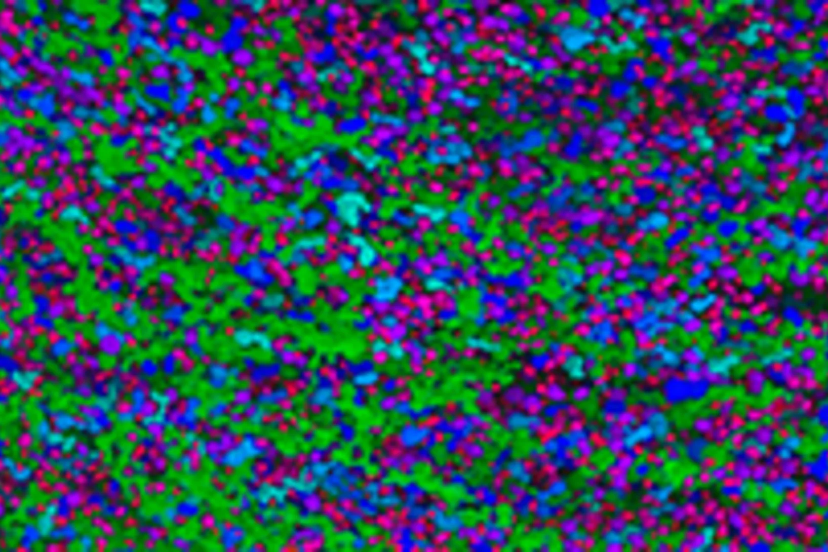
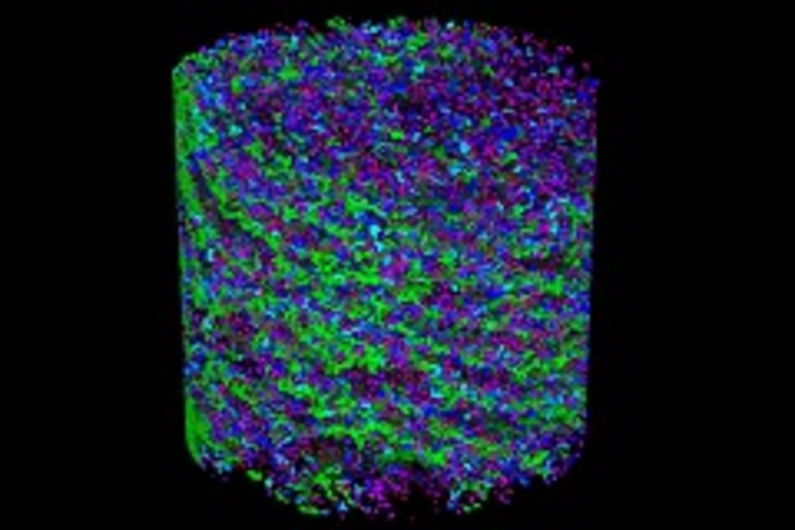
Tiny cavities inside eggshells supply the materials that stimulate and control the shell’s growth. Using a novel imaging technique, researchers from the Paul Scherrer Institute (PSI), ETH Zurich and the Dutch FOM Institute AMOLF have succeeded in depicting these voids in 3D for the first time. In doing so, they lift an old limitation of tomographic images and hope that one day medicine will also benefit from their method.
Research geared towards the future
Interview with Gabriel AeppliGabriel Aeppli has been head of synchrotron radiation and nanotechnology research at PSI since 2014. Previously, the Swiss-born scientist set up a leading research centre for nanotechnology in London. In this interview, Aeppli explains how the research approaches of the future can be implemented at PSI's large research facilities and talks about his view of Switzerland.
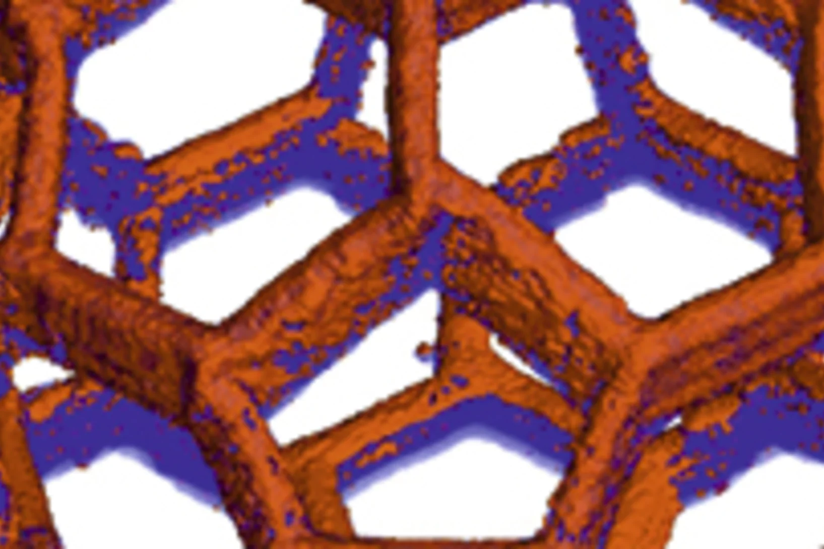
Multiresolution X-ray tomography, getting a clear view of the interior
Researchers at PSI have developed a technique that combines tomography measurements at different resolution levels to allow quantitative interpretation for nanoscale tomography on an interior region of interest of the sample. In collaboration with researchers of the institute AMOLF in the Netherlands and ETH Zurich in Switzerland they showcase their technique by studying the porous structure within a section of an avian eggshell. The detailed measurements of the interior of the sample allowed the researchers to quantify the ordering and distribution of an intricate network of pores within the shell.
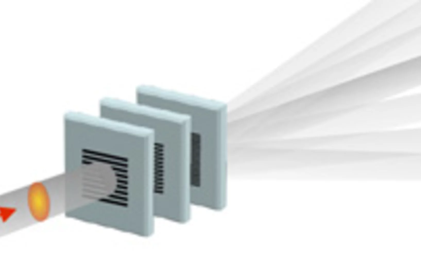
Split x-ray flash shows rapid processes
SwissFEL, PSI’s x-ray laser, is to render the individual steps of very rapid processes visible. A new method will facilitate especially precise experiments: the individual x-ray flashes are split into several parts that arrive at the object under examination one by one. The principle of the method harks back to the ideas of the earliest high-speed photography.
Nanometres in 3D
Scientists at the Paul Scherrer Institute and ETH Zurich have created 3D images of tiny objects showing details down to 25 nanometres. In addition to the shape, the scientists determined how particular chemical elements were distributed in their sample and whether these elements were in a chemical compound or in their pure state.
Prepared for the SwissFEL
For many years, PSI researchers have been testing experimental methods that will provide insights into novel materials for electronic devices. Using a special trick to make the Swiss Light Source (SLS) at PSI generate light with similar properties to that of PSI’s x-ray laser SwissFEL, the researchers were able to demonstrate that the experiments planned for SwissFEL are possible and they are now building an experimental station at SwissFEL.
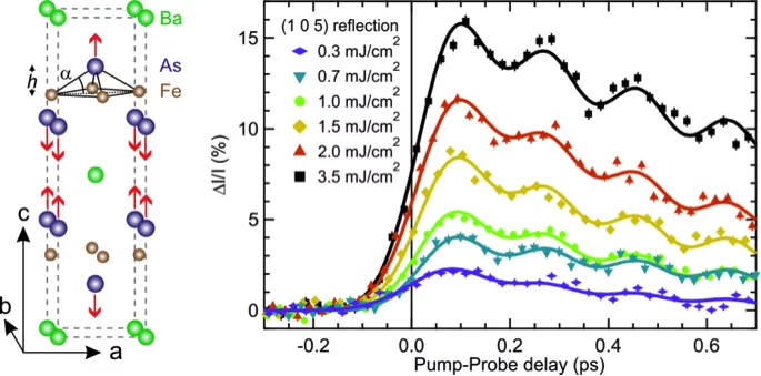
Ultrafast structural dynamics of the Fe-pnictide parent compound BaFe2As2
Understanding the interplay of the various degrees of freedom such as the electrons, spins and lattice is essential for many complex materials, including the high-temperature superconductors.
Nanoscale sub-100 picosecond all-optical magnetization switching in GdFeCo microstructure
Ultrafast magnetization reversal driven by femtosecond laser pulses has been shown to be a promising way to write information. Seeking to improve the recording density has raised intriguing fundamental questions about the feasibility of combining ultrafast temporal resolution with sub-wavelength spatial resolution for magnetic recording. Here we report on the experimental demonstration of nanoscale sub-100 ps all-optical magnetization switching, providing a path to sub-wavelength magnetic recording.
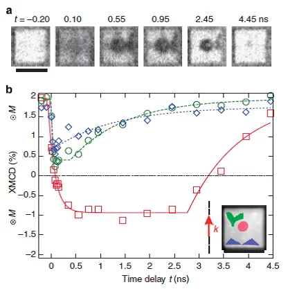
Batman lights the way to compact data storage
Researchers at the Paul Scherrer Institute (PSI) have succeeded in switching tiny, magnetic structures using laser light and tracking the change over time. In the process, a nanometre-sized area bizarrely reminiscent of the Batman logo appeared. The research results could render data storage on hard drives faster, more compact and more efficient.