At the frontier of research in novel engineering materials is the understanding of the relationship between microstructure and materials properties. In order to design improved structural materials we need to understand what happens on the atomic scale when the material is pushed into extreme conditions (temperature, stress, pressure, corrosion). It is well known that the dynamics of dislocation and other defect structures play an important role in the mechanism of metals plasticity and failure under stress. Diffraction (x-ray, neutron) is very sensitive to the strain fields related to the defect structures and is therefore the method of choice to study the evolution of dislocation densities. Experimental methods based on x-ray diffraction at synchrotron sources have made giant steps, so that it is currently possible to acquire excellent data sets in situ at or below the millisecond scale. On the opposite, for interpreting this wealth of data, theoretical methods have not been significantly advanced since the ‘70s - with the outstanding (but nowadays too limited) works of Krivoglaz and Wilkens.
White beam Laue diffraction is one of the oldest X-ray diffraction methods and was predominantly used as a crystallographic method to determine the orientation of the crystal. It is however well known that the presence of dislocations in a crystal volume results in streaking, broadening and splitting of the Laue peaks. It has major advantages over traditional monochromatic diffraction for micron scale spatial resolution because no sample rotations are required; furthermore, the depth at which the scattering originates can be resolved trough a differential aperture microscopy technique. With new technological developments at synchrotron sources, it is now possible to use micro-focused polychromatic beams to study small crystal volumes or single grains in polycrystalline materials (APS; Soleil, SLS, ALS). During the last ten years an increasing number of authors have been using this technique in studying the mesoscale dynamics of materials by means of in situ Laue methods combined with microcompression or indentation techniques.
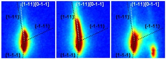
A Laue diffraction pattern taken with a polychromatic X-ray beam is characterized by individual spots (see Fig.) each related to a different hkl-family. The position of such spots depends on the crystal orientation, and the shape of the unit cell. Therefore any crystal rotation and/or change in the shape of the unit cell will result in peak movements. Moreover, strain gradients appearing in the sample during deformation are manifested trough continuous/discontinuous streaking of Laue spots. The possibility of measuring the spots continuously during mechanical testing has lead to an in situ peak profile analysis capability, allowing studying the microstructure evolution in real time.
The aim of the current research is to go beyond the current models used for the interpretation of the Laue patterns in terms of dislocation ensembles by development of a code allowing to calculate Laue patterns from dislocation ensembles simulated using 3 dimensional dislocation dynamics schemes.
Funding
The Swiss National Science Foundation finances this project with a postdoc position (SNF 132699)
