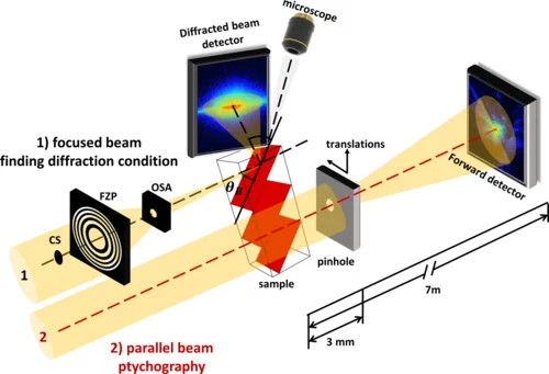
Strain and defects in crystalline materials are responsible for the distinct mechanical, electric, and magnetic properties of a desired material, making their study an essential task in material characterization, fabrication, and design. Existing techniques for the visualization of strain fields, such as transmission electron microscopy and diffraction, are destructive and limited to thin slices of the materials. On the other hand, nondestructive x-ray imaging methods either have a reduced resolution or are not robust enough for a broad range of applications. Here we present x-ray ptychographic topography, a method for strain imaging, and demonstrate its use on an InSb micropillar after microcompression, where the strained region is visualized with a spatial resolution of 30 nm. Thereby, x-ray ptychographic topography proves itself as a robust nondestructive approach for the imaging of strain fields within bulk crystalline specimens with a spatial resolution of a few tens of nanometers.
Contact
Steven Van Petegem
Paul Scherrer Institut, Villigen, Switzerland
Telephone: +41 56 310 2537
E-mail: steven.vanpetegem@psi.ch
Ana Diaz
Paul Scherrer Institut, Villigen, Switzerland
Telephone: +41 56 310 5626
E-mail: ana.diaz@psi.ch
Original Publication
X-ray ptychographic topography: A robust nondestructive tool for strain imaging
Mariana Verezhak, Steven Van Petegem, Angel Rodriguez-Fernandez, Pierre Godard, Klaus Wakonig, Dmitry Karpov, Vincent L. R. Jacques, Andreas Menzel, Ludovic Thilly, and Ana Diaz
Phys. Rev. B 103 (2021) 144107
DOI: https://doi.org/10.1103/PhysRevB.103.144107
