Magnetic vortices come full circle
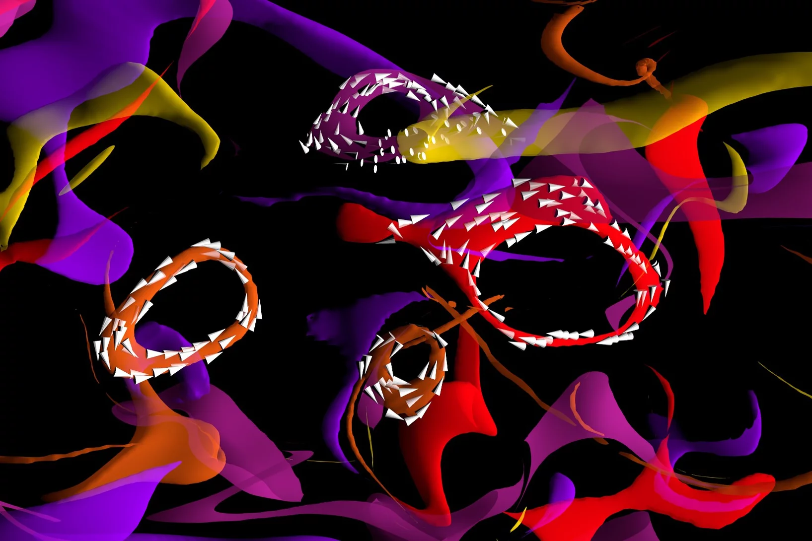
The first experimental observation of three-dimensional magnetic ‘vortex rings’ provides fundamental insight into intricate nanoscale structures inside bulk magnets, and offers fresh perspectives for magnetic devices.
World Record: 7 nm Resolution in Scanning Soft X-ray Microscopy
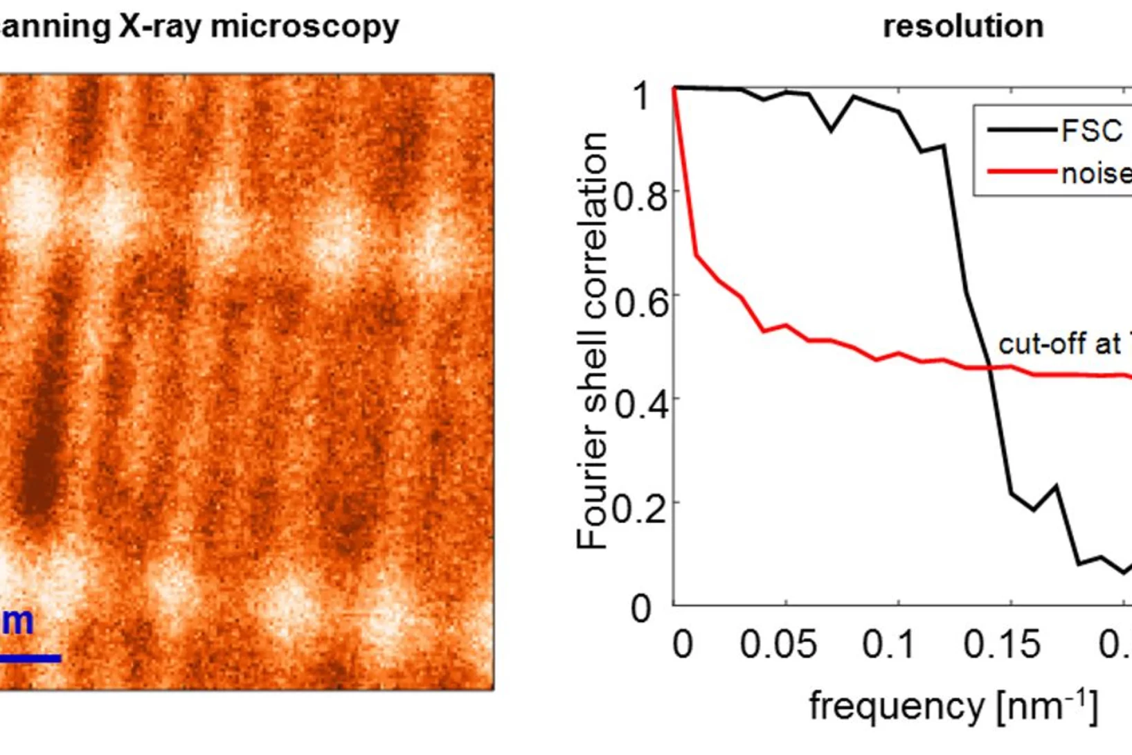
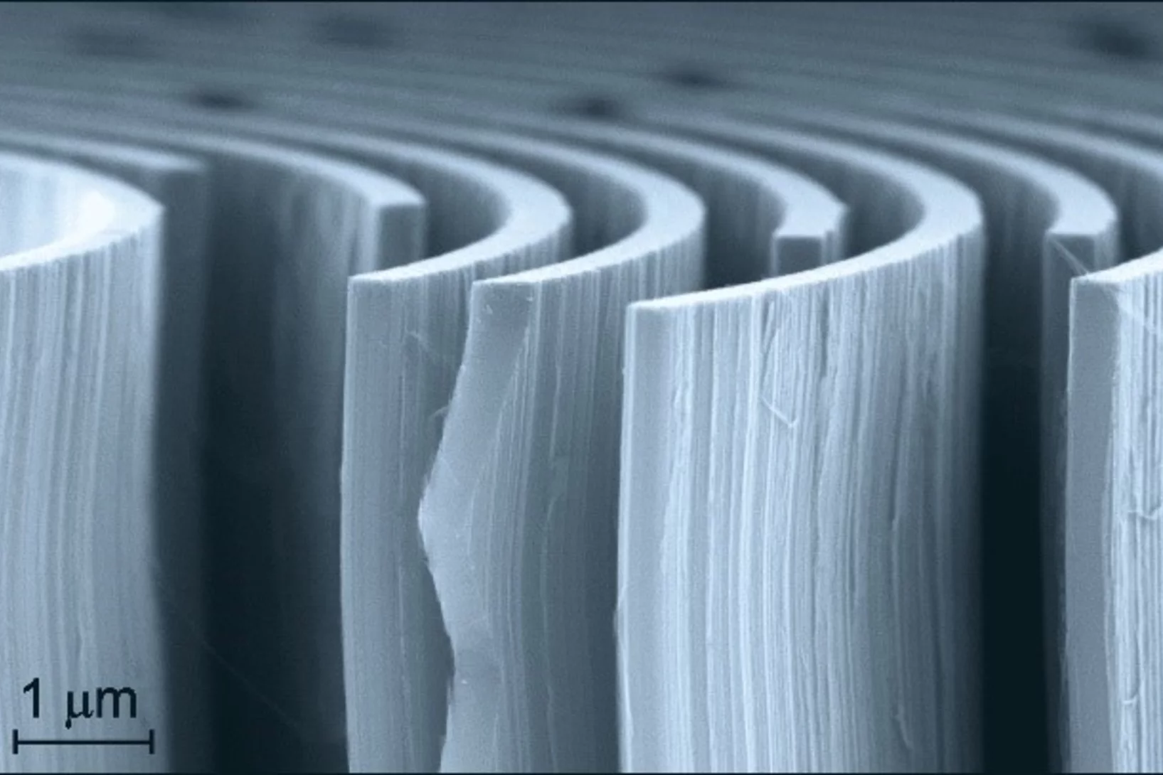
During the past decade, scientists have put high effort to achieve sub-10 nm resolution in X-ray microscopy. Recent developments in high-resolution lithography-based diffractive optics, combined with the extreme stability and precision of the PolLux and HERMES scanning X-ray microscopes, resulted now in a so far unreached resolution of seven nanometers in scanning soft X-ray microscopy. Utilizing this highly precise microscopy technique with the X-ray magnetic circular dichroism effect, dimensionality effects in an ensemble of interacting magnetic nanoparticles can be revealed.
Enhanced Stability of a Pyrophosphate cathode for Na-ion batteries
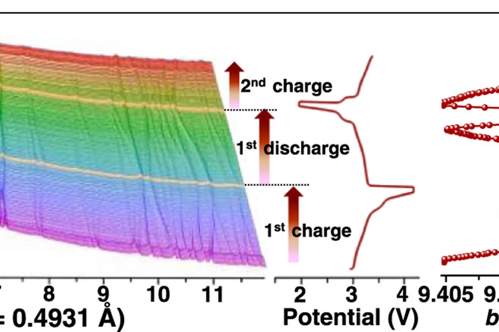
The structural changes of Na3.32Fe2.11Ca0.23(P2O7)2 during several charge discharge cycles is viewed by its powder pattern and selected cell parameter evolution.
Dr. Manuel Guizar-Sicairos elected as Fellow member of The Optical Society (OSA)
Dr. Manuel Guizar-Sicairos, beamline scientist at the cSAXS beamline, was elected as a Fellow Member of The Optical Society (OSA) for seminal contributions to methods and applications of coherent lensless imaging, ptychography, x-ray nanotomography, and new modalities of x-ray microscopy.
Two-color snapshots of ultrafast charge and spin dynamics
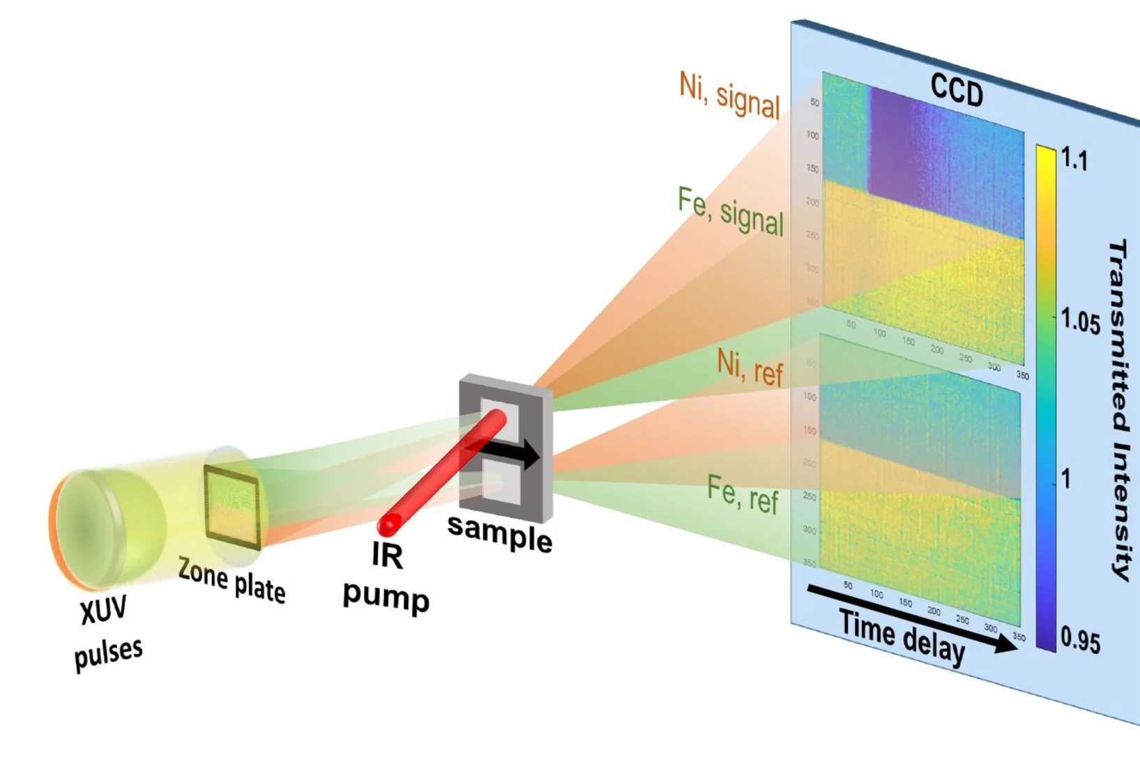
In a joint research effort, an international team of scientists lead by Emmanuelle Jal (Sorbonne Université) performed a time-resolved experiment at the FERMI free-electron laser to disclose the dynamic behavior of two magnetic element of a compount material in only one snapshot. The X-ray Optics and Applications group developed a dedicated optical element for this experiment that is usable with two different photon energies (colors) simultaneously.
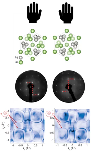
Réaliser un matériau électronique sur mesure
Des chercheurs du PSI ont analysé un matériau qui pourrait entrer en ligne de compte pour de futures applications dans le domaine du stockage de données. Une astuce leur a permis de déformer de manière ciblée la structure cristalline de leur échantillon et de mesurer la manière dont cette déformation influençait les propriétés magnétiques et électroniques.
Question de liaison
Au PSI, des chercheurs passent au crible des fragments de molécules pour voir si ces derniers se lient à certaines protéines importantes du coronavirus SARS-CoV-2 afin de les neutraliser. A partir de ces informations, ils espèrent trouver une réponse sur le profil potentiel d’un médicament efficace.
«Nous préparons la SLS à l’avenir»
La Source de Lumière Suisse SLS doit faire l’objet d’une mise à jour afin qu’elle puisse aussi permettre une recherche d’excellence au cours des prochaine décennies. Dans cette interview, Hans Braun, chef du projet SLS 2.0, explique en quoi consiste cet upgrade.
3D printing silica aerogels at the micrometer scale
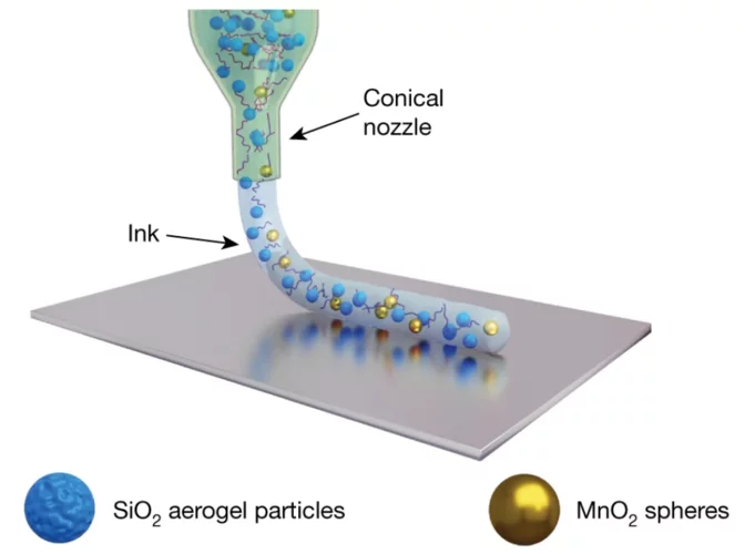
A group of EMPA and ETH Zürich researchers have developed a new method to directly write ink made of silica aerogels in 3D. Thanks to X-ray phase contrast tomography at the TOMCAT beamline they characterized the resulting printed material with different compositions. Their results were published in Nature on August 18, 2020.
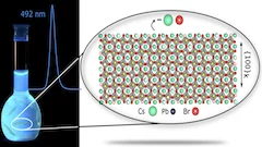
X-rays illuminate the particle atomic structure of cyan light emitting 6-monolayers CsPbBr3 nanoplatelets by Total Scattering
A cyan light (492 nm) emitting colloidal suspension of CsPbBr3 nanoplatelets in a flask, together with the high-quality XRPD Total Scattering pattern of the suspension measured at the X04SA-MS beamline and the full-nanoparticle structure thereby inferred.
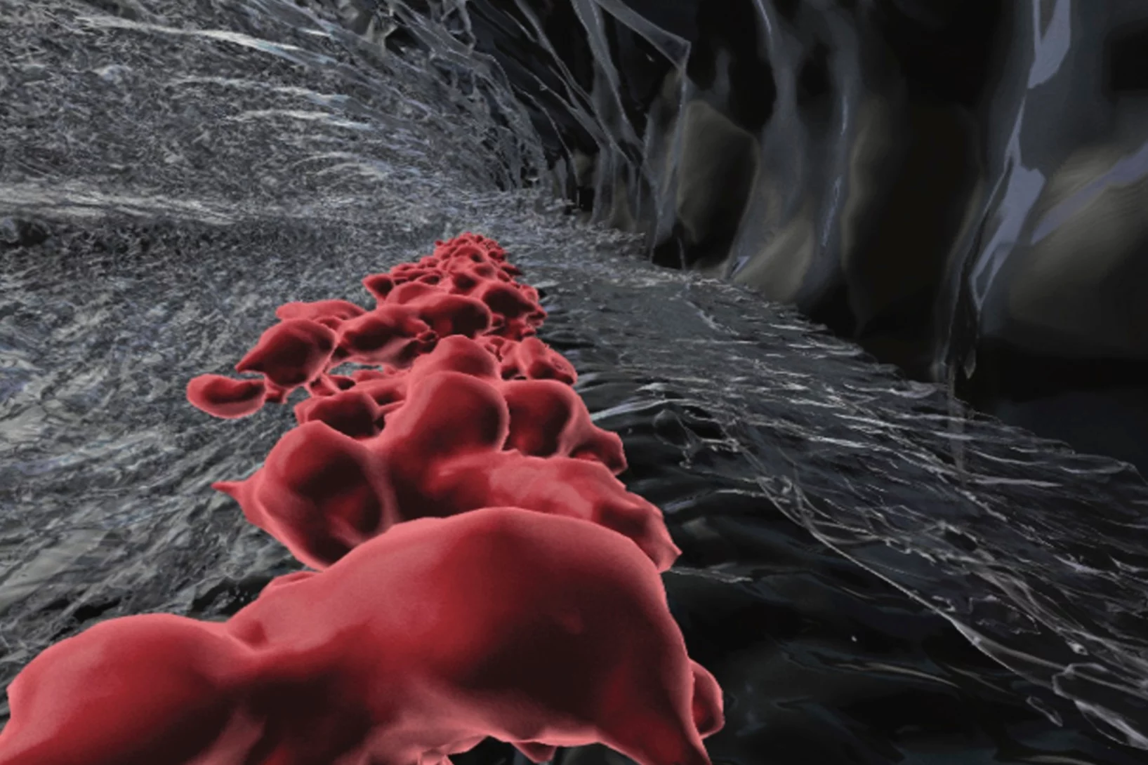
Phase contrast microtomography reveals nanoparticle accumulation in zebrafish
Metal-based nanoparticles are a promising tool in medicine – as a contrast agent, transporter of active substances, or to thermally kill tumor cells. Up to now, it has been hardly possible to study their distribution inside an organism. Researchers at the University of Basel in collaboration with the TOMCAT team have used phase contrast X-ray tomographic microscopy to take high-resolution captures of the nanoparticle aggregation inside zebrafish embryos.
The study was published in the journal Small and featured on the cover of its current issue.
Recherche sur le Covid-19: stratégie antivirale à double effet
Des scientifiques de Francfort ont identifié un éventuel point faible du virus SARS-CoV-2. Ils ont effectué une partie de leurs mesures à la Source de Lumière Suisse SLS du PSI. Leurs résultats de recherche paraissent cette semaine dans la revue spécialisée Nature.
Cherned up to the maximum
In topological materials, electrons can display behaviour that is fundamentally different from that in ‘conventional’ matter, and the magnitude of many such ‘exotic’ phenomena is directly proportional to an entity known as the Chern number. New experiments establish for the first time that the theoretically predicted maximum Chern number can be reached — and controlled — in a real material.
Grosser Rat bewilligt 2,4 Millionen für Technologiezentrum Anaxam
Der Kanton Aargau unterstützt das Technologietransferzentrum Anaxam in Villigen für die Dauer von vier Jahren mit insgesamt 2,4 Millionen Franken. Der Grosse Rat hat am Dienstag in Spreitenbach den entsprechenden Kredit mit 124 zu 3 Stimmen bewilligt.
First MX results of the priority COVID-19 call
The Dikic group at the Goethe University in Frankfurt am Main, Germany has published the first results following the opening of the "PRIORITY COVID-19 Call” at SLS.
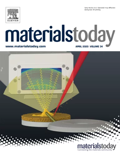
Operando X-ray diffraction during laser 3D printing
Ultra-fast operando X-ray diffraction experiments reveal the temporal evolution of low and high temperature phases and the formation of residual stresses during laser 3D printing of a Ti-6Al-4V alloy. The profound influence of the length of the laser-scanning vector on the evolving microstructure is revealed and elucidated.
Miniaturized fluidic circuitry observed in 3D
The team of Prof. Thomas Hermans at the University of Strasbourg in France managed to create wall-less aqueous liquid channels called anti-tubes. Thanks to X-ray phase contrast tomography at the TOMCAT beamline those anti-tubes could be observed in 3D. The exciting results were published in Nature on May 6, 2020.
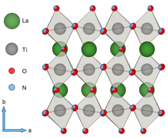
Examining the surface evolution of LaTiOxNy an oxynitride solar water splitting photocatalyst
LaTiOxNy oxynitride thin films are employed to study the surface modifications at the solid- liquid interface that occur during photoelectrocatalytic water splitting. Neutron reflectometry and grazing incidence x-ray absorption spectroscopy were utilised to distinguish between the surface and bulk signals, with a surface sensitivity of 3 nm.
SLS MX beamtime update
Update of the SLS MX beamline operation during the COVID-19 period
X-ray Imaging for Biomedicine: Imaging Large Volumes of Fresh Tissue at High Resolution
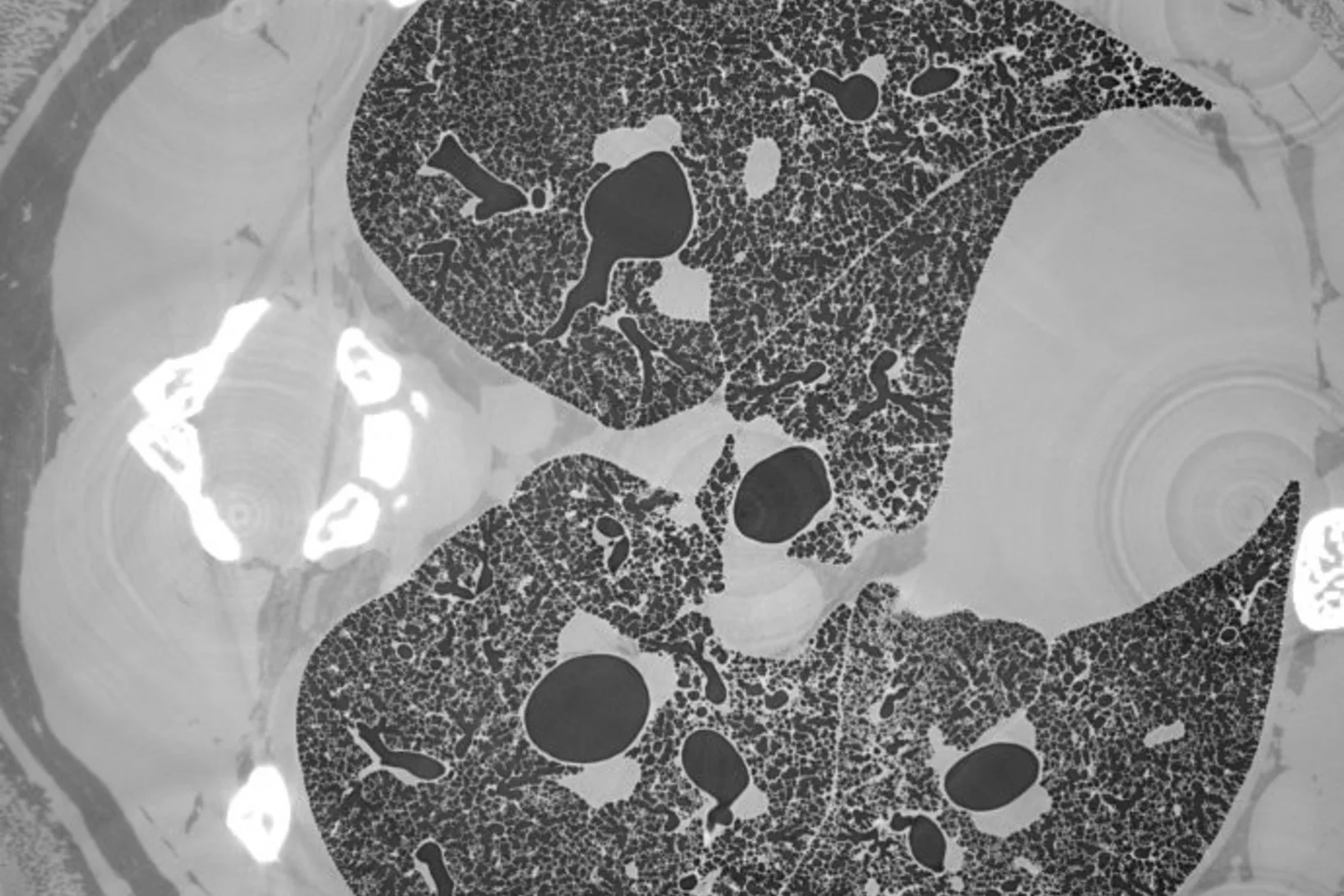
The TOMCAT beamline at the Swiss Light Source specializes in rapid high-resolution 3-dimensional tomographic microscopy measurements with a strong focus on biomedical imaging. The team has recently developed a technique to acquire micrometer-scale resolution datasets on the entire lung structure of a juvenile rat in its fresh natural state within the animal’s body and without the need for any fixation, staining or other alteration that would affect the observed structure (E. Borisova et al., 2020, Histochem Cell Biol).
Logic operations with domain walls
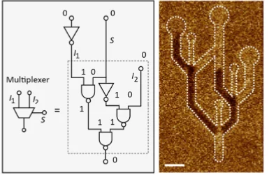
A collaboration of scientists from the ETH Zürich and the Paul Scherrer Institute successfully demonstrated the all-electric operation of a magnetic domain-wall based NAND logic gate, paving the way towards the development of logic applications beyond the conventional metal-oxide semiconductor technology. The work has been published in the journal Nature.
Optics for spins
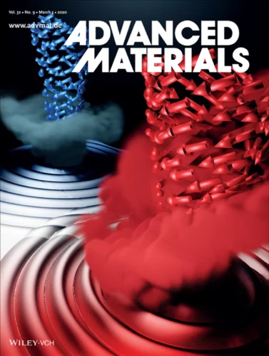
In this work, published on the front cover page of Advanced Materials, an international collaboration of Italian, American, and Swiss scientists demonstrated a novel concept for the generation and manipulation of spin waves, paving the way towards the development of magnonic nano-processors.
Can skyrmions read?
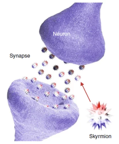
Can a skyrmion-based device be used to read a handwritten text? In this work, an international scientist collaboration led by the Korea Institute of Technology and the IBM Watson research center could provide a first answer to this question by fabricating a proof-of-principle single-neuron artificial neural network, using X-ray magnetic microscopy at the Swiss Light Source to investigate its performances.
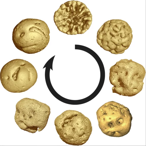
Court-métrage d’un nano-tourbillon magnétique
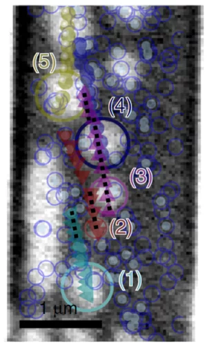
Des chercheurs ont développé une nouvelle méthode d’analyse qui leur a permis de visualiser la structure magnétique à l’intérieur d’un matériau à l’échelle du nanomètre. Ils ont réussi à réaliser un petit «film» de sept images qui montre pour la première fois en 3D les changements que de minuscules tourbillons magnétiques subissent au cœur du matériau.
Many skyrmions, one angle
Employing a tailored multilayered magnetic film, optimized for the zero-field stabilization of magnetic skyrmions, researchers have investigated the influence of the skyrmion diameter on its current-induced sideways motion, uncovering mechanisms that allow for this topological property to be controlled.
MacEtch in gas phase: a new nanofabrication technology at PSI
The grating fabrication team of the X-ray tomography group has scored another record in etching technology of silicon by realizing a MacEtch process in gas phase. Ultra-high aspect ratios (up to 10 000 : 1) in the nanoscale regime (down to 10 nm) were achieved by platinum assisted chemical etching of silicon in the gas phase. The results were published in Nanoscale Horizons on February 17, 2020.
Soft X-ray Laminography: 3D imaging with powerful contrast mechanisms
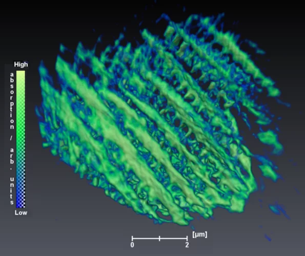
3D imaging using synchrotron radiation is a widely used tool that allows access to the inner structure of complex objects. An international and interdisciplinary consortium of scientists from the Swiss Light Source (PolLux and cSAXs), the Friedrich-Alexander-Universität Erlangen-Nürnberg, and the University of Cambridge developed the new 3D imaging technique of Soft X-ray Laminography (SoXL). SoXL allows for the investigation of thin and extended samples while taking advantage of the characteristic absorption contrast mechanisms in the soft X-ray range, providing 3D information with nm spatial resolution.
Animal embryos evolved before animals
Detailed characterization of cellular structure and development of exceptionally preserved ancient tiny fossils from South China by synchrotron based X-ray tomographic microscopy at TOMCAT led an international team of researchers from the University of Bristol and Nanjing Institute of Geology and Palaeontology to the discovery that animal-like embryos evolved long before the first animals appear in the fossil record.
Radiographier rapidement et précisément des matériaux composites renforcés de fibres
Des chercheurs de l’Institut Paul Scherrer PSI ont mis au point une méthode de diffusion des rayons X aux petits angles qui peut être utilisée pour le développement ou le contrôle qualité de matériaux composites novateurs renforcés de fibres. Grâce à elle, les analyses de ces matériaux pourraient se faire à l’avenir non seulement par recours aux rayons X issus de sources puissantes comme la Source de Lumière Suisse SLS, mais aussi avec le rayonnement issu de tubes à rayons X conventionnels.
3D imaging for planar samples with zooming
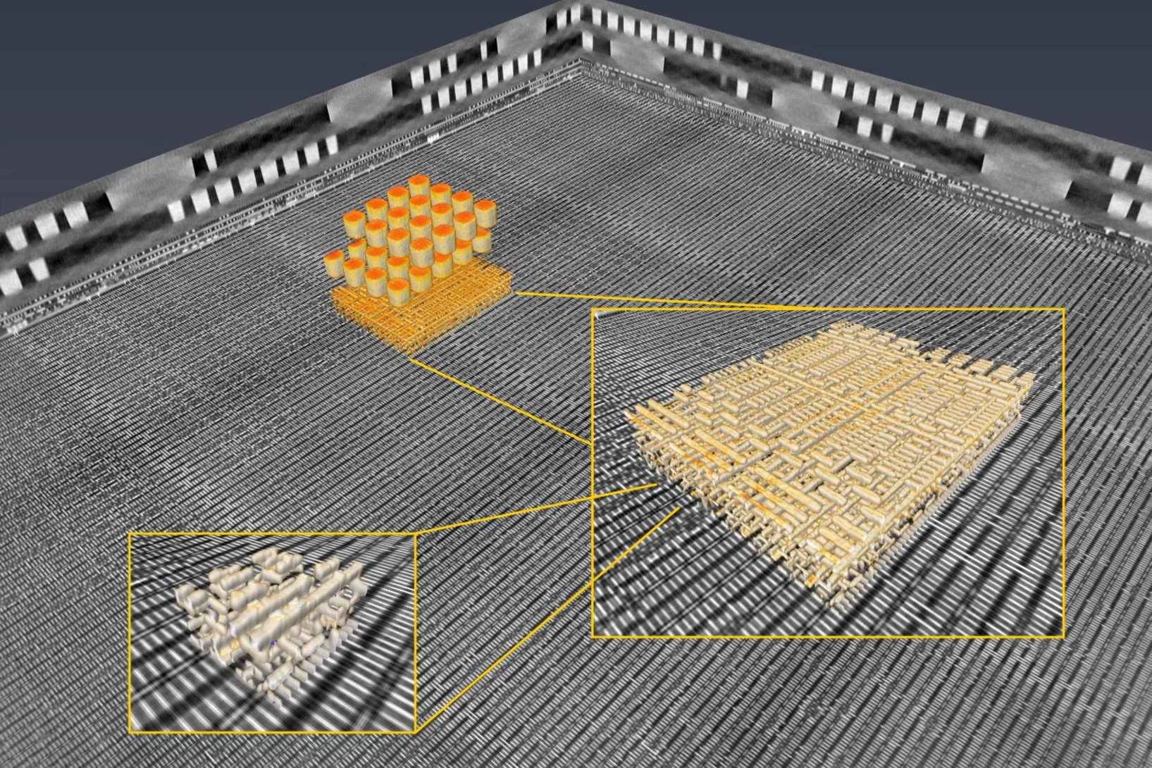
Researchers of the Paul Scherrer Institut have previously generated 3-D images of a commercially available computer chip. This was achieved using a high-resolution tomography method. Now they extended their imaging approach to a so-called laminography geometry to remove the requirement of preparing isolated samples, also enabling imaging at various magnification. For ptychographic X-ray laminography (PyXL) a new instrument was developed and built, and new data reconstruction algorithms were implemented to align the projections and reconstruct a 3D dataset. The new capabilities were demonstrated by imaging a 16 nm FinFET integrated circuit at 18.9 nm 3D resolution at the Swiss Light Source. The results are reported in the latest edition of the journal Nature Electronics. The imaging technique is not limited to integrated circuits, but can be used for high-resolution 3D imaging of flat extended samples. Thus the researchers start now to exploit other areas of science ranging from biology to magnetism.
