
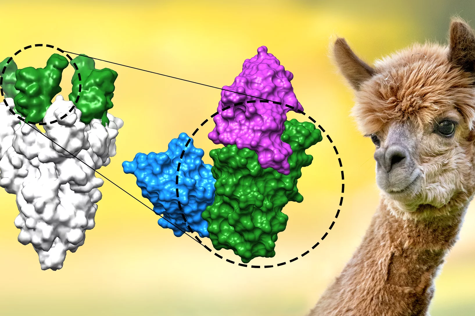
Nanobodies against SARS-CoV-2
In a study published in EMBO Journal, researchers at the Max Planck Institute for Biophysical Chemistry, Göttingen, Germany, developed nanobodies that efficiently block the coronavirus SARS-CoV-2 and its variants. The high resolution structural characterization was performed at the X10SA crystallography beamline at the Swiss Light Source.
Imaging strain with high resolution
Imaging strain in crystalline materials with high resolution can be a challenging task. Researchers demonstrate an original use of X-ray ptychography for this purpose: ptychographic topography.
How ethane-consuming archaea pick up their favorite dish
Scientists decode the structure of the enzyme responsible for the ethane fixation by – beside others – using the SLS.
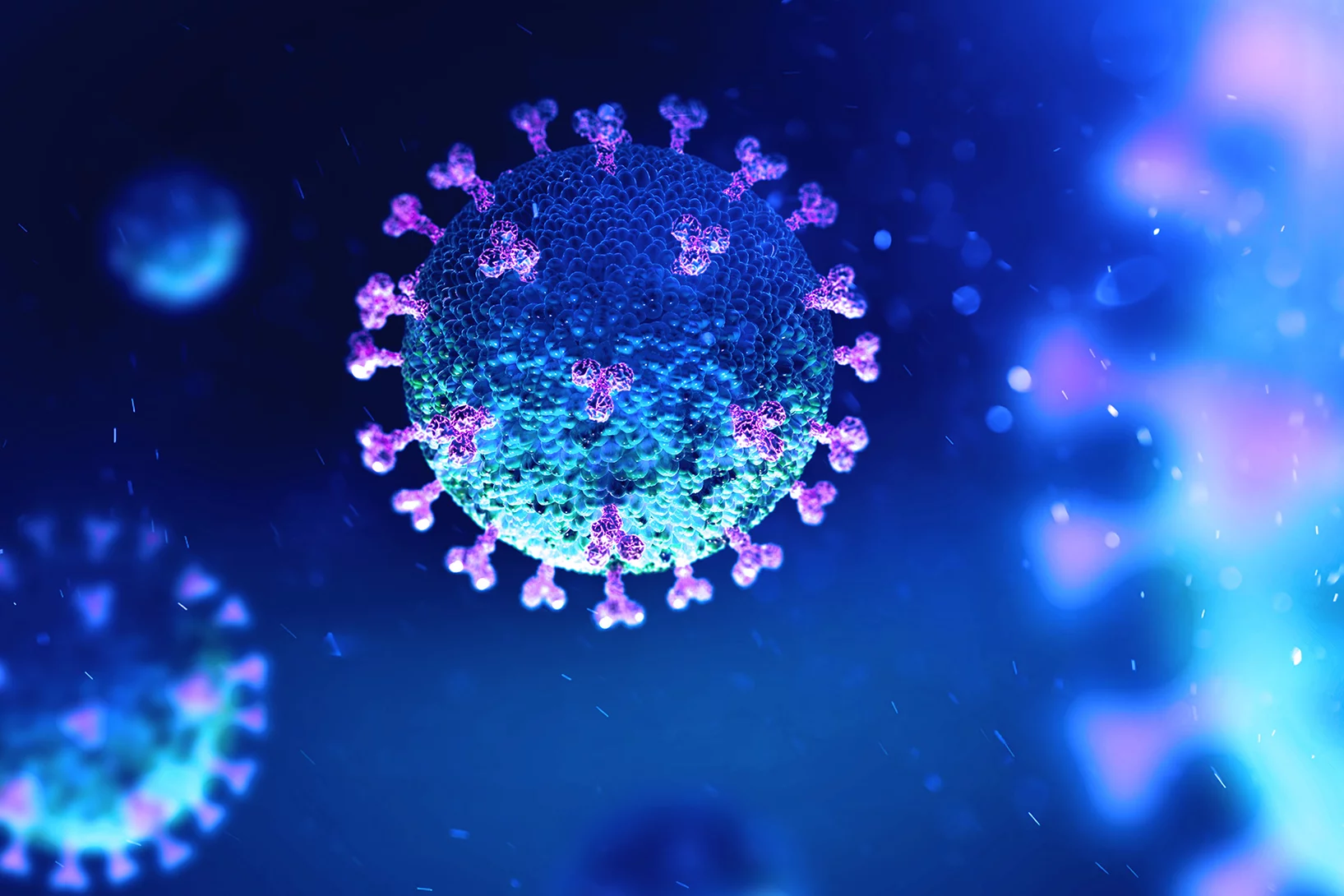
PSI: advancing in the fight against Covid-19
Crystal structure analysis, computer models, cell cultures – to pursue research on Sars-CoV-2, PSI is exploring many avenues. An overview.
Stoneflies: Youth influences adulthood
Evolutionary biologists at the University of Bonn scan 219 species in different particle accelerators, beside others the Swiss Light Source SLS.
How catalysts age
Catalysts used in industry change their material structure over the years. Using a new method, PSI researchers have now studied this on the nanoscale.

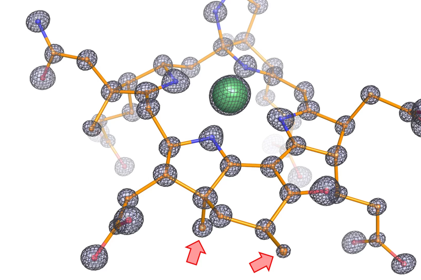
Crystal structure of SARS-CoV-2 Orf9b in complex with human TOM70 suggests unusual virus-host interactions
In a study published in Nature Communications, researchers at the NHC Key Laboratory of Systems Biology of Pathogens in Beijing, China, in collaboration with the Paul Scherrer Institut characterize the interactions of SARS-CoV-2 orf9b and human TOM70 biochemically, and they determine the 2.2 Å crystal structure of the TOM70 cytosolic domain with a bound SARS-CoV-2 orf9b peptide.

Quantifying oriented myelin in mouse and human brain
Myelin 'insulates' our neurons enabling fast signal transduction in our brain. Myelin levels, integrity, and neuron orientations are important determinants of brain development and disease. Small-angle X-ray scattering tensor tomography (SAXS-TT) is a promising technique for non-destructive, stain-free imaging of brain samples, enabling quantitative studies of myelination and neuron orientations, i.e. of nano-scale properties imaged over centimeter-sized samples.

How remdesivir works against the coronavirus
Researchers at Goethe University Frankfurt, in cooperation with the PSI have probably discovered another, previously unknown mechanism of action of the antiviral remdesivir.

Combating antimicrobial resistance
In a study addressing the global health threat of drug resistance, researchers at the Biozentrum, University of Basel, have revealed how a new antibiotic, Darobactin, binds to the external membrane of gram-negative bacteria.
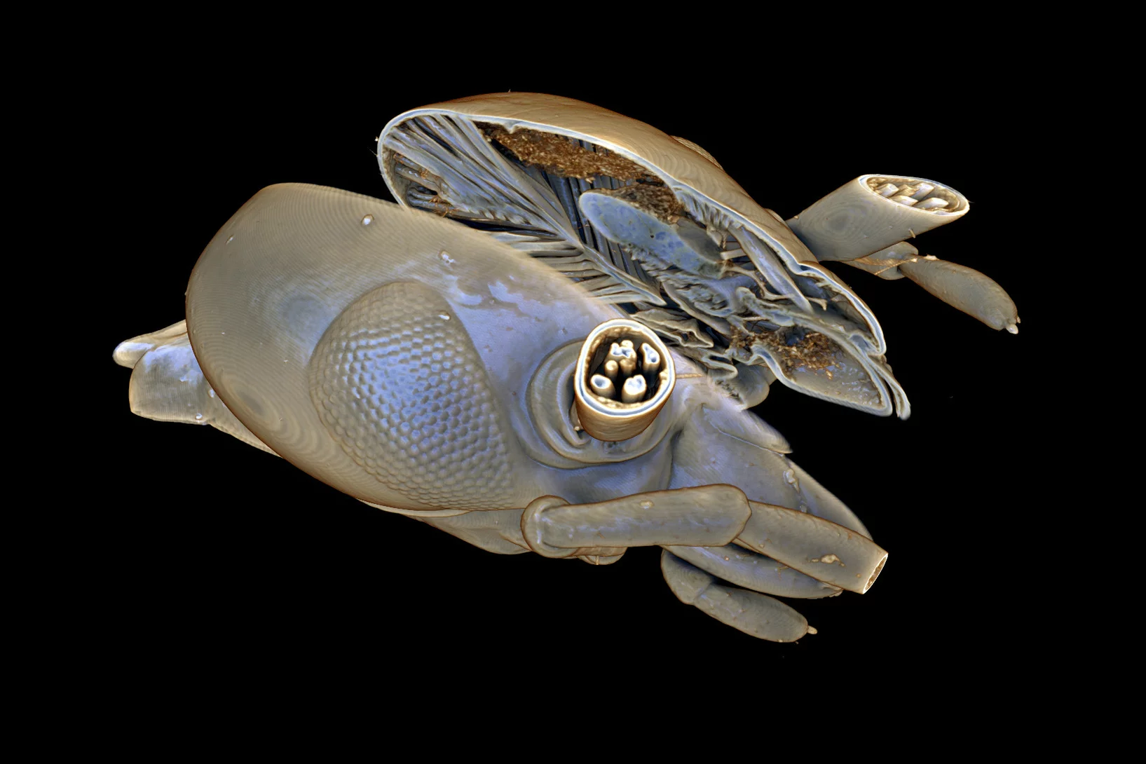
Deep evolutionary origins of the human smile
Detailed characterization of the tooth and jaw structure and development among shark ancestors by synchrotron based X-ray tomographic microscopy at TOMCAT led an international team of researchers from the Naturalis Biodiversity Center in Leiden and the University of Bristol to the discovery that while teeth evolved once, complex dentitions have been gained and lost many times in evolutionary history.
Women in Engineering Materials highlights Dr. Lucia Romano's work
The paper "High aspect ratio grating microfabrication by Pt assisted chemical etching and Au electroplating” by Dr. Lucia Romano & coauthors has been highlighted in the "Women in Engineering Materials" Virtual Issue published on Advanced Engineering Materials. This Virtual Issue draws attention to outstanding works within Materials Science created under the lead of women as principal investigators.
Congratulations to Lucia and the x-ray optics design and fabrication team!
HERCULES SCHOOL 2021 AT PSI
During the week of March 15 – 19, we had the pleasure to welcome 20 international PhD students, PostDocs and assistant professors at PSI, taking part in the first virtual HERCULES SCHOOL on Neutrons & Synchrotron Radiation.
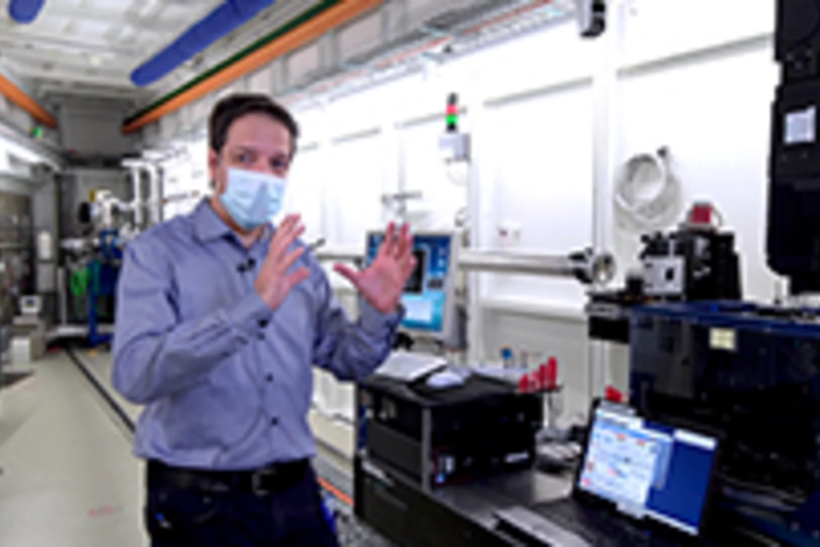
Forschung zu Covid-19 am Paul Scherrer Institut
Während viele Bereiche des Lebens eingeschränkt sind, bleiben wichtige Forschungsanlagen am PSI in Betrieb.

SLS 2.0 approved - TOMCAT 2.0 cleared for takeoff!
In December 2020 the Swiss parliament approved the Swiss Dispatch on Promotion of Education, Research and Innovation (ERI) for 2021 to 2024 which includes funding for the planned SLS 2.0 upgrade. The new machine will lead to significantly increased brightness, thus providing a firm basis for keeping the SLS and its beamlines state-of-the-art for the decades to come. The TOMCAT crew is very excited that the TOMCAT 2.0 plans (deployment of the S- and I-TOMCAT branches, see SLS 2.0 CDR, p. 353ff) have been included in the Phase-I beamline upgrade portfolio. These beamlines will receive first light right after the commissioning of the SLS 2.0 machine around mid 2025. A first milestone towards this goal has just been achieved, with the successful installation of the S-TOMCAT optics hutch during W1 of 2021. The TOMCAT scientific and technical staff would like to thank Mr. Nolte and his Innospec crew for delivering perfectly on schedule.
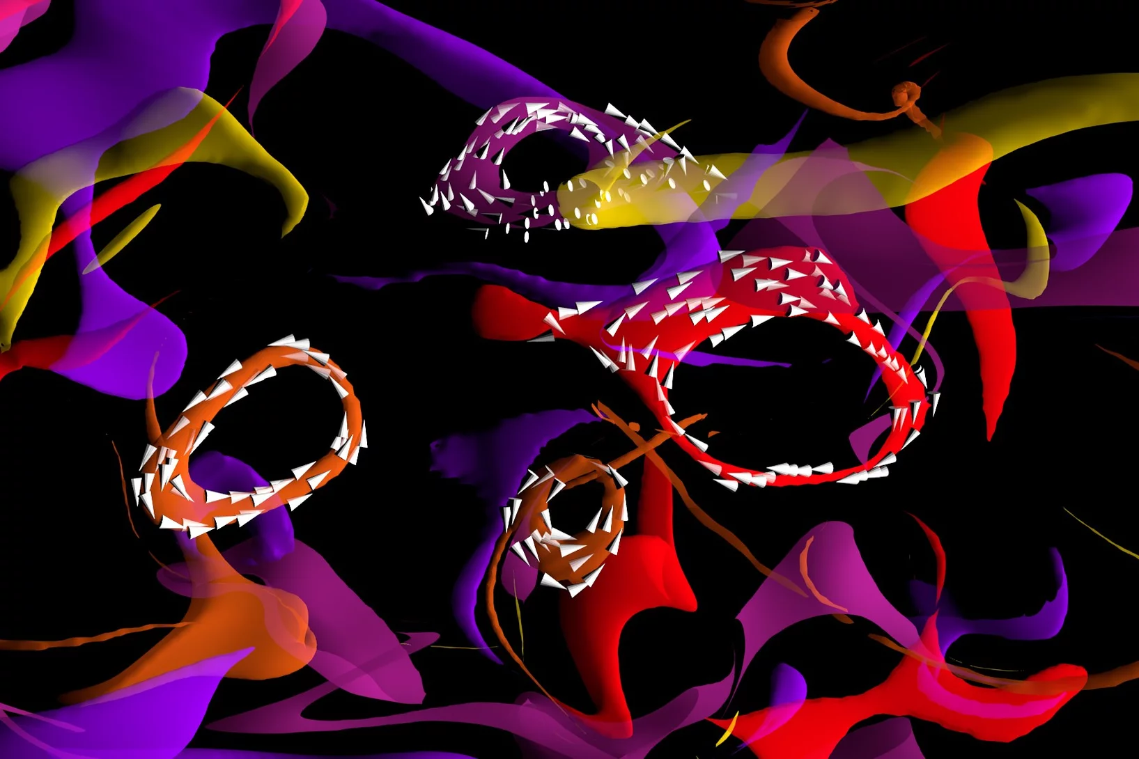
Magnetic vortices come full circle
The first experimental observation of three-dimensional magnetic ‘vortex rings’ provides fundamental insight into intricate nanoscale structures inside bulk magnets, and offers fresh perspectives for magnetic devices.
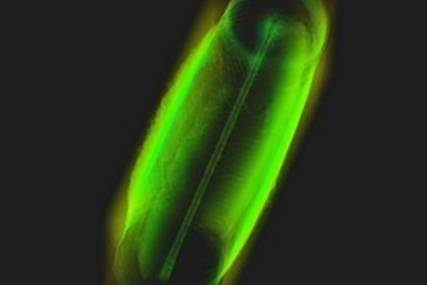
Scientists determine the structure of glass-shaping protein in sponges
Measurements at the Swiss Light Source SLS have helped to understand how the only known natural protein-mineral crystal is formed. It is part of the fascinating glass skeleton of sponges.

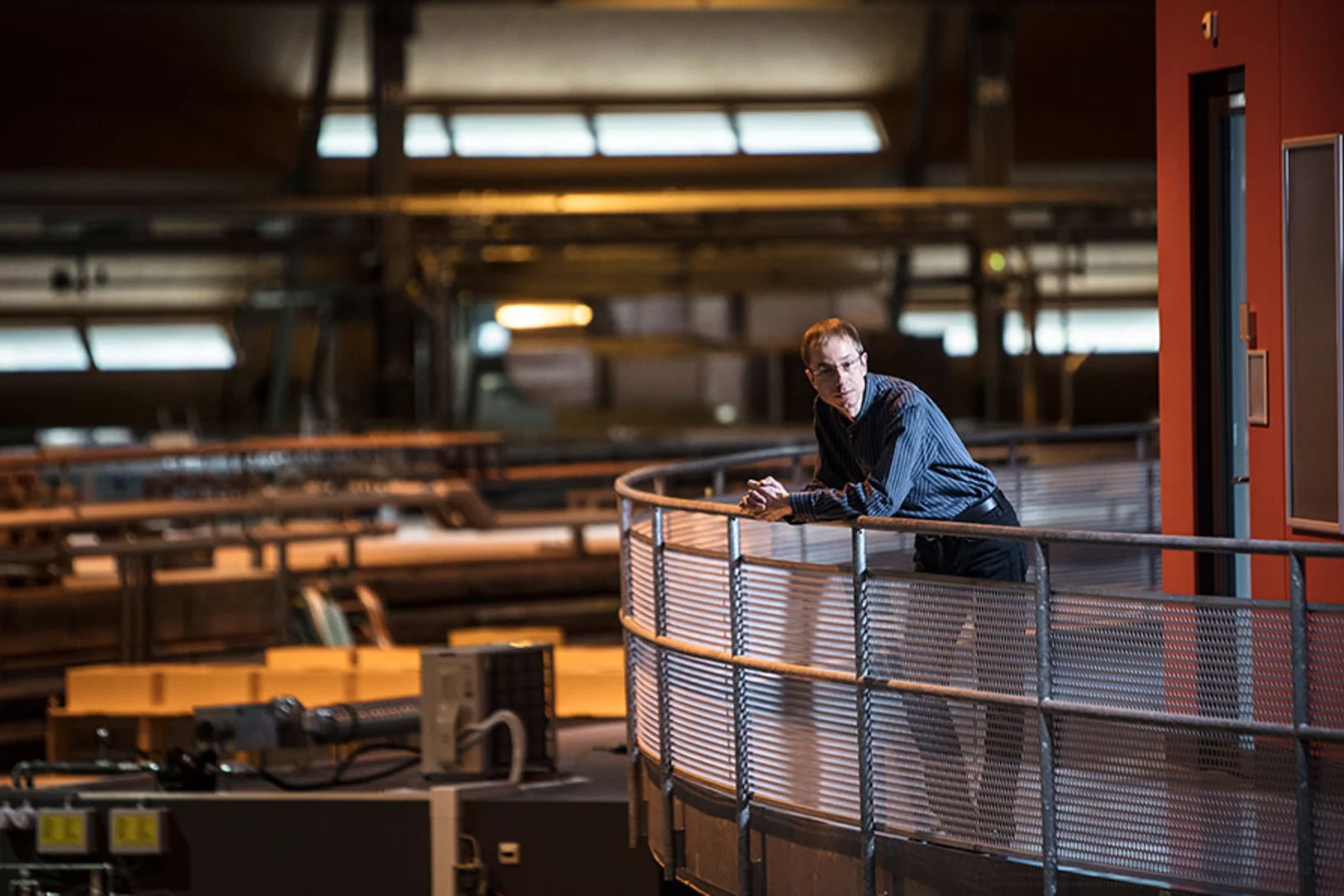
Wait and see, and grow crystals
At PSI, researchers decipher the structure of the proteins in bacteria and viruses. This knowledge can aid, for example, in the development of drugs against infectious diseases. But before the investigation can begin, an extremely tricky problem has to be solved: the crystallisation of the molecules.
Dr. Manuel Guizar-Sicairos elected as Fellow member of The Optical Society (OSA)
Dr. Manuel Guizar-Sicairos, beamline scientist at the cSAXS beamline, was elected as a Fellow Member of The Optical Society (OSA) for seminal contributions to methods and applications of coherent lensless imaging, ptychography, x-ray nanotomography, and new modalities of x-ray microscopy.
Measurements at PSI enabled detailed understanding of genetic scissors
PSI congratulates Emmanuelle Charpentier and Jennifer Doudna on winning this year's Nobel Prize in Chemistry. Experiments at the Swiss Light Source SLS in 2013 made it possible to elucidate the structure of the protein complex CRISPR-Cas9.
A question of binding
At PSI, researchers are screening molecule fragments to see if these bind to important proteins of the coronavirus SARS-CoV-2 and thus have the potential to disable it. They are hoping the many individual pieces of information will yield an answer as to what an effective drug might look like.

3D printing silica aerogels at the micrometer scale
A group of EMPA and ETH Zürich researchers have developed a new method to directly write ink made of silica aerogels in 3D. Thanks to X-ray phase contrast tomography at the TOMCAT beamline they characterized the resulting printed material with different compositions. Their results were published in Nature on August 18, 2020.
")
Phase contrast microtomography reveals nanoparticle accumulation in zebrafish
Metal-based nanoparticles are a promising tool in medicine – as a contrast agent, transporter of active substances, or to thermally kill tumor cells. Up to now, it has been hardly possible to study their distribution inside an organism. Researchers at the University of Basel in collaboration with the TOMCAT team have used phase contrast X-ray tomographic microscopy to take high-resolution captures of the nanoparticle aggregation inside zebrafish embryos.
The study was published in the journal Small and featured on the cover of its current issue.
Covid-19 research: Anti-viral strategy with double effect
Frankfurt scientists identify a possible weakness of the SARS-CoV-2 virus. They carried out part of their measurements at PSI's Swiss Light Source SLS. The research results are published this week in the scientific journal Nature.

First MX results of the priority COVID-19 call
The Dikic group at the Goethe University in Frankfurt am Main, Germany has published the first results following the opening of the "PRIORITY COVID-19 Call” at SLS.
Miniaturized fluidic circuitry observed in 3D
The team of Prof. Thomas Hermans at the University of Strasbourg in France managed to create wall-less aqueous liquid channels called anti-tubes. Thanks to X-ray phase contrast tomography at the TOMCAT beamline those anti-tubes could be observed in 3D. The exciting results were published in Nature on May 6, 2020.

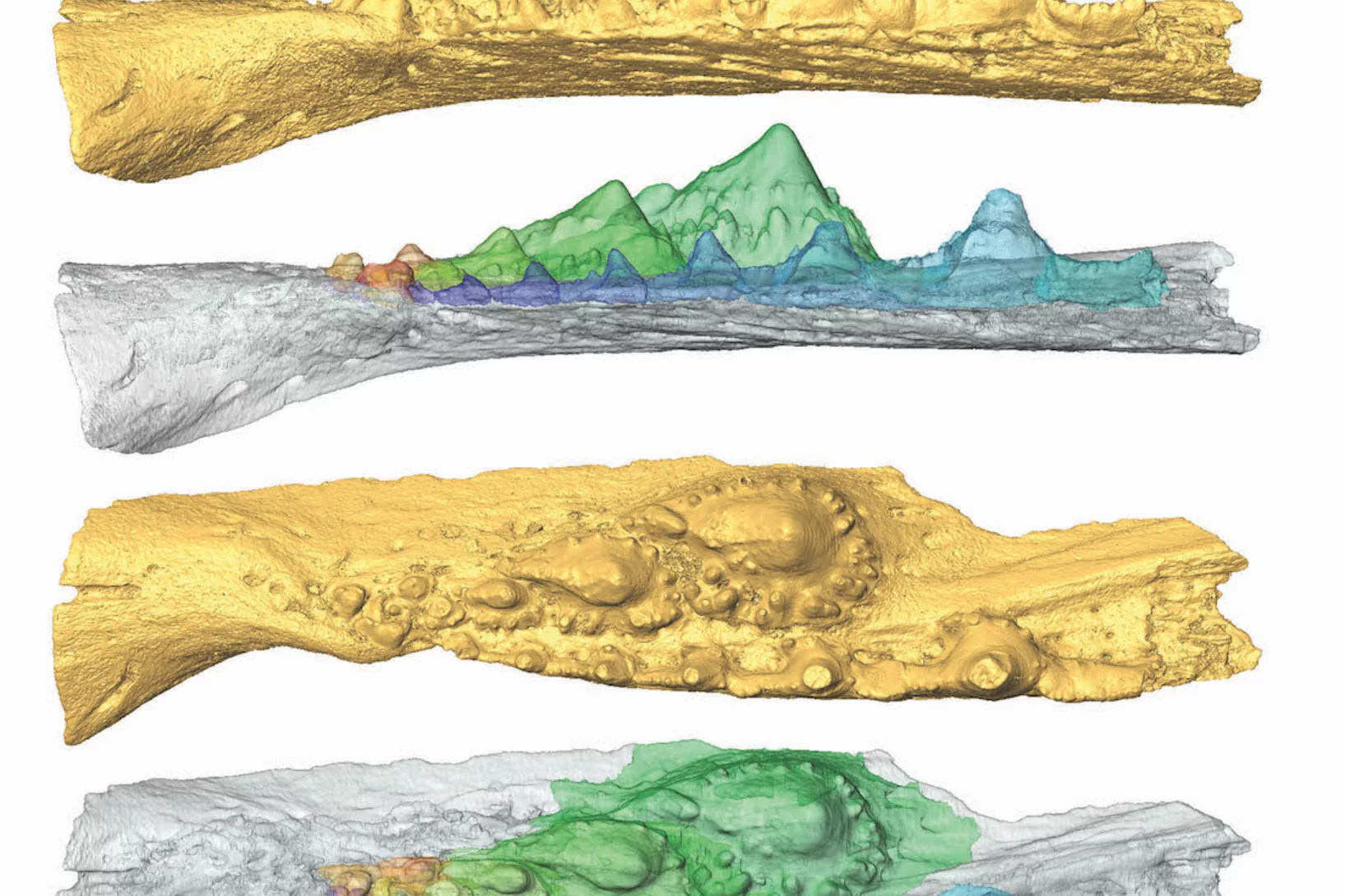
Microcalcifications in breast tissue: Paving the way for future diagnostic solutions?
Microcalcifications are a common sign in mammography. For example, in 90% of ductal carcinoma in situ microcalcifications are the first and unique sign indicating the presence of the lesion. Nevertheless, up to around 50% of the resulting biopsies reveal a benign lesion, a 'false positive'. Researchers were able to show now that breast microcalcifications detected in tumors have specific chemical and crystalline features different from those observed in benign samples. Moreover, microcalcifications detected in tumors but located outside the lesion show malignant features too. This indicates that cancer influences the surrounding tissue even if it exhibits apparently healthy morphological features. These results indicate that the biochemical differences between benign and malignant microcalcifications can be potentially identified by light-based tools, able to investigate microcalcifications inside the breast without performing biopsies.

SLS MX beamtime update
Update of the SLS MX beamline operation during the COVID-19 period

X-ray Imaging for Biomedicine: Imaging Large Volumes of Fresh Tissue at High Resolution
The TOMCAT beamline at the Swiss Light Source specializes in rapid high-resolution 3-dimensional tomographic microscopy measurements with a strong focus on biomedical imaging. The team has recently developed a technique to acquire micrometer-scale resolution datasets on the entire lung structure of a juvenile rat in its fresh natural state within the animal’s body and without the need for any fixation, staining or other alteration that would affect the observed structure (E. Borisova et al., 2020, Histochem Cell Biol).
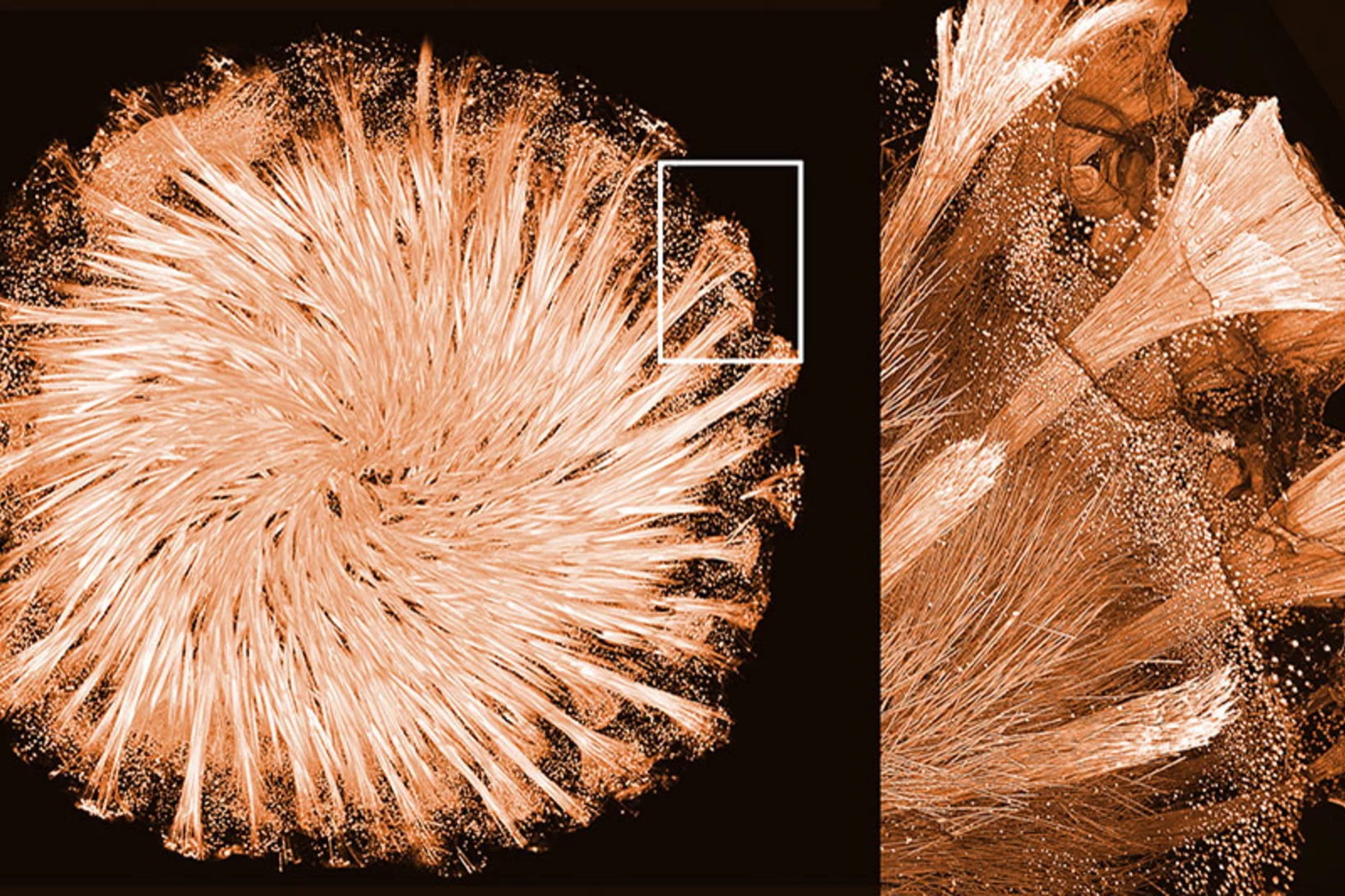
Nanoworlds in 3-D
Tomographic images from the interior of fossils, brain cells, or computer chips are yielding new insights into the finest of structures. These 3-D images are made possible by the X-ray beams of the Swiss Light Source SLS, together with detectors and sophisticated computer algorithms developed at PSI.