
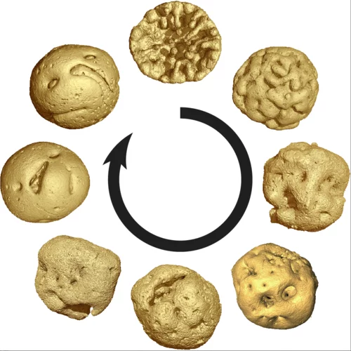
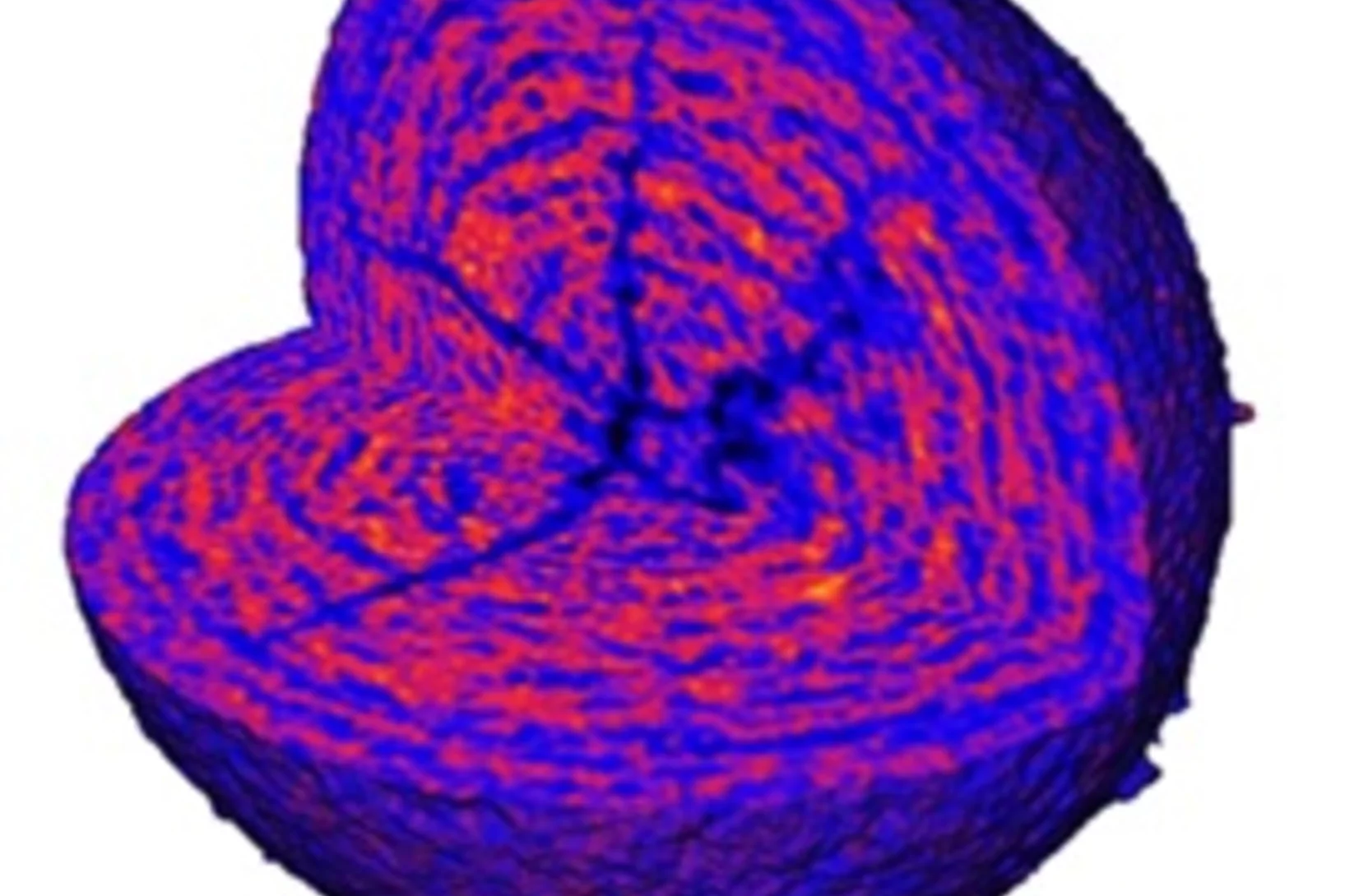
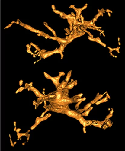
Short film of a magnetic nano-vortex
Using a newly developed imaging method, researchers were able to visualise the magnetic structure inside a material with nanoscale resolution. They succeeded in creating a short "film" consisting of seven movie frames that shows, for the first time in 3D, how tiny vortices of the magnetisation deep within a material change over time.

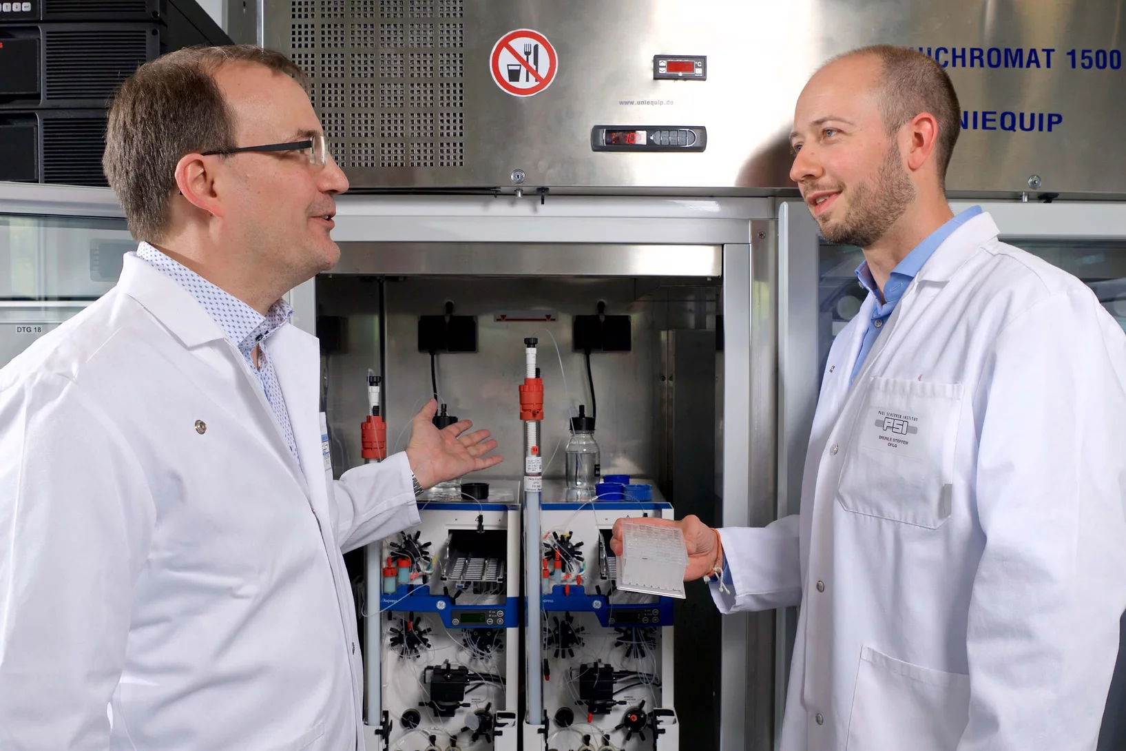
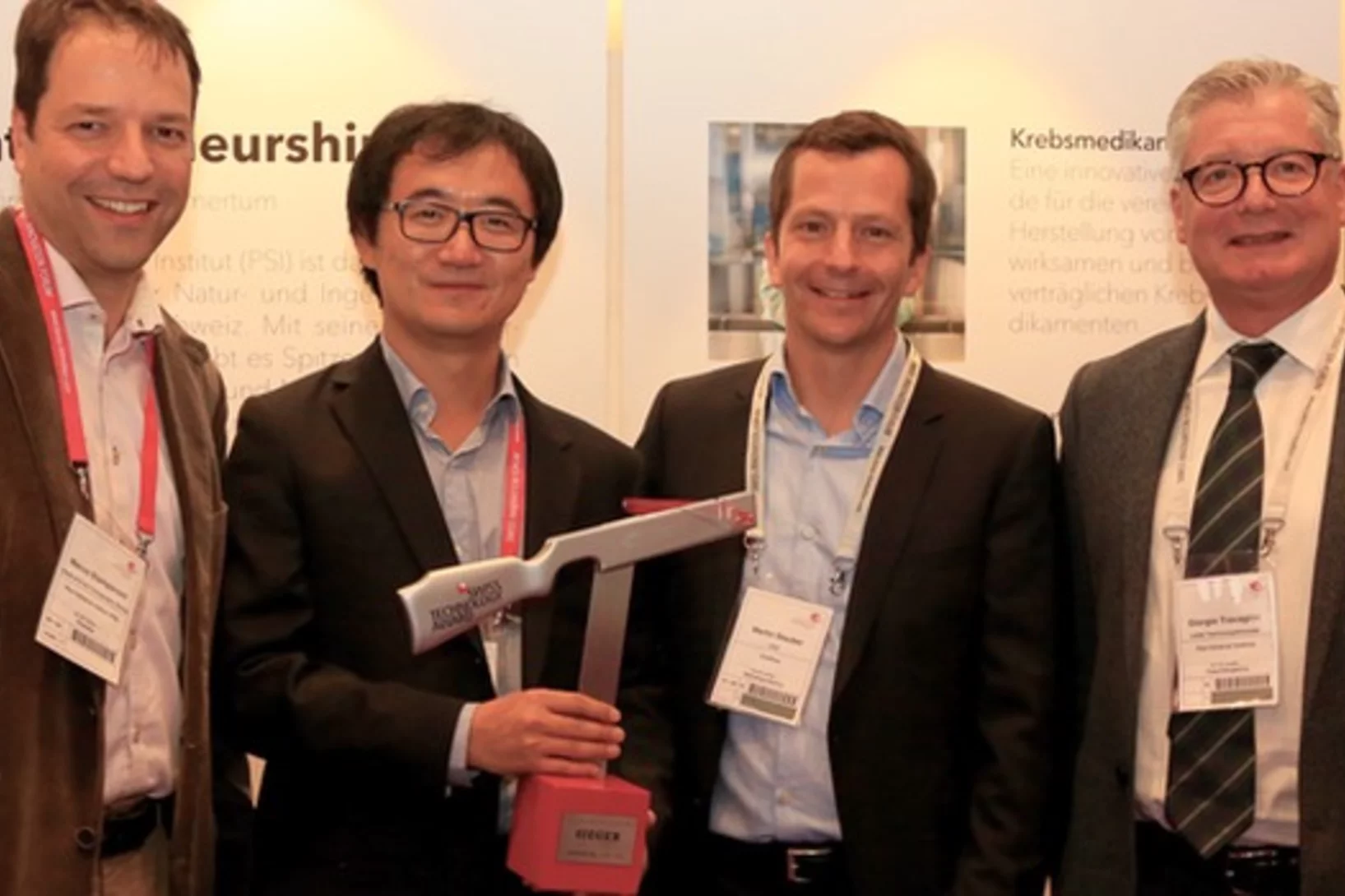
Innovation Award on Synchrotron Radiation 2019 for the development of XFEL detectors using the adaptive gain principle
The Innovation Award on Synchrotron Radiation 2019 was given to the researchers Prof. Heinz Graafsma from Desy and Dr. Aldo Mozzanica and Dr. Bernd Schmitt both from the Paul Scherrer Institute. The three physicists were honored for their contributions to the development of detectors for XFEL applications based on the dynamic gain switching principle enabling simultaneously single photon resolution and a large dynamic range. The laudation was held by Prof. Edgar Weckert from Desy. The Synchrotron Radiation Innovation Award is sponsored by SPECS GmbH and BESTEC GmbH.

MacEtch in gas phase: a new nanofabrication technology at PSI
The grating fabrication team of the X-ray tomography group has scored another record in etching technology of silicon by realizing a MacEtch process in gas phase. Ultra-high aspect ratios (up to 10 000 : 1) in the nanoscale regime (down to 10 nm) were achieved by platinum assisted chemical etching of silicon in the gas phase. The results were published in Nanoscale Horizons on February 17, 2020.
Animal embryos evolved before animals
Detailed characterization of cellular structure and development of exceptionally preserved ancient tiny fossils from South China by synchrotron based X-ray tomographic microscopy at TOMCAT led an international team of researchers from the University of Bristol and Nanjing Institute of Geology and Palaeontology to the discovery that animal-like embryos evolved long before the first animals appear in the fossil record.
A fast and precise look into fibre-reinforced composites
Researchers at the Paul Scherrer Institute PSI have developed a new process with which fibre-reinforced composite materials can be precisely X-rayed. This could help to develop better materials with novel properties.
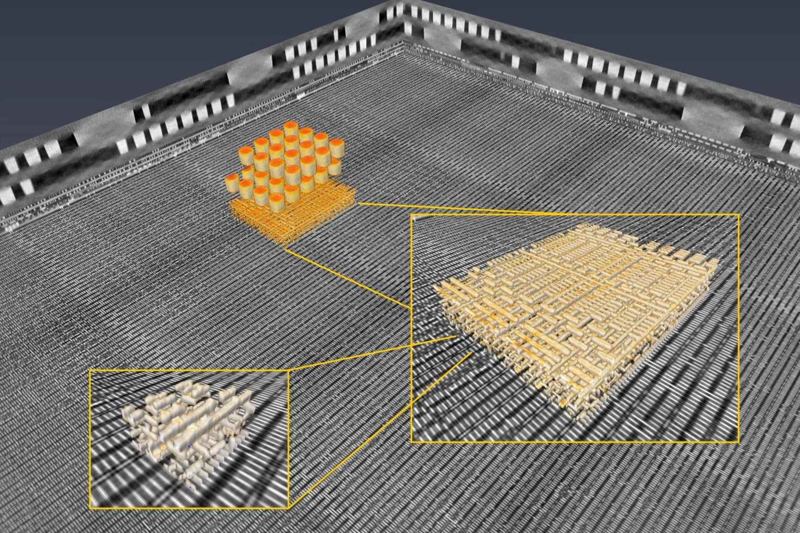
3D imaging for planar samples with zooming
Researchers of the Paul Scherrer Institut have previously generated 3-D images of a commercially available computer chip. This was achieved using a high-resolution tomography method. Now they extended their imaging approach to a so-called laminography geometry to remove the requirement of preparing isolated samples, also enabling imaging at various magnification. For ptychographic X-ray laminography (PyXL) a new instrument was developed and built, and new data reconstruction algorithms were implemented to align the projections and reconstruct a 3D dataset. The new capabilities were demonstrated by imaging a 16 nm FinFET integrated circuit at 18.9 nm 3D resolution at the Swiss Light Source. The results are reported in the latest edition of the journal Nature Electronics. The imaging technique is not limited to integrated circuits, but can be used for high-resolution 3D imaging of flat extended samples. Thus the researchers start now to exploit other areas of science ranging from biology to magnetism.
Preventing tumour metastasis
Researchers at the Paul Scherrer Institute PSI, together with colleagues from the pharmaceutical company F. Hoffmann-La Roche AG, have taken an important step towards the development of an active substance against the metastasis of certain cancers. Using the Swiss Light Source SLS, they deciphered the structure of a receptor that plays a crucial role in the migration of cancer cells.

World record in time-resolved tomography
Researchers from the Helmholtz Zentrum Berlin (HZB) and the TOMCAT beamline have achieved a new world record in time-resolved tomography by measuring over 200 tomographies per second during heating of an evolving aluminium metal foam.
Observing solid-state batteries during deformation
PSI researchers have observed mechanical processes in solid-state batteries with unprecedented precision. Using X-ray tomography at the Swiss Light Source SLS, they discovered how fissures inside the batteries propagate. These insights can help to make batteries for electric cars or smartphones safer and more efficient.

Glycation of collagen in decellularized pericardium tissue: pilot study
Aging populations and diabetics suffer from the effects of the glycation of collagen, the non-enzymatic formation of glucose bridges. While the secondary effects have been studied intensely, comparatively little is known on the direct effect of glycation on the structure of collagen. It has been demonstrated in this study, that the direct impact of glycation can be determined with sub-atomic precision, and that a model system based on abundantly available connective tissue of farm animals can be used for such studies. This opens new avenues for inspecting the effects of diabetes mellitus on connective tissues and the influence of therapies on the resulting secondary disorders.
HERCULES school 2019 at SLS
In the week of April 1-5 PSI welcomes 20 PhD students and postdocs taking part in the European HERCULES 2019 school on Neutron and Synchrotron Radiation. They will attend lectures and perform two days of practical courses at several beam lines of the Swiss Light Source.
Inside Batteries
Lithium ion batteries (LIB) are essential in modern everyday life, with increasing interest in enhancing their performance and lifetime. Secondary particles of Li-rich cathode material were examined with correlated ptychographic X-ray tomography and diffraction microscopy at different stages of cycling to probe the aging mechanism.
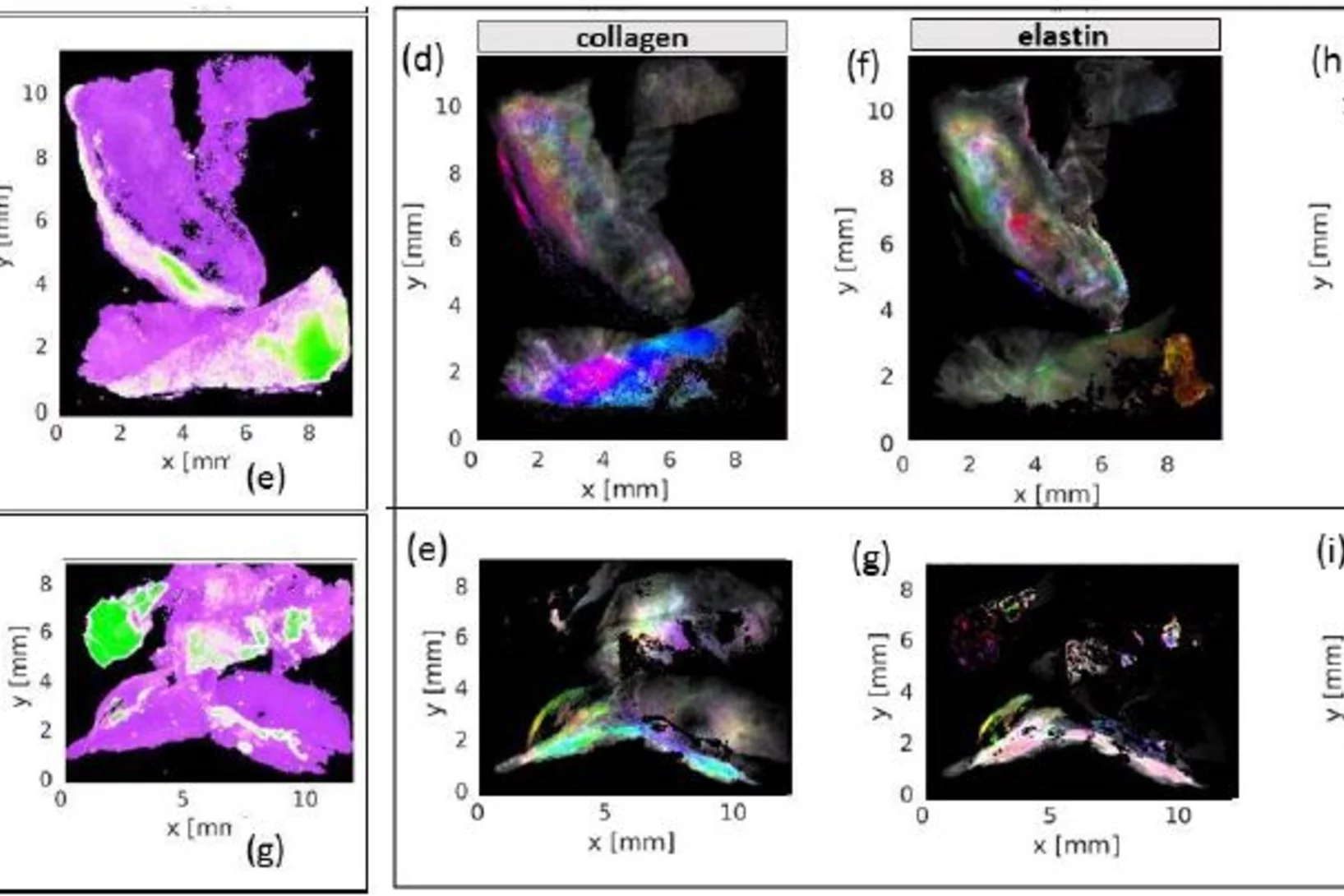
Insights into a well-known disease in ageing populations: Abdominal and popliteal aneurysm
Abdominal aortic aneurysm, an enlargement of the abdominal aorta, may lead to rupture and thus acute health issues and death. Scanning X-ray imaging enabled new insights in the nano-structure of calcifications associated with abdonimal and popliteal aneurysm and allowed mapping the distribution of nano- and micro-calcifications as well as of collagen, elastin and myofilament as building blocks of connective tissue across samples from human donors.
Virtual lens improves X-ray microscopy
A method developed by PSI researchers makes X-ray images of materials even better. The researchers took a number of individual images while moving an optical lens. From these, with the help of computer algorithms, they generated one overall image.
Soft-tissue evidence for homeothermy and crypsis in a Jurassic ichthyosaur
Synchrotron-based X-ray tomographic microscopy of melanophores (skin pigment cells) of an amazingly well preserved 180 million years old ichtyosaur (extinct marine reptile similar to whales) contributed in a multidisciplinary investigation to the new findings published today in Nature.
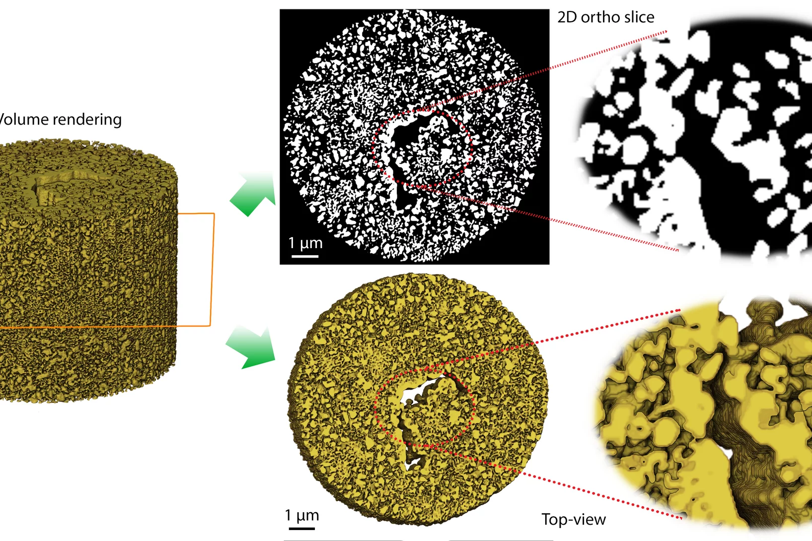
Helping chemists to understand degradation and stabilization of catalytic nanoporous gold structures
Catalytic materials are ubiquitously used in industrial processes to perform chemical reactions efficiently and in a sustainable manner. Nanoporous gold (npAu) is a monolithic sponge-like catalyst exhibiting a hierarchical structure with pores and connecting ligaments of typically 10 to 50 nm.
A crystal-clear picture
Fast and accurate data collection for macromolecular crystallography using the JUNGFRAU detector.
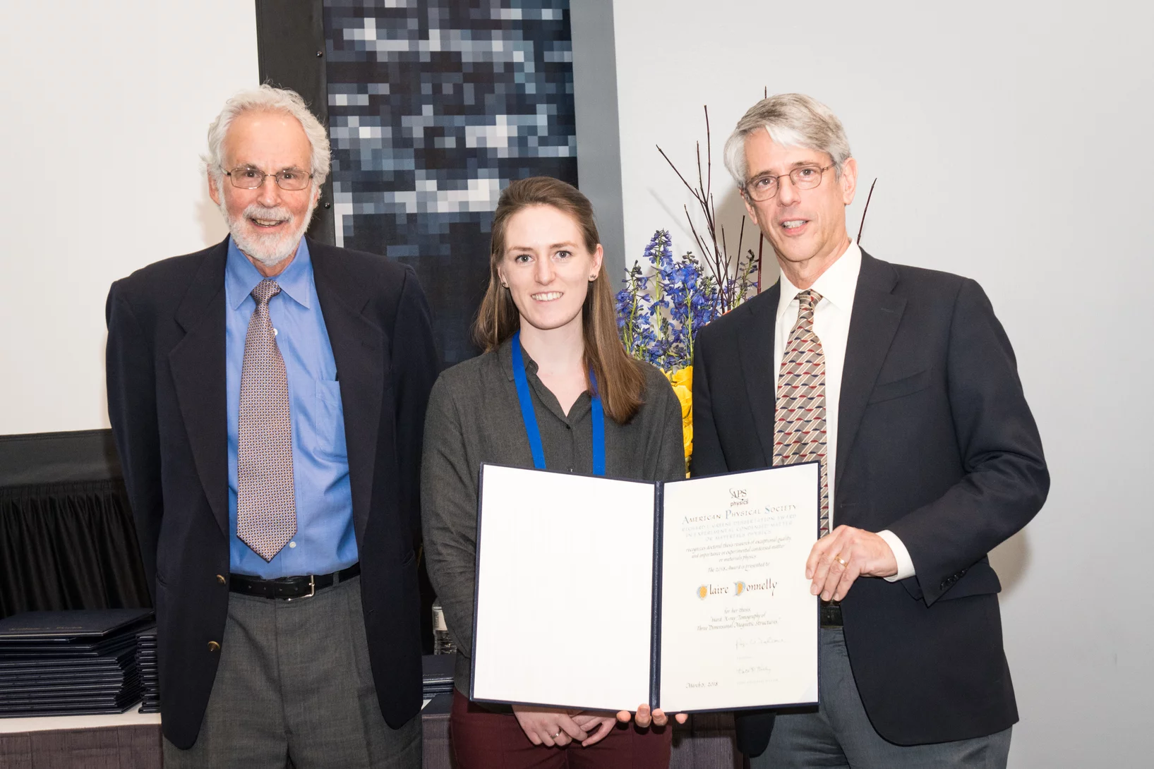
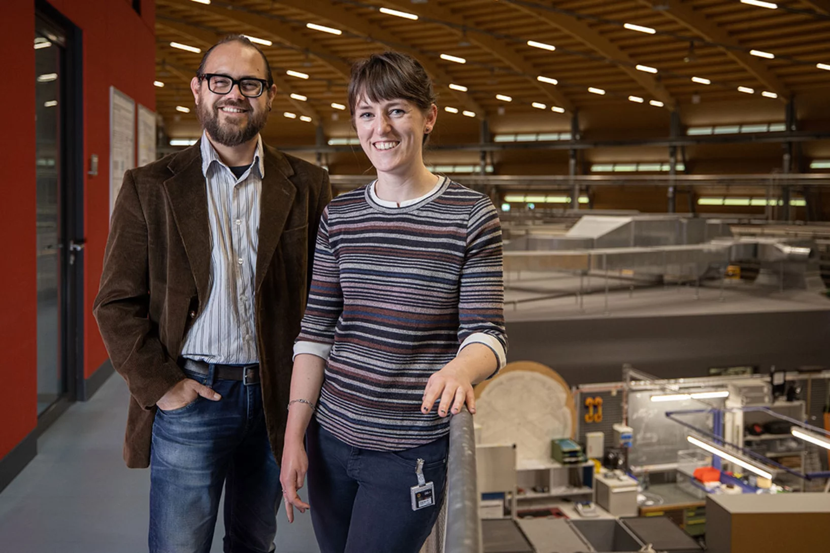
Claire Donnelly dissertation research awards
Claire Donnelly, Mesoscopic Systems (ETH Zurich - PSI), was awarded the COMSOL SPS Award in Computational Physics, the Werner Meyer-Ilse Memorial Award, the ETH Medal for an outstanding doctoral thesis, and the American Physical Society Richard L. Greene Dissertation Award.
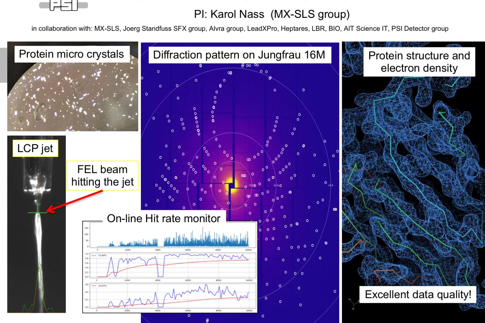
First serial femtosecond crystallography (SFX) pilot user experiment at SwissFEL
On the 7th to 12th of August 2018, a collaborative group of scientists from the Paul Scherrer Institute and members of the LeadXpro and Heptares pharmaceutical companies led by Karol Nass (PSI macromolecular crystallography MX-SLS group) performed the first serial femtosecond crystallography (SFX) pilot user experiment at the SwissFEL X-ray free electron laser (XFEL).
Rhine-Knee Regiomeeting 2018
Since it was first established in 1987, the annual Regio-Meeting has been instrumental in facilitating interactions in the structural biology community in southwestern Germany, the eastern region of France and an expanding area of Switzerland. It is set as an informal event to foster young scientists to discuss their research results in an international context. The 2018 edition will take place in the heart of Switzerland in Emmetten from September 26 to 28, 2018. Registration and abstract submission deadline: September 7, 2018.
HERCULES at the Swiss Light Source
In the week of March 18-23 PSI welcomes 20 PhD students and postdocs taking part in the HERCULES 2018 school on Neutron and Synchrotron Radiation. They will attend lectures and perform two days of practical courses at several beam lines of the Swiss Light Source.
PSI spin-off GratXray wins Swiss Technology Award 2017
A spin-off from PSI has received this year's Swiss Technology Award: The young company GratXray is developing a new method for early diagnosis of breast cancer.

Making the world go round - a look into the structure of a prominent heterogeneous catalyst
Fluid catalytic cracking catalysts, which are composite particles of hierarchical porosity, were examined using ptychographic X-ray tomography. These particles are essential to the conversion of crude oil into gasoline. Examination of catalysts at decreasing levels of catalytic conversion efficacy allowed the detection of possible deactivation causes.
In Situ Serial Crystallography 2 Workshop at the SLS
A workshop dedicated to the presentation of the in meso in situ serial crystallography (IMISX) method (Huang et al. 2015, 2016 ActaD) for the X-ray structure determination of membrane proteins is organised at the Swiss Light Source at PSI for the second time. It will be held between November 27 and 29, 2017.
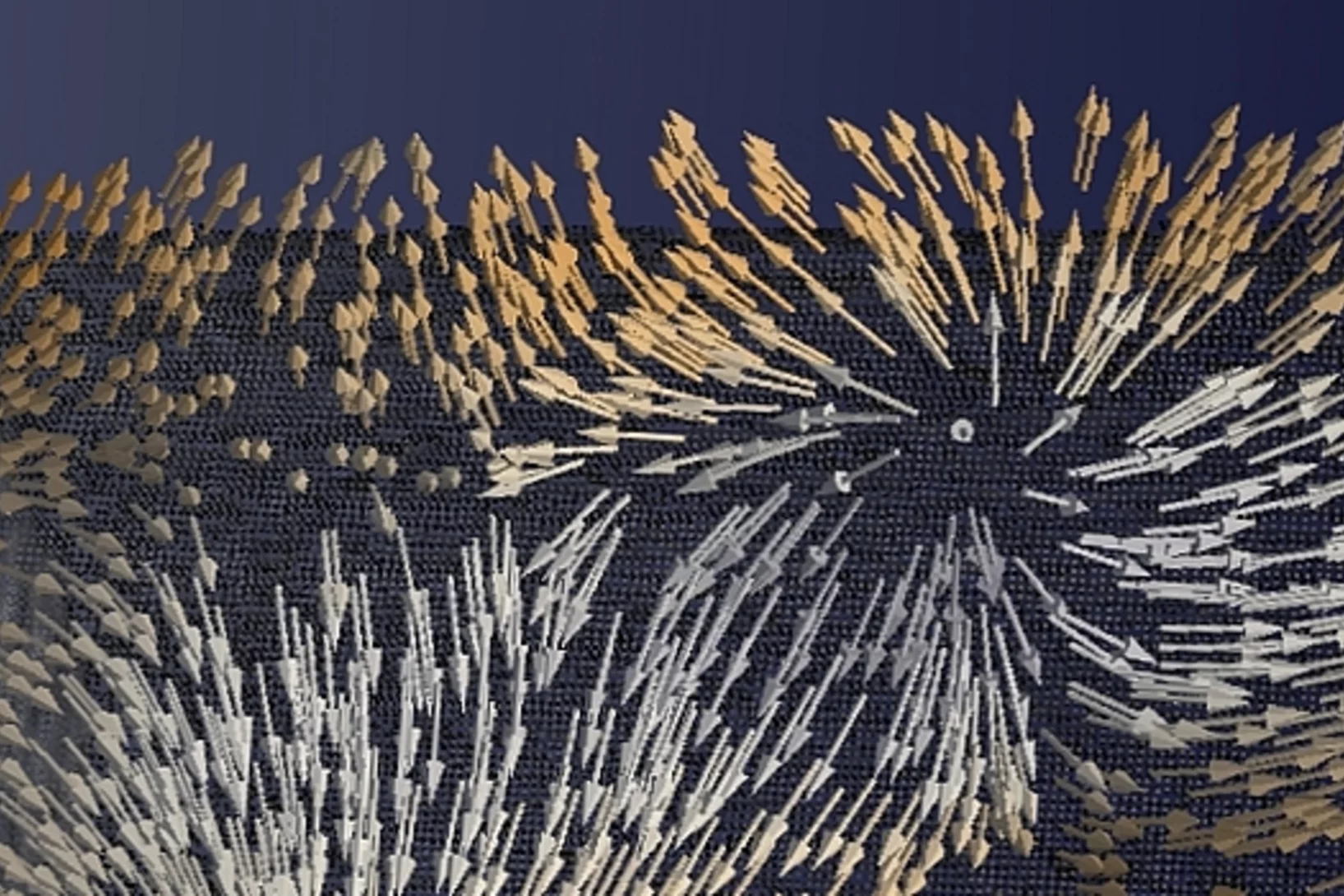
Diving into magnets
For the first time, scientists have made visible the directions of the magnetisation inside a 3D magnetic object. The smallest details in their visualisation were ten thousand times smaller than a millimetre. Among others, the magnetic structure contained one outstanding kind of pattern: magnetic singularities called Bloch points, which up to now were only known in theory.
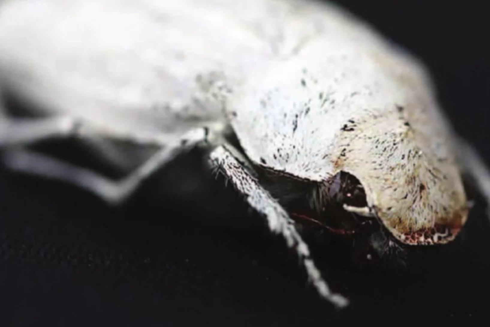
Photonic structure of white beetle wing scales: optimized by evolution
A very thin layer on this beetle’s wings exhibits a complicated structure on the nanoscale that gives them a bright white color. X-ray nanotomography acquired at the Swiss Light Source provides a faithful image of this structure in three dimensions with which scientists can confirm its evolutionary optimization: just enough material for an efficient reflection of white light.
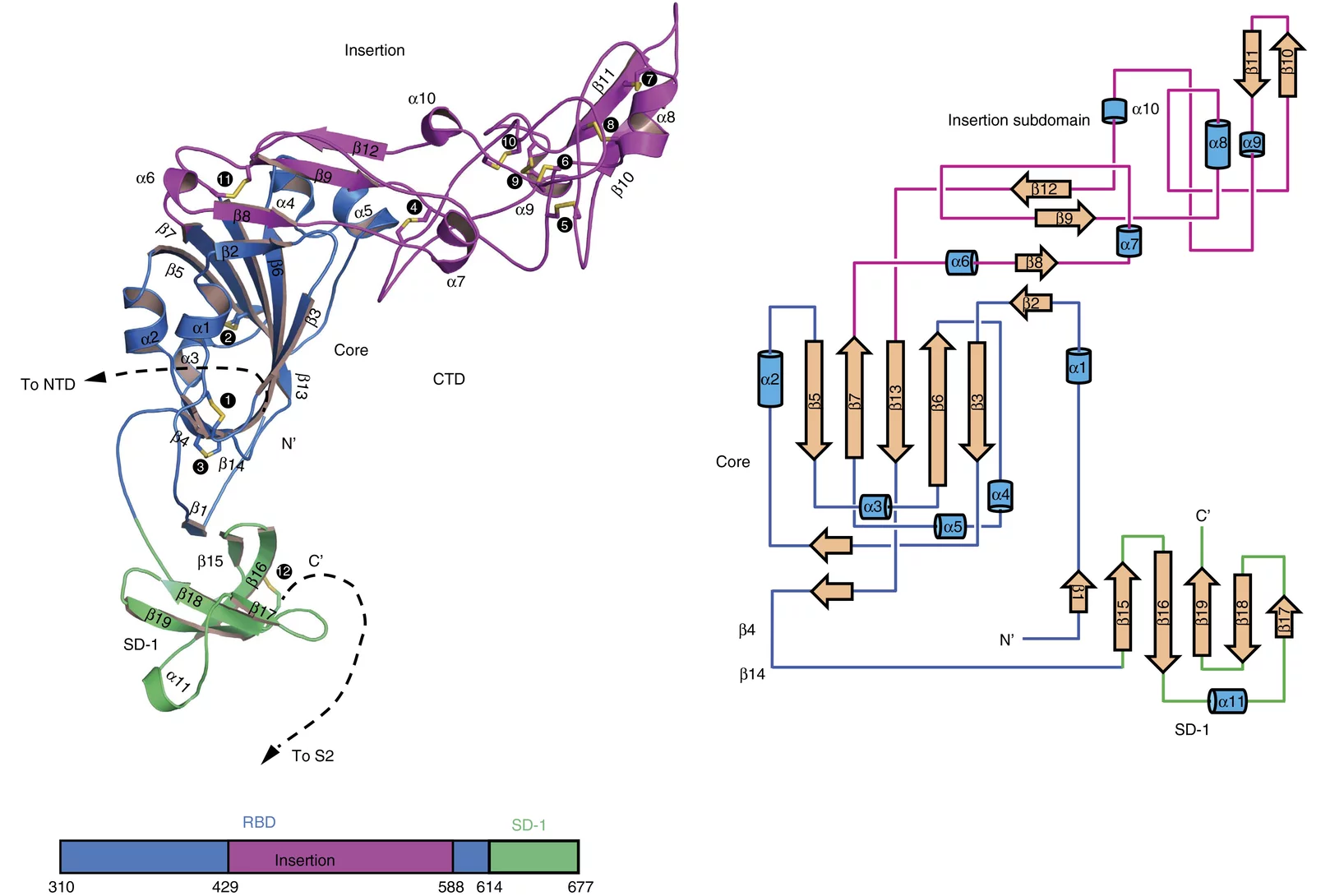
Towards understanding of human betacoronavirus HKU1 life cycle
Researchers from China and USA join forces with Swiss Light Source (SLS) macromolecular crystallography (MX) beamline scientists in a study, which aims at understanding an important step in the life cycle of the human betacoronavirus HKU1.

3-D X-ray imaging makes the finest details of a computer chip visible
Researchers at the PSI have made detailed 3-D X-ray images of a commercially available computer chip. In their experiment, they examined a small piece that they had cut out of the chip beforehand. This sample remained undamaged throughout the measurement. It is a major challenge for manufacturers to determine if, in the end, the structure of their chips conforms to the specifications. Thus these results represent one important application of an X-ray tomography method that the PSI researchers have been developing for several years.
1000 Structures solved at X06DA-PXIII
The macromolecular crystallography beamline X06DA-PXIII has reached 1,000 structures in the Protein Data Bank (PDB) on February 22, 2017.
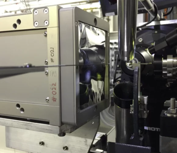
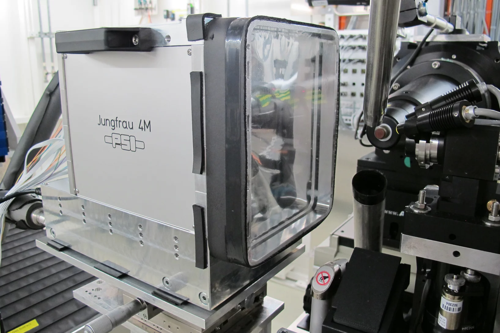
First protein structure solved using the JUNGFRAU detector!
JUNGFRAU is a charge-integrating, two-dimensional pixel detector developed at the Paul Scherrer Institut for use at free-electron lasers, in particular SwissFEL, and synchrotron light sources. On the 10th October, the first protein crystallography experiment using the JUNGFRAU detector, was performed at the beamline X06SA (PXI) of the Swiss Light Source by the members of the Protein Crystallography and Detectors groups at PSI.