Researchers reveal in detail what is happening in the retina during the process of sight
During the process of sight, light passes into the eye and triggers a whole series of chemical reactions. At the end of this process, a nerve pulse is generated that carries the visual information to the brain. At the beginning of the process, the light interacts with a protein molecule called Rhodopsin. This molecule contains the actual light sensor that is stimulated by the incoming light and changes its form, in order to trigger the rest of the process.
Researchers from the Paul Scherrer Institute, together with colleagues from the UK and the USA, have now managed to determine the exact structure of the Rhodopsin molecule in its short-lived, excited state. From this, they have obtained a precise picture of the first step of the process of sight. This result may provide the basis for understanding the hereditary eye disease Retinitis Pigmentosa (a group of genetic eye conditions that lead to incurable blindness) and indicate ways of treating it, or of slowing down its progress. Simultaneously, the result provides a foundation for understanding many other processes in the body that are based on a similar mechanism, such as the perception of smells or the control of biological processes by hormones. The research results have been reported in the latest issue of the scientific journal Nature.
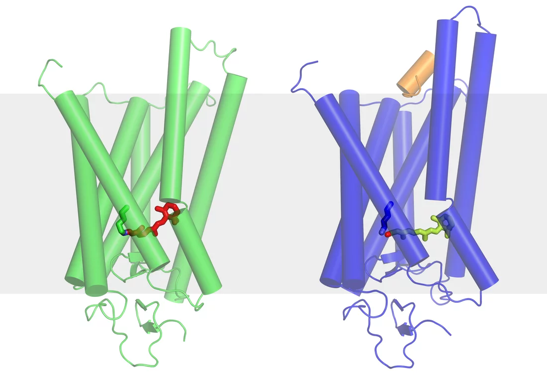
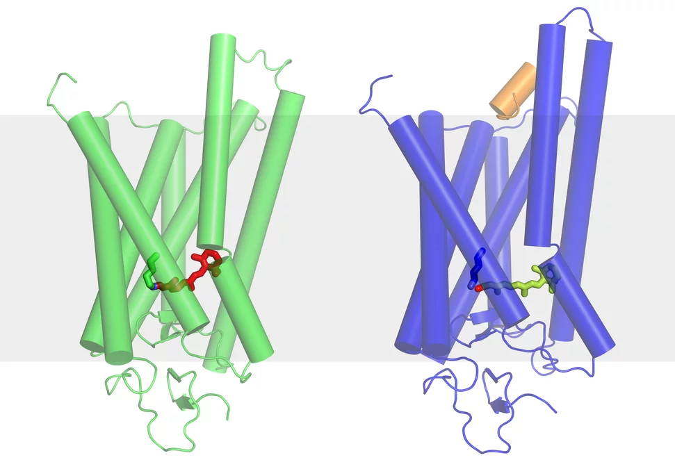
Sight is a highly complex process. A whole series of chemical reactions has to take place before the impression of what has been seen reaches our consciousness. At the very beginning of the process, light reaches photoreceptor cells – the cones or rods – in the retina of the eye. In the cell membranes of the rods in the retina – responsible for seeing under low-light conditions –are Rhodopsin molecules, the actual light sensors. Each sensor is made up of a total of seven rod-like molecular components that extend through the membrane from the outside of the cell into its interior. When light from outside the eye falls on the Rhodopsin, its rod-like components are reorganised throughout its whole structure, including the region inside the cell. This allows a so-called G-protein molecule in the cell to connect to the Rhodopsin. The docking of the G-protein then triggers a whole cascade of events, which in the end generates a nerve pulse.
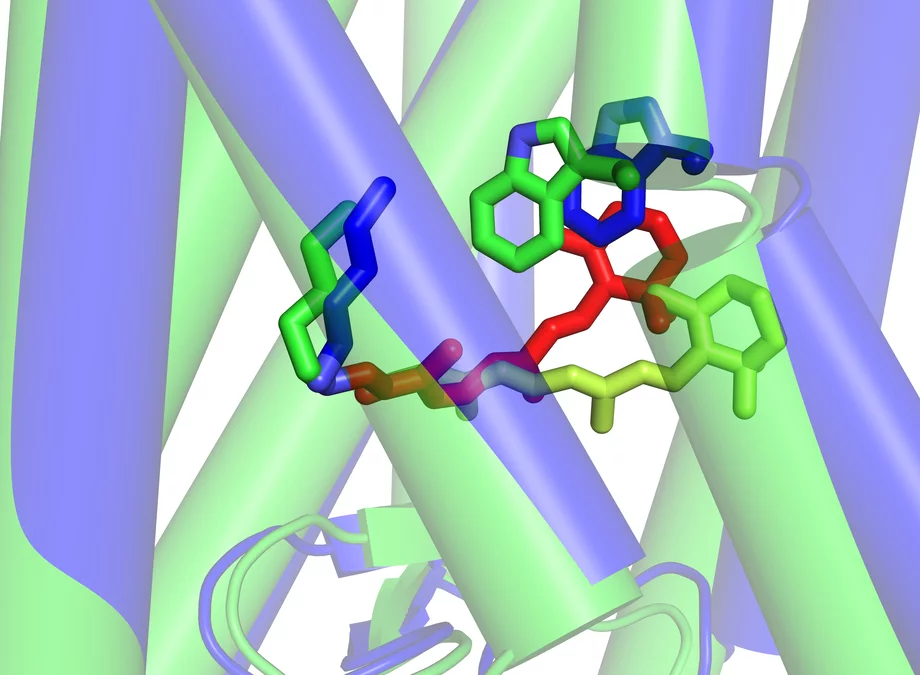
The actual light-sensitive pigment is retinal, a form of vitamin A that is located between the seven parts of the Rhodopsin in the form of a small, bent molecule. As soon as light falls on the retinal, this small molecule stretches and pushes the parts of the Rhodopsin apart, creating room for the G-protein. Now, scientists from the Paul Scherrer Institute have been able to determine the structure of the Rhodopsin molecule in its activated state, i.e. in the distorted form with the stretched retinal that was produced by the light. This state is actually very short-lived, as the Rhodopsin has to return as quickly as possible to the original state in which it is receptive to incoming light. The PSI researchers have found a way to modify the molecule slightly so that it remains in its activated state for a longer period of time, thus allowing its structure to be determined. Together with the known structure of the inactive form of Rhodopsin as found in the absence of light, this enables the scientists to understand exactly how the early stages of sight begin at the molecular level.
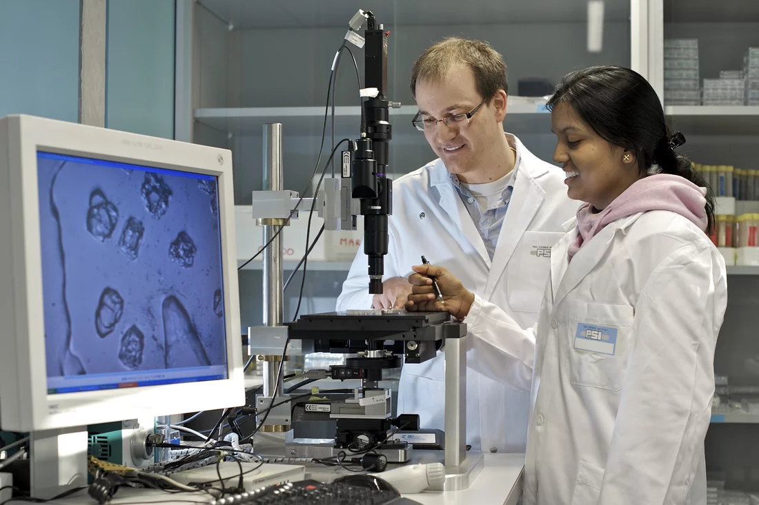
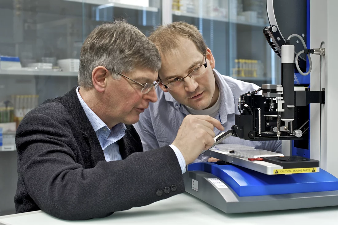
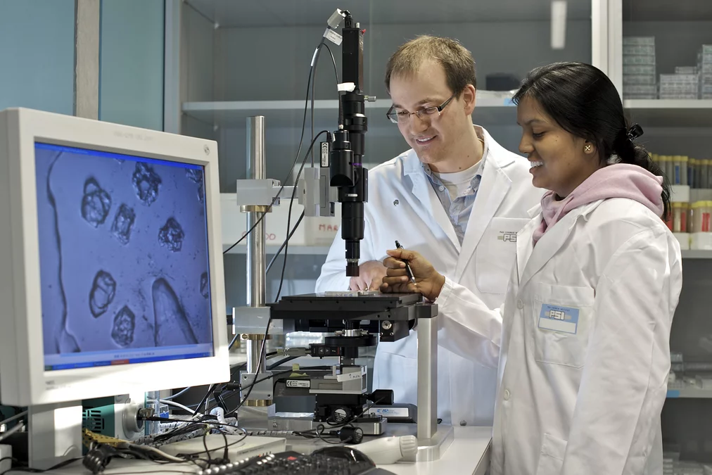
For the investigation, a large quantity of the appropriate molecules was created and arranged in a regular crystalline order. Fortunately, Rhodopsin is one of the very few membrane proteins of this class that can be crystallized. In the experiment, these crystals were illuminated with synchrotron light and, from the deflection of the light by the crystal, the scientists were able to deduce the structure of the molecule. The experiments were performed at the Swiss Light Source (SLS) at the Paul Scherrer Institute and also at two other, similar, facilities in other countries.
Understanding the fundamental mechanisms of life
“Our studies of the Rhodopsin molecule also help us to understand a large class of similar molecules, of which there are more than 800 present in the human body,” explains Jörg Standfuss, head of the research project. “Most of these are not responsive to light but to other stimuli, and thus perform the widest variety of tasks. For the sense of smell, they respond to substances present in the inhaled air; or they act as receptors for hormones within the body – for example, as beta receptors in the heart that are responsible for controlling blood pressure”. These are the receptors which are manipulated by the medicines, known as Beta Blockers, used against hypertension. Overall, these molecules are of great interest in pharmaceutical research because one can influence, or even block, many processes in the body through them. For example, many medicines used against cardiac arrhythmia, migraine or various allergies interact with these receptors. The detailed structure of the beta receptor was the topic of another article published in Nature by scientists from PSI, together with colleagues from the University of Cambridge.
Optimized therapy for eye disease
“Currently, we are using the experience we have gained from investigating the structure of modified Rhodopsin molecules for studying the common eye disease Retinitis Pigmentosa”, explains Standfuss. In this hereditary disease, the Rhodopsin in the cones of the retina is often altered. Instead of being regularly renewed, as is the case in a healthy eye, parts of “old” Rhodopsin molecules remain in the cells and slowly ‘poison’ them. At first, this leads to night blindness and then, with the passage of time, to a greatly reduced field of vision. Standfuss says: “In the future, we will be able to determine in detail how Rhodopsin is changed in this disease and then investigate how small molecules used as medication to halt the disease are incorporated into the Rhodopsin”. Based on this knowledge, one might then be able to use a computer to optimize the structure of the medication.
International research
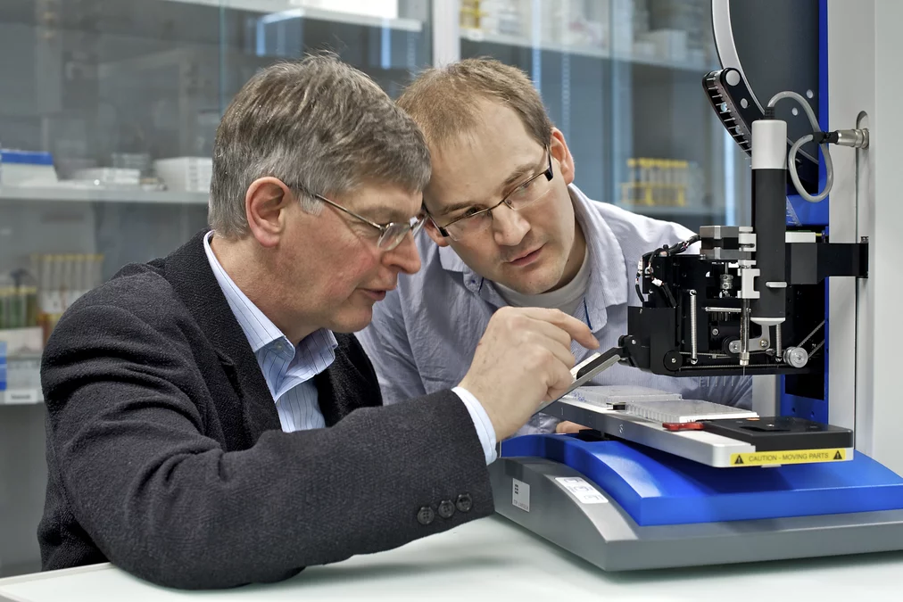
Jörg Standfuss and Prof. Gebhard Schertler, head of the Laboratory for Biomolecular Research at PSI, began this research project while working at the MRC Laboratory of Molecular Biology in Cambridge (UK) and completed it after their relocation to PSI. Throughout, they cooperated closely with colleagues from the Brandeis University in the USA.
Text: Paul Piwnicki
About PSI
The Paul Scherrer Institute develops, builds and operates large-scale, complex research facilities, and makes these facilities available to the national and international research community. The Institute”s own research focuses on solid-state physics and the materials sciences, elementary particle physics, biology and medicine, as well as research involving energy and the environment. With a workforce of 1400 and an annual budget of about 300 million CHF, PSI is the largest research institution in Switzerland.
Contact
Dr. Jörg Standfuss, Laboratory of Biomolecular Research, Paul Scherrer Institute, 5232 Villigen PSI, SwitzerlandPhone: +41 56 310 2586, E-mail: joerg.standfuss@psi.ch
Prof. Dr. Gebhard Schertler, Laboratory of Biomolecular Research, Paul Scherrer Institute, 5232 Villigen PSI, Switzerland
Phone: +41 56 310 4265, E-mail: gebhard.schertler@psi.ch
Original Publication
The structural basis of agonist induced activation in constitutively active RhodopsinJörg Standfuss, Patricia C. Edwards, Aaron D’Antona, Maikel Fransen, Guifu Xie, Daniel D. Oprian, Gebhard F. X. Schertler
Nature Advance Online Publication 9 March 2011;
doi: 10.1038/nature09795

