
A new method forms the basis for the widespread use of an X-ray technique which distinguishing types of tissue that normally appear the same in conventional X-ray images.
Traditional X-ray images can clearly distinguish between bones and soft tissue, with muscles, cartilage, tendons and soft-tissue tumours all look virtually identical. The phase-contrast technique developed a few years ago at the Paul Scherrer Institute enables X-ray images to be produced that clearly distinguish between these tissue types. Researchers at the Paul Scherrer Institute and the Chinese Academy of Science have now further developed the technique to such an extent that, in the future, it will be as simple to use as conventional X-rays. They anticipate that the process will help tumours to be detected in medical practices and could also help identify hazardous objects in luggage at airports. The researchers are reporting their findings this week in the online edition of the Proceedings of the National Academy of Sciences of the United States of America (PNAS).
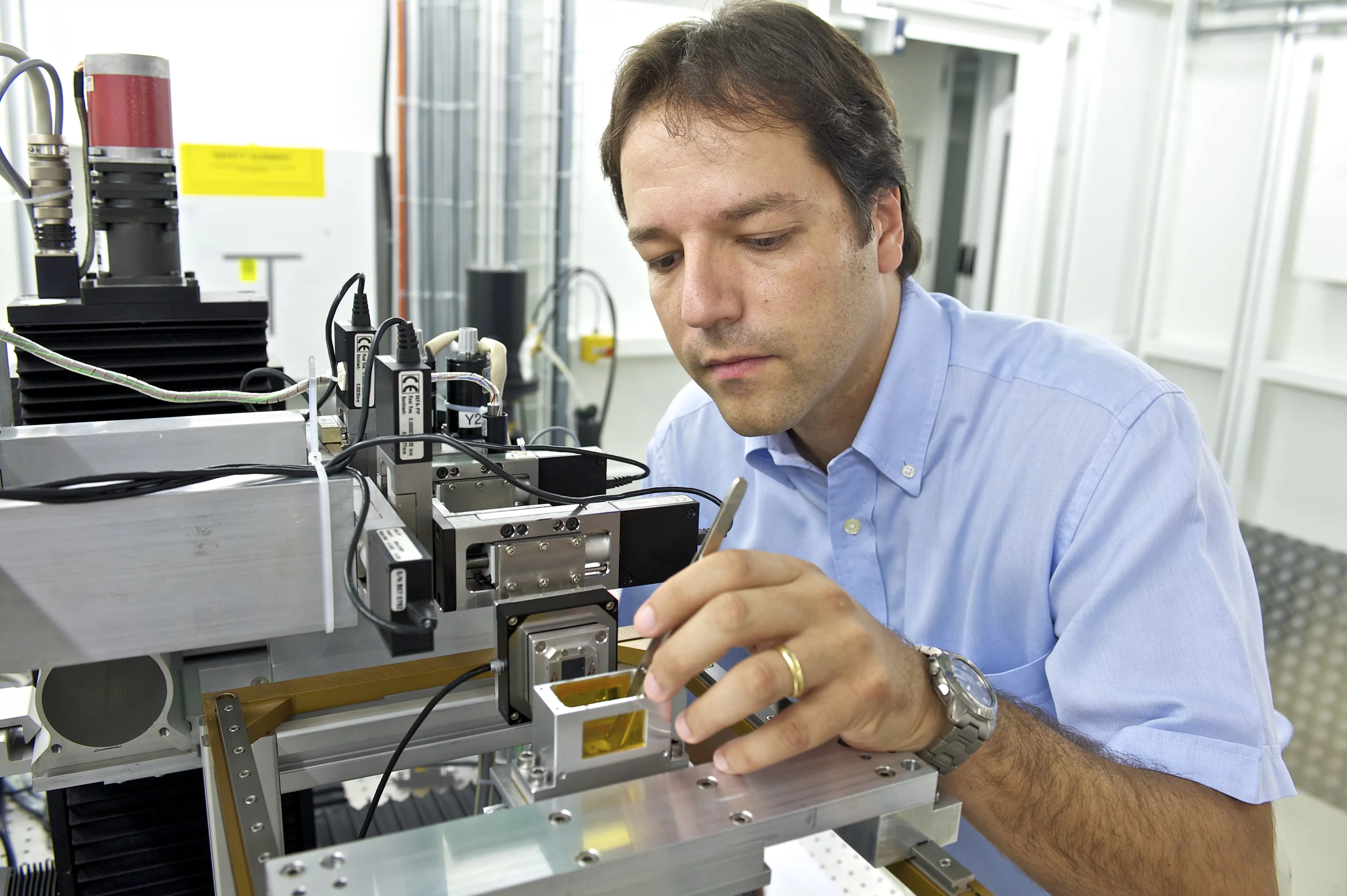
Reverse Projection Method(RP) was tested. (PSI/M. Fischer)
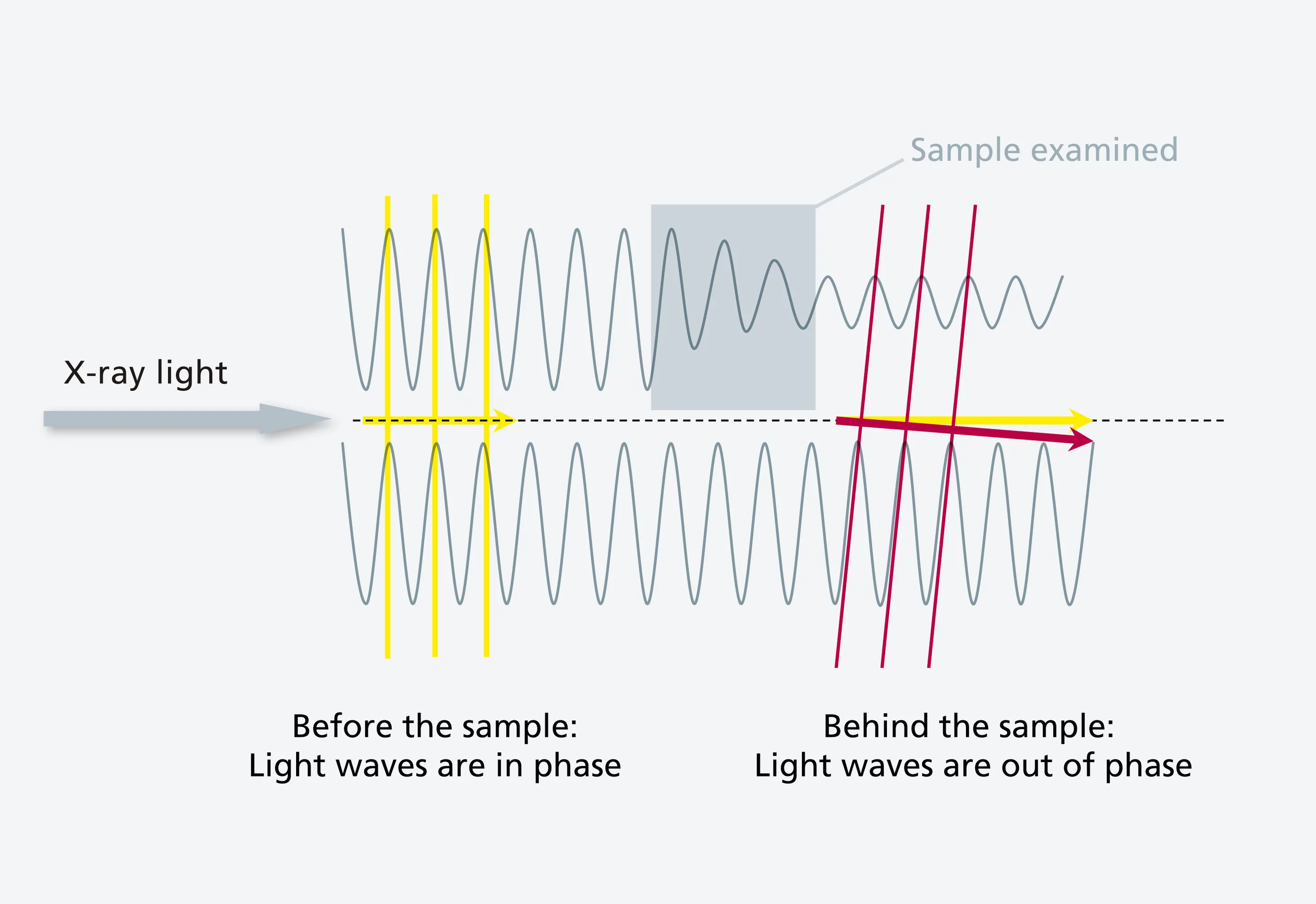
in phase(meaning the peaks of both waves are adjacent) and then pass through different materials are subsequently phase-shifted. The difference in phase contains important information about the structure of the object being examined, which can be utilised using the phase-contrast method. (PSI/M Stampanoni and M. Fischer)
In conventional X-ray images, the bones are particularly visible because they attenuate X-rays more strongly than the tissue around them. Thus, to a certain extent, an X-ray image is a shadow image of the inside of the body. However, various kinds of soft tissue attenuate X-rays in approximately the same way, which makes it difficult to distinguish between them.
Phase shift makes structures visible
In order to distinguish between these tissue types, researchers are utilising the fact that various tissues often differ from one another with respect to density, which also affects how fast the X-rays pass through them. These different densities cause what is known as a phase shift of the X-ray light.
Light is a wave: one can picture a ray of light as a constantly changing succession of wave peaks and troughs. Now imagine that several rays of light emerge in parallel from an X-ray source – "in phase", i.e. so that the peaks of all the waves are adjacent. If these rays now pass through tissue which has different densities in different places, they will no longer be in phase afterwards, because they will have moved through the tissue at different speeds. This phase difference can then be measured to determine the structure of the tissue.
A new technique for medical practices, as well
In order to create an image of tissue structure using phase differences, the researchers must make the different rays overlap. Thus, they pass the light through a fine grating that has spacing distances of a few thousandths of a millimetre. The overlap then allows the structure to be determined with a level of accuracy that was previously unattainable. The Swiss-Chinese team has now developed the "Reverse Projection Method” (RP), which allows phase shifts to be determined in a very simple way.
"This will make phase-contrast images just as easy to take as normal X-ray images are today," explains Marco Stampanoni, Professor of X-Ray Microscopy at the Institute of Biomedical Technology at the ETH Zürich and Project Manager at PSI. "These findings are an important step towards the broad application of the phase-contrast method in areas such as medicine, non-destructive materials testing and security technology because they use examination conditions and algorithms similar to those applied in existing systems."
Text: Paul Piwnicki
About PSI
The Paul Scherrer Institute develops, builds and operates large-scale, complex research facilities, and makes these facilities available to the national and international research community. The Institute’s own research focuses on solid-state physics and the materials sciences, elementary particle physics, biology and medicine, as well as research involving energy and the environment. With a workforce of 1300 and an annual budget of about 260 million CHF, PSI is the largest research institution in Switzerland.
Contact
Prof. Marco Stampanoni, Institute of Biomedical Technology at the ETH Zürich and Laboratory for Macromolecules and Bioimaging at the Paul Scherrer Institute, 5323 Villigen PSI; Telephone: +41 56 310 4724 or +41 79 874 92 22; e-mail: marco.stampanoni@psi.ch [German, English, Italian, French]Original publication
Low-dose, simple, and fast grating-based X-ray phase-contrast imaging Peiping Zhu, Kai Zhang, Zhili Wang, Yinjin Liu, Xiaosong Liu, Ziyu Wu, Samuel A. McDonald, Federica Marone, and Marco Stampanoni PNAS Early Edition, July 19, 2010 DOI: 10.1073/pnas.1003198107Images