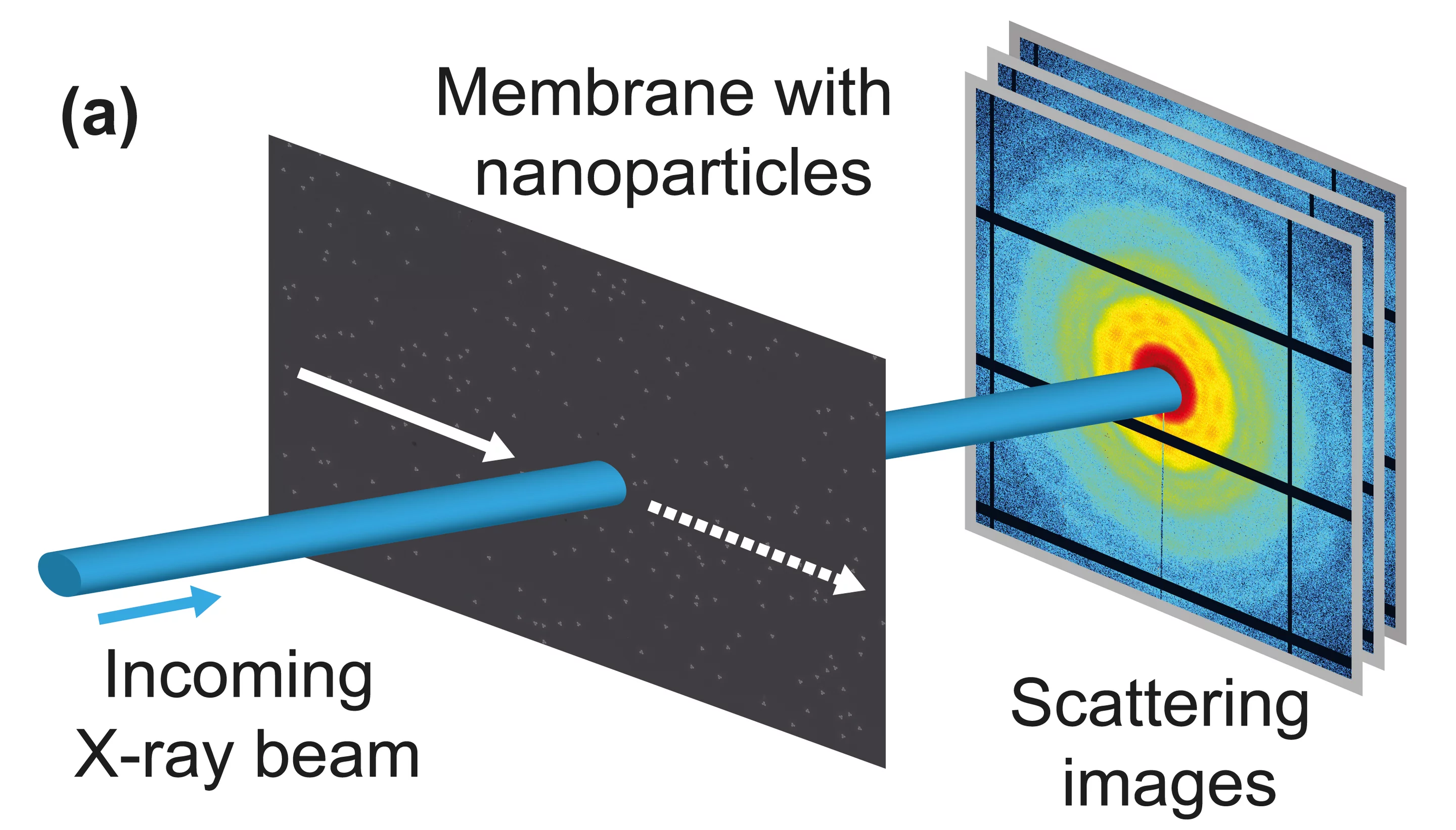
Development of analysis method for X-ray laser scattering data.
Prominent among the planned applications of X-ray free electron laser facilities, such as the future SwissFEL at the Paul Scherrer Institute, PSI, are structural studies of complex nano-particles, down to the scale of individual bio-molecules. A major challenge for such investigations is the mathematical reconstruction of the particle form from the measured scattering data. The experiment consists of exposing the nano-particles to the X-ray laser pulses and of registering the resulting scattered rays. To guarantee sufficient statistical accuracy, many repeated exposures are required – each one on a different collection of identical, but randomly-oriented particles. Researchers at PSI have now demonstrated an optimized mathematical procedure for treating such data, which yields a dramatically improved single-particle structural resolution. The procedure was successfully tested at the Swiss Light Source synchrotron at PSI, with custom-fabricated, two-dimensional nano-structures. With the inclusion of some additional information, such as the particle symmetry, the method can be extended to real three-dimensional objects. The researchers report their results on-line in the current issue of Nature Communications.
There is a strong scientific impetus to determine the three-dimensional structure of nanometer-scale particles. This is particularly true for the architecture of complex bio-molecules, the knowledge of which is necessary both for the understanding of vital processes and for the custom design of new pharmaceuticals. The structures of such molecules are generally determined using X-rays from synchrotrons, such as the Swiss Light Source at PSI. The advent of X-ray Free Electron Lasers (XFEL), such as the SwissFEL, currently under construction at PSI, will allow more detailed information on the structure of important bio-molecules than is currently available.
Structure revealed by intense X-ray pulses
In an XFEL structure-determination experiment, a stream of nano-particles will intersect the X-ray pulses. Each pulse will be so intense that the illuminated particles will scatter a significant amount of X-ray light into the detector, and it will be of such a short duration that during the illumination, no significant particle rotation will occur. However, the information obtained from a single XFEL pulse will not be sufficient to determine the structure, so the experiment will be repeated many, many times, each time with a different collection of particles with new orientations.
Single-particle structure from many-particle scattering
It is a challenging problem to extract the structure of a single particle from many in-equivalent scattering patterns, in particular when the number of illuminated particles is neither known nor constant. With their work, PSI physicist Bill Pedrini and his colleagues have taken an important step toward the solution of this problem, by further developing a method proposed in 1977 by the Israeli physicist Zvi Kam. In mathematical terms, one computes the cross-correlations of the measured scattering intensities, from which the structural information can be extracted. “In order to produce observable correlations, each investigated particle must scatter on average at least two photons per X-ray pulse. With the extremely intense pulses from an XFEL, this requirement will be met, even for very small particles”, explains Pedrini.
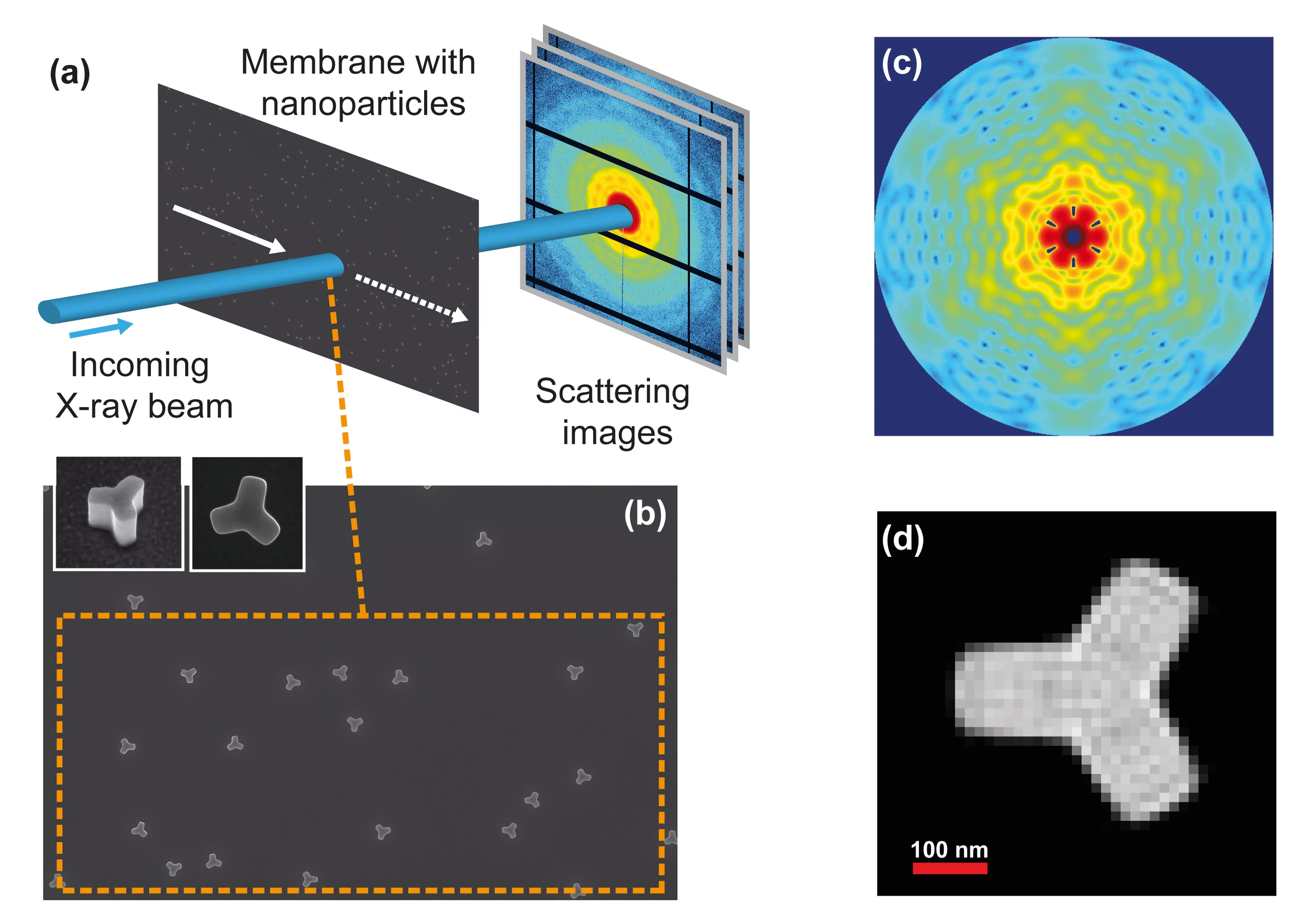
Tested at the SLS
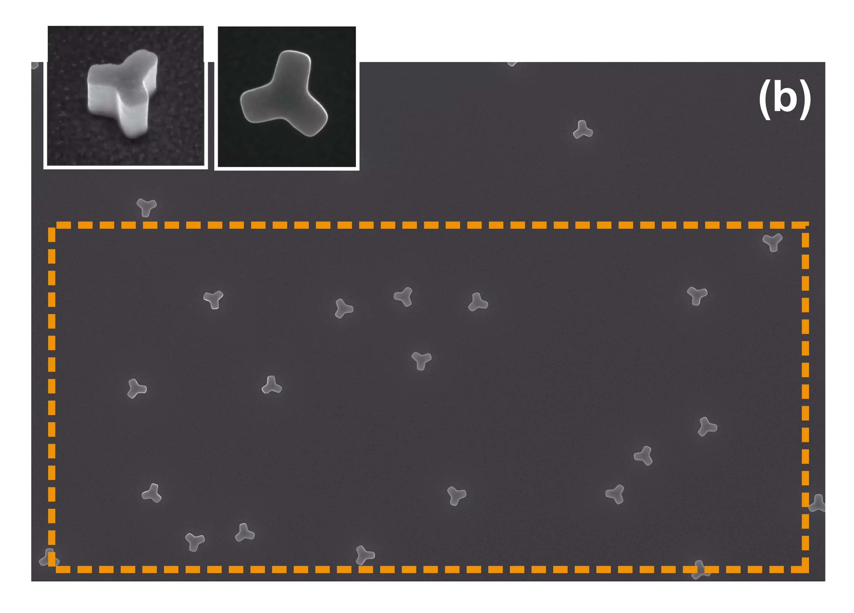
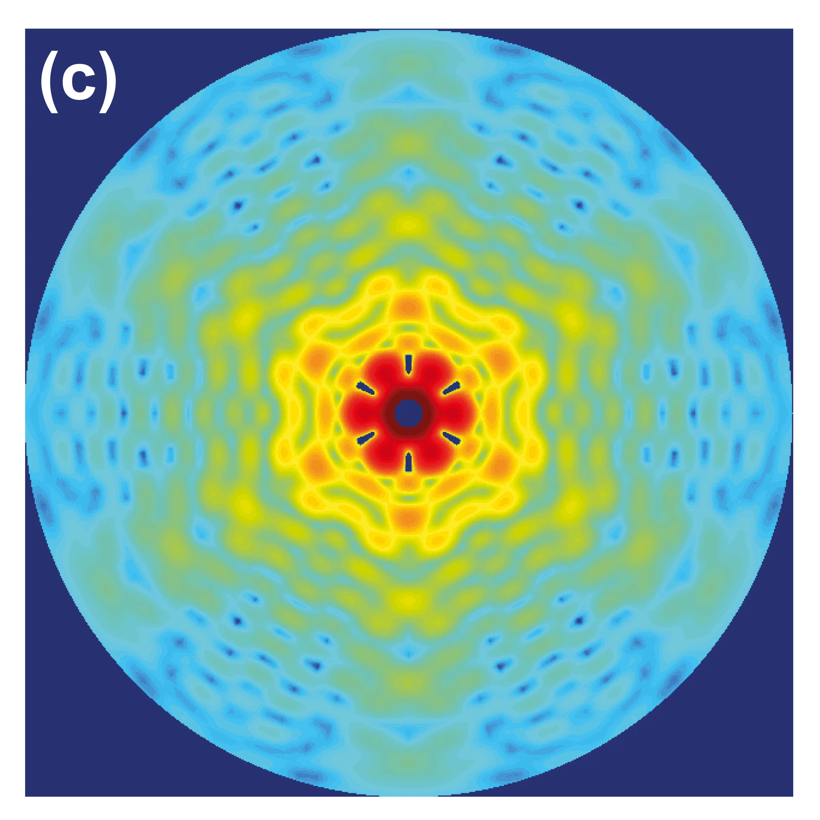
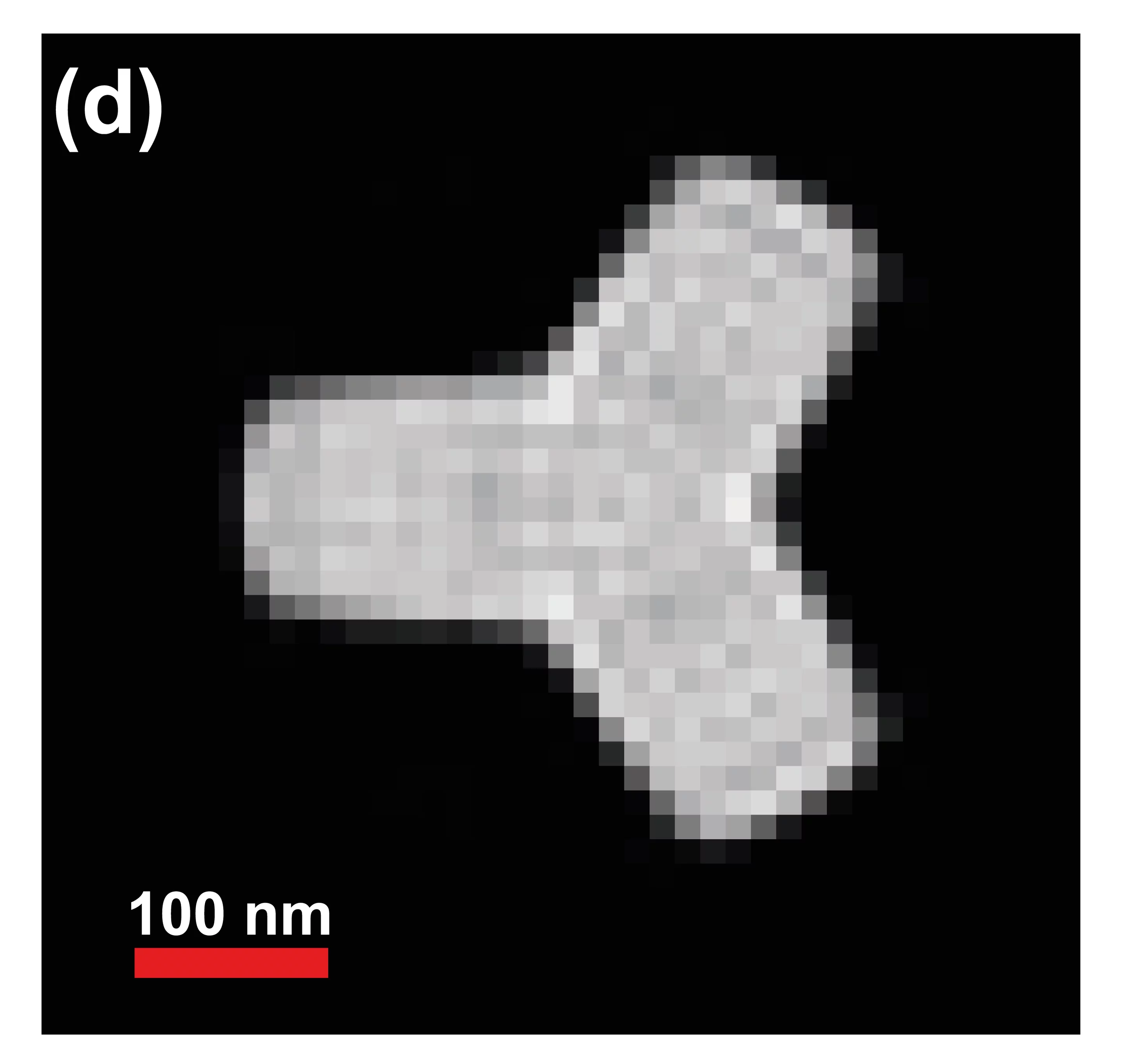
A test of the method was performed in an experiment at the cSAXS X-ray beamline of the SLS. As scattering particles, identical, star-shaped objects, approximately 300 nanometers across, were fabricated at the PSI Laboratory for Micro- and Nano-Technology. They were placed on a membrane at random positions and in random orientations. “We illuminated such a sample at various positions with the SLS X-ray beam, in this way simulating an experiment at an X-ray laser, in which each exposure samples a different configuration of several stationary particles”, according to Pedrini. With their method, the researchers could accurately determine the form of the nano-star, using approximately 4000 scattering images. “The shape reconstruction succeeded, in spite of the fact that each illumination covered an unknown number of particles. This corresponds to the situation in a real experiment”, comments Pedrini further.
The future is 3-D
The next step will be to generalize the method to three-dimensional objects, for example molecules. In spite of the fact that fundamental principles require additional information, such as the particle symmetry, to obtain a complete 3-D structure, the cross-correlation method offers important advantages over conventional techniques. In particular, it entails a computationally simple analysis of large numbers of scattering images. The test experiments at the SLS demonstrate the power of the method, and they provide important information for future experiments at an XFEL. “We now understand, for example, the effects of interference between the scattered rays from two different particles, and we have learned to account for the fact that the cross-section of the incoming X-ray beam is not everywhere of equal intensity”, says Pedrini.
One of several structural methods
In spite of the intense X-ray pulses that an XFEL will provide, the cross-correlation method alone will probably not be capable of yielding the position of each individual atom in a large bio-molecule with „atomic resolution“. Such resolution is achievable using the technique of protein crystallography, a standard method at synchrotrons such as the SLS. Protein crystallography demands that many identical molecules are isolated, purified and ordered in a three-dimensional lattice – a crystal. But many important bio-molecules, for example membrane proteins, cannot be crystallized, with the result that X-ray scattering experiments are only possible on molecules in solution. Nowadays, such experiments yield information only on the outer form of the molecules. The application at an XFEL of the cross-correlation method to dissolved samples promises significant improvement, namely a resolution of a few nanometers, which is sufficient to identify important structural properties.
Text: Paul Piwnicki
About PSI
The Paul Scherrer Institute develops, builds and operates large, complex research facilities, and makes them available to the national and international research community. The Institute's own key research priorities are in the investigation of matter and material, energy and the environment; and human health. PSI is Switzerland's largest research institution, with 1500 members of staff and an annual budget of approximately 300 million CHF.
Contact
Dr. Bill Pedrini, SwissFEL-Projekt, Paul Scherrer Institut, 5232 Villigen PSI, SchweizTelephone: +41 56 310 3371; E-mail: bill.pedrini@psi.ch
Original Publication
Two-dimensional structure from random multiparticle X-ray scattering images using cross-correlations.Pedrini, B. et al.
Nat. Commun. 4:1647 (2013)
DOI: 10.1038/ncomms2622
Additional Information
SwissFEL OverviewSwissFEL Project
Images