X-ray lasers belong to a modern generation of light sources from which scientists in widely different disciplines expect to obtain new knowledge about the structure and function of materials at the atomic level. On the basis of this new knowledge, it could then be possible one day to develop better medicines, more powerful computers or more efficient catalysts for energy transformation. The scientific value of an X-ray laser stands or falls on the quality of the ultra-short X-ray pulses it produces and which researchers use to illuminate their samples. An international team led by scientists from the Paul Scherer Institute, PSI, has now precisely measured these pulses. In so doing, they have laid the foundation for a scientifically optimal utilisation of X-ray lasers – not least, of the planned SwissFEL at PSI. The results of this work have recently been published in the scientific journal Nature Communications.
The research team led by graduate student Simon Rutishauser and his group leader Christian David was privileged to be the first users ever of the first hard X-ray laser in the world. This facility, at Stanford in the USA, known as LCLS, has produced laser pulses of hard X-rays for selected experiments since 2010.
The unique characteristics of a hard X-ray laser – short wavelengths, of the size of individual atoms, combined with an ultra-short pulse duration of the order of 100 femtoseconds (0.1 billionths of a second) and an extremely high intensity – enable many new and interesting experiments in biology, chemistry and physics. Similar X-ray lasers are therefore being built, or are at the planning stage, at various research institutes around the world. Since the middle of last year, the second hard X-ray laser, called SACLA, has been in operation in Japan. Additional projects, such as the European X-ray laser in Hamburg and SwissFEL at the Paul Scherrer Institute, should follow within the next few years.
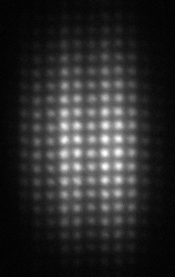
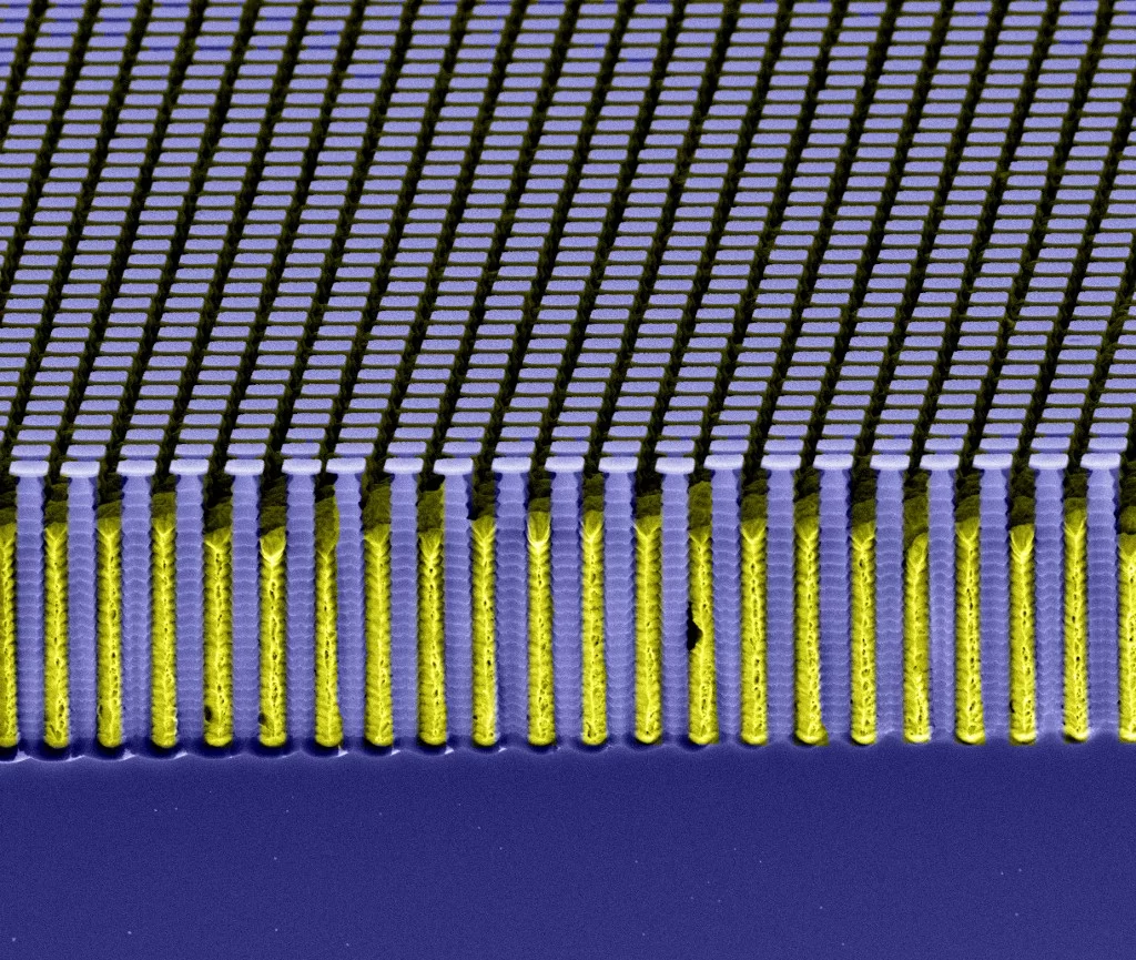
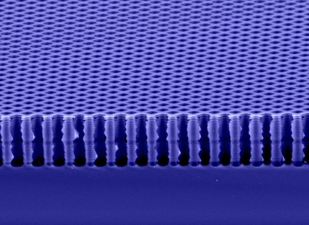
Mantaining the quality of the pulses from an X-ray laser places a huge demand on the X ray mirrors with which these pulses are formed and directed into the experimental stations. In order to test these mirrors under experimental conditions, the research team used the principle of grating interferometry. In this technique developed at PSI a pulse of light is sent through two very fine gratings. The fine grating pattern is produced on a carrier material by means of an extremely fine electron beam, thereby enabling the grating structures to be precisely positioned to an accuracy of a few nanometres. This, in turn, enables an otherwise unattainable accuracy of angular measurement of about 0.5 millionths of a degree (10 nanoradians).
Improvement of X-ray optics and experimental data
X-ray pulses are electromagnetic waves – exactly the same as visible light, whose propagation can be described in terms of a wave front. As with a water wave in a pond, the wave front at any point in time is given by the position of the wave peak. The particular challenge here lies in the fact that one is dealing with extremely short X-ray pulses. The researchers have nevertheless been able to measure each pulse separately, so that they could deduce their original shape.
A key discovery of the observations made by means of grating interferometry is that the X-ray mirrors used do not reflect the beam without modifying it, but (unintentionally) slightly focus it, though only in the horizontal plane. As a result of this distortion, the focusing of the X ray beam on an object being investigated is impaired. These mirrors cannot be dispensed with, however, because they guide the X-ray beam to the experimental stations and filter out unwanted, but unavoidable, gamma radiation. On the basis of the new results, however, it is now possible to adapt the X-ray mirrors in such a way that the adverse effects of the unidirectional focusing can be compensated for and the full potential of the X-ray laser can be exploited.
In addition, complex deformations of the wave front were also observed. These only slightly altered the size of the focal point, but fluctuated from pulse to pulse. This type of variation in intensity and phase of the X-ray pulses also affects the measured data that is for instance being acquired to determine the structure of a biological molecule. In such experiments, data is collected over a number of X-ray pulses and then averaged. The average value is, however, only reliable under the assumption that the characteristics of the X-ray beam remain unchanged for each pulse. If there are fluctuations between the pulses, as observed here, the quality of the measured data could nevertheless be improved by the use of grating interferometry. To that purpose, the wavefront of each pulse would be measured and this information could be used to correct the acquired experimental data for the fluctuations.
Better understanding of X-ray lasing
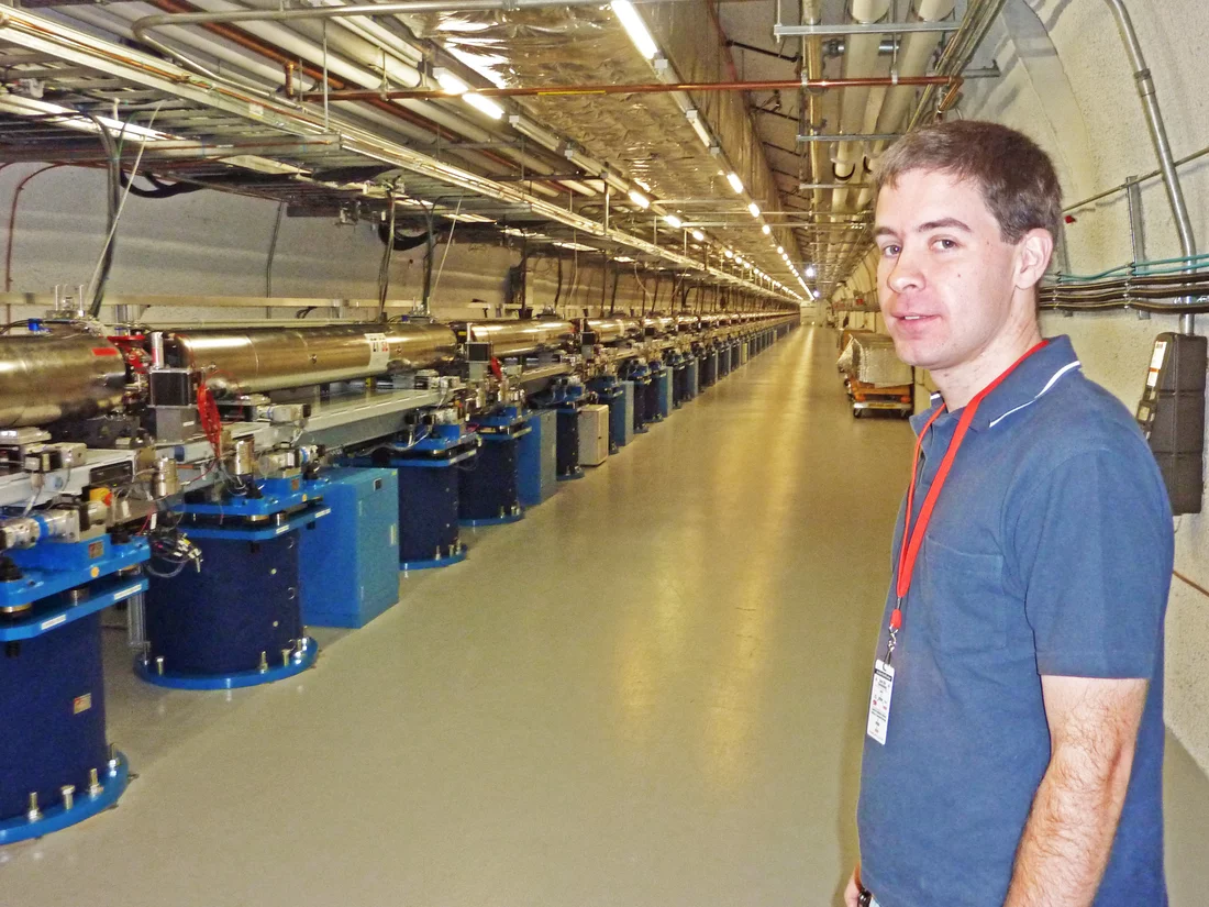
All X-ray lasers planned or built so far are based on the principle of the Free-Electron Laser. Here, electron bunches are first accelerated to almost the speed of light and then sent along an undulating path, which causes them to emit ultra-short, high-intensity X-ray pulses. The deflection of the electrons onto an undulating path takes place in the undulator, a series of magnets arranged one after the other with alternating polarity. The undulators in LCLS cover a length of 130 metres, and it was not initially clear where the light source actually was; that is, the point at which the electrons emitted X-ray light. The researchers have now been able, for the first time, to determine experimentally the exact position of the origin of each individual X-ray pulse. This demanded the highest precision, as only measurements over a tiny spot of light were used. To be more precise, the X-ray pulses have a diameter of only 0.5 mm at the experimental station, a distance of 200 metres from the source.
The experimental determination of the position of the source point in a 130-metre-long laser, as well as its displacement from one pulse to the next, is of great interest because, until now, this was only approximately known from simulations. The scientists were also able to find the location of the source position as a function of the accelerator settings. This should, in the future, lead to improved performance of X-ray lasers.
The authors of this study believe that this technique, which was developed at PSI, will soon become a standard technique at X-ray lasers.
About PSI
The Paul Scherrer Institute develops, builds and operates large, complex research facilities, and makes them available to the national and international research community. The Institute's own key research priorities are in the investigation of matter and material, energy and the environment; and human health. PSI is Switzerland's largest research institution, with 1500 members of staff and an annual budget of approximately 300 million CHF.
Contact
Simon Rutishauser, Laboratory for Micro- and NanotechnologyPaul Scherrer Institute, 5232 Villigen, Switzerland;
Telephone: +41 56 310 25 02, E-Mail: simon.rutishauser@psi.ch
Dr. Christian David, Group Leader X-ray optics and applications
Laboratory for Micro- and Technology
Paul Scherrer Institute, 5232 Villigen, Switzerland;
Telephone: +41 56 310 37 53, E-Mail: christian.david@psi.ch
Original Publication
Exploring the wavefront of hard X-ray free electron laser radiationSimon Rutishauser, Liubov Samoylova, Jacek Krzywinski, Oliver Bunk, Jan Grünert,Harald Sinn, Marco Cammarata, David M. Fritz, Christian David
Nature Communications DOI: 10.1038/ncomms1950

