A measurement method developed at the Paul Scherrer Institute creates a three-dimensional nano-view
An international team of researchers from Denmark, Germany, Switzerland and France has developed a new method for making detailed X-ray images of brain tissue, which has been used to make the myelin sheaths of nerve fibres visible. Damage to these protective sheaths can lead to various disorders, such as multiple sclerosis. The facility for creating these images of the protective sheaths of nerve cells is being operated at the Swiss Light Source (SLS), at the Paul Scherrer Institute. The research team has reported on its work in the online version of the scientific journal NeuroImage.
The so-called myelin sheath is a protective covering around the axons of nerve cells – long, thin fibres which carry electrical impulses between nerve cells or from nerve cells to other cells. These myelin layers ensure the rapid transfer of signals from nerves. Changes or a breakdown in their function are associated with degenerative brain disorders, such as multiple sclerosis.
“The detailed development of these diseases is not yet understood,” says Prof. Franz Pfeiffer, from the Technische Universität München, “but it is increasingly being thought to be related to changes in the myelin layers that are responsible for the interruption of the transfer of signals between nerve cells. Thinking of it in a simplified way, this is similar to what happens when the insulation of an electrical cable is damaged, leading to either a short circuit or current leakage.”
Images in 3D
“We have combined two well-known research methods of scanning SAXS (Small-Angle X-ray Scattering) and CT (Computed Tomography) with a specially developed program for data processing. In this way, we have been able to make variations in the myelin sheaths of a rat’s brain visible without having to prepare small brain samples”, explains Dr. Torben Haugaard Jensen, from the Niels Bohr Institute at the University of Copenhagen.
The experiments have been carried out at the Paul Scherrer Institute (PSI) in Switzerland. Here, the SLS provides the high-intensity X-rays needed for performing SAXS experiments. “In this method, a narrow X-ray beam is directed at the sample under investigation. From the way the beam is deflected, we can determine the structure of the sample at the point being scanned by the beam. We repeat this process for many thousands of points and subsequently obtain a detailed picture of the interior of the sample. The PILATUS X-ray detector used to monitor the deflected beams allows us to make optimal use of the X-ray beams produced at the SLS, thus providing outstanding conditions for such investigations”, explains staff scientist Oliver Bunk, from PSI. This detector uses a technology that was originally developed at PSI for application at one of the new experiments at CERN, but is now being further developed for numerous other applications.
Computed Tomography has been established for a long time and is used throughout the world in clinical applications. In a CT examination, a body is X-rayed from different directions and an image detector produces a two-dimensional shadowgraph of the human body each time. These detailed images are then processed to create a three-dimensional picture of the interior of the body. The researchers followed this same procedure with scanning SAXS. The detailed images produced by this technique contain no information on depth but, in a combined process, a sample is scanned from different angles so that detailed 3D images can subsequently be produced. In order to generate these extremely accurate images, 800,000 individual 2D images had to be processed, which required the development of special software.
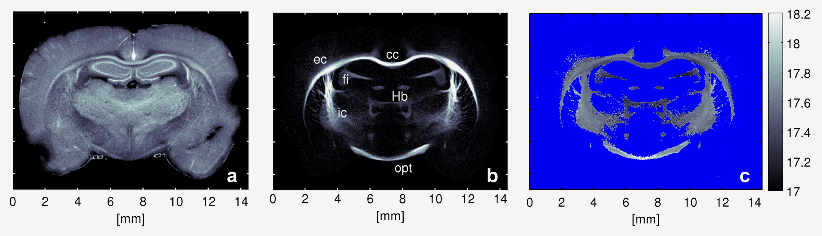
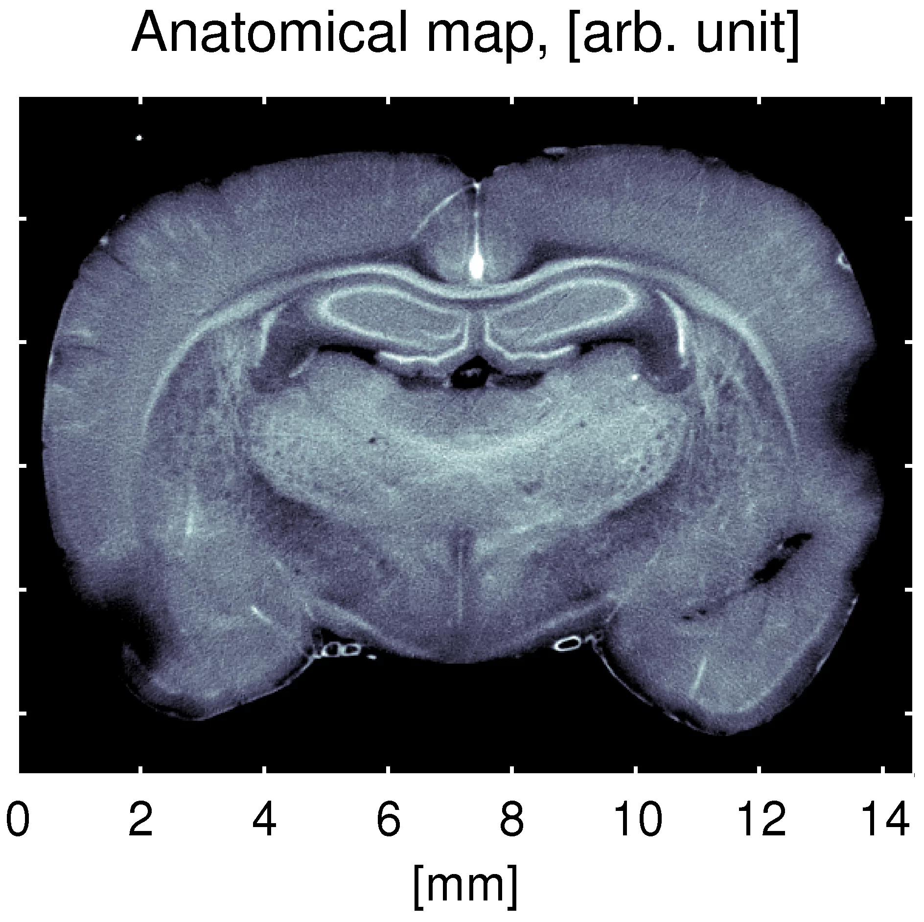
a) This image shows the results of scanning Small-Angle X-ray Scattering, which can be used as an anatomical map of the rat’s brain.
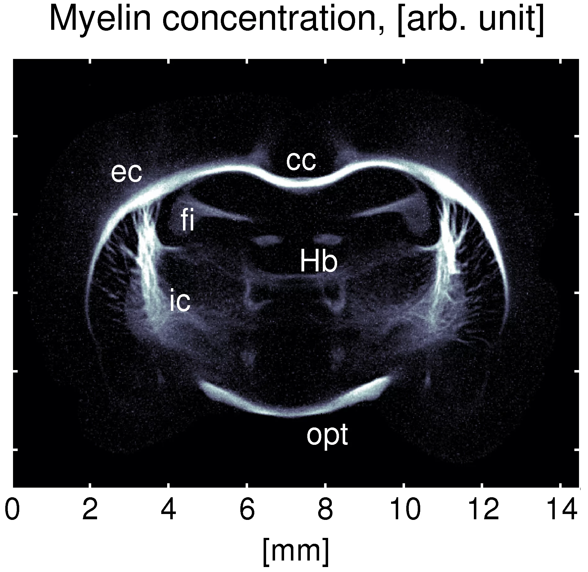
b) The myelin concentration in the rat’s brain is seen here, with the highest concentration occurring around the corpus callosum (the bundle of nerve fibres connecting the two hemispheres of the brain) and at the internal and external capsules. The myelin concentration could be determined without having to dissect the brain.
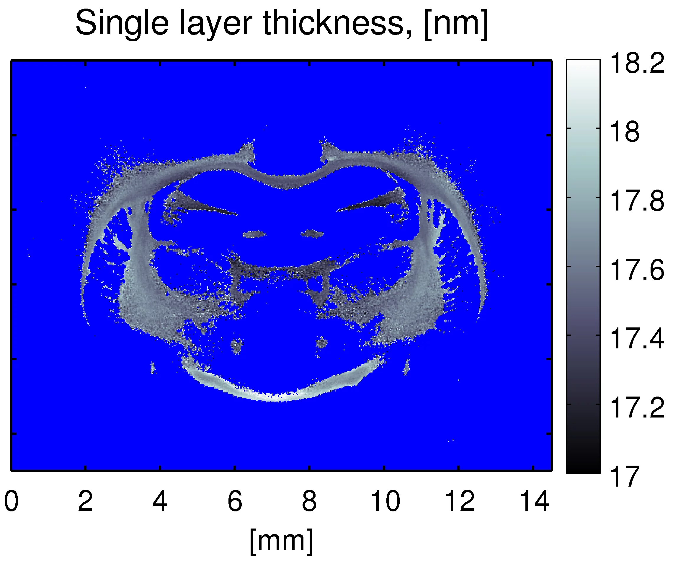
c) The image shows that the thickness of the myelin layer in a rat’s brain varies between 17.0 and 18.2 nanometres. Using this new process, these variations can now be demonstrated. This showed that it is now possible to determine the exact thickness of the myelin sheath, which could, in turn, help to determine the relationship between specific disorders and changes in the myelin sheath.
250,000 points at one stroke
An example of an application that has been made with this method is a study of the brain of a laboratory rat, producing some surprising insights. “We can see details of the myelin sheaths of the nerve fibres and distinguish layers with a thickness of only 17.6 nanometres”, explains Prof. Robert Feidenhans’l, from the Niels Bohr Institute. “Up until now, small pieces had to be dissected from a brain and examined, in order to obtain similar information. With the new method, we can examine 250,000 points at one stroke, without having to cut small pieces from the brain in advance. This will give us the possibility of carrying out series examinations on the thickness and concentration of the myelin in relation to different patterns of illness”.
This research is providing new opportunities for collaboration with doctors, and close contact already exists with the University Hospital and the Panum Institute at Copenhagen. The new method cannot be used on living patients, but can provide decisive knowledge about the diseases, and help to answer questions such as: “What kind of damage takes place in a specific illness?” and “Where does it happen?” It will now be possible to follow the development of the diseases and find out how the brain will be damaged. With this knowledge, new therapies for various disorders could then be developed.
About PSI
The Paul Scherrer Institute develops, builds and operates large-scale, complex research facilities, and makes these facilities available to the national and international research community. The Institute”s own research focuses on solid-state physics and the materials sciences, elementary particle physics, biology and medicine, as well as research involving energy and the environment. With a workforce of 1400 and an annual budget of about 300 million CHF, PSI is the largest research institution in Switzerland.
Contact
Dr. Oliver Bunk, Labor für Makromoleküle und Bioimaging,Paul Scherrer Institute, 5232 Villigen PSI, Switzerland,
Phone: (+41) 56 310 3077, E-mail: oliver.bunk@psi.ch
Dr. Torben Haugaard Jensen, Niels Bohr Institute,
Universität Kopenhagen, Dänemark
Phone: (+45) 2097-3682, E-mail: torbenj@fys.ku.dk
Prof. Robert Feidenhans’l, Niels-Bohr-Institut,
Universität Kopenhagen, Dänemark,
Phone: (+45) 3532-0397, (+45) 2875-0397, E-mail: robert@fys.ku.dk
Prof. Franz Pfeiffer, Lehrstuhl für Biomedizinische Physik,
Technische Universität München, 85748 Garching, Deutschland
Phone: (+49) 89 289 12551, E-mail: franz.pfeiffer@tum.de
Original Publication
Molecular X-ray computed tomography of myelin in a rat brain.T.H. Jensen, M. Bech, O. Bunk, A. Menzel, A. Bouchet, G. Le Duc, R. Feidenhans'l, F. Pfeiffer.
NeuroImage, 2011;
DOI: 10.1016/j.neuroimage.2011.04.013
Images