Thanks to the analysis of protein samples at the PSI, Lausanne researchers have managed to demonstrate which instrument bacteria use to transmit diseases
Researchers from ETH Lausanne EPFL have described how a particular strain of bacteria transmits diseases with unprecedented precision. The team of scientists headed by Petr Leiman, an assistant professor at the EPFL’s Laboratory of Structural Biology and Biophysics, demonstrated that the tip of a bacterial infection tool consists of a PAAR protein, which envelops a metal atom and tapers off to a sharp point. The findings are based on measurements carried out at the Swiss Light Source (SLS), one of the three large research facilities at the Paul Scherrer Institute (PSI).
Scientists often not only know how viruses or bacteria transmit diseases; they also understand with increasing accuracy the way by which these pathogens cause damage if a bacterium such as Vibrio cholerae, for instance, attacks a human cell and triggers the dreaded cholera infection. Nowadays, it is possible to understand this infection process with fascinating precision and thus track how the disease is transmitted to people at the single-molecule level.
Infection tool identified
A team of researchers headed by Petr Leiman, an assistant professor at the EPFL, has now managed to do just that. In a Nature publication, the scientists write that a certain class of proteins – the PAAR-repeat proteins – play a key role in bacterial virulence. In their experiments, the researchers focused on bacteria that use a particular tool known as T6SS (type VI secretion system) for the infection process. T6SS is found in about 25% of all known bacterial genomes. It can best be envisaged as a long tubular structure embedded in the bacterial cytoplasm with a spike at its tip. The tube can contract driving the spike into a neighbouring bacteria or a human cell causing infection.
The T6SS spike plays a key role in the infection. Now, for the first time, the EPFL researchers were able to reveal that the tip of the spike consists of a PAAR-repeat protein folded into a pointed cone. Many PAAR-repeat proteins contain up to three instances of the Proline-Alanine-Alanine-aRginine amino acid sequence motif, which is responsible for giving these proteins a pointed cone shape. The protein polypeptide chain folds around a zinc ion that is positioned close to the cone’s vertex. The Lausanne researchers believe that the zinc ion plays an important role in disrupting the membrane of the target cell, which is pierced by the spike. “This improves our understanding of how bacterial infections take place. In the medium term, however, it also gives us and many other researchers working in this field new research directions for rendering many bacterial pathogens harmless,” says Leiman.
Evidence at the SLS in Villigen
Leiman found explanation for the role of PAAR proteins in bacterial pathogens on the Swiss Light Source (SLS), a large research facility at the Paul Scherrer Institute (PSI) where the Russian-born and US-educated researcher has already studied around fifty different protein samples. Studies of PAAR repeat proteins were complicated by their unusual biochemical properties – these proteins cannot be isolated alone, but instead require a binding partner, which by itself represents a difficult sample. Leiman and his team were able to “paste” a PAAR-binding site onto a well-behaving protein, thus making it possible to isolate several PAAR proteins. With the aid of the SLS, the 37-seven-year-old researcher was able to determine the atomic structure of several PAAR-repeat proteins in complex with a chimerical partner. In doing so, Leiman also found evidence that PAAR proteins contain a “metallic core”, which is composed of a zinc atom.
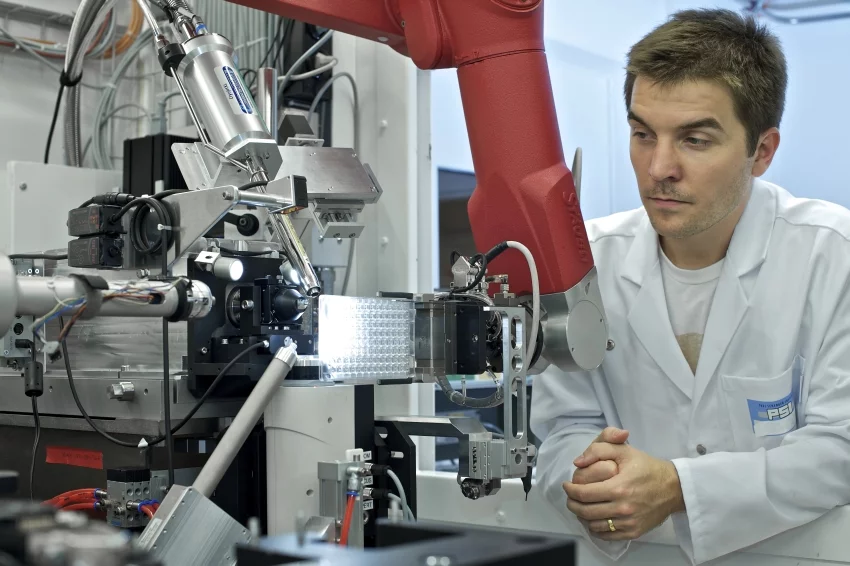
The SLS provides a platform for researchers to study biological and other samples on a miniscule scale right down to a resolution of single atoms. The facility generates a highly focused beam of x-ray light, no more than 50 micrometres across, which is directed towards the sample and generates its diffraction image. The method used is known as x-ray crystallography. In order to be able to produce such “photography”, the sample must be in crystalline form, which involves arranging a cluster of a large number of molecules at regular intervals in a crystal lattice. In Petr Leiman’s case, the crystals comprised PAAR proteins and their binding partners. When x-ray hits a crystal, it is reflected off the crystal lattice. In several intermediate steps and with painstaking data analysis, the structure of the crystallized proteins can then be determined with high precision from the X-ray diffraction pattern.
High demand for PSI research facilities
Petr Leiman is one of 5,782 external users of the large research facilitieswho visited the Paul Scherrer Institute for research purposes in 2012. Like Leiman, around two thirds of them (3,825) worked on the Swiss Light Source (SLS), conducting 1,187 experiments in all. However, the PSI’s other two large research facilities were also in high demand. 1,001 guest scientists conducted a total of 474 experiments on the Swiss Spallation Neutron Source (SINQ) and 359 external researchers used the Swiss Muon Source (SμS) for 214 experiments.
Users from the four corners of the globe flock to work on the PSI’s three large research facilities, where they conduct basic research or study the fundamentals for later applications, such as in the pharmaceutical industry, microelectronics or the automobile sector. The demand for experimentation time at the SLS is very high: sometimes as much as twice the time available. This necessitates a selection procedure that evaluates scientific and technological merit of applications for experimentation time. The selection is performed by an international panel of renowned scientists. In order to satisfy the demand as fairly as possible, the facility operates around the clock on a three-shift basis, seven days a week. During an eight-hour visit, scientists can collect x-ray diffraction data for one hundred crystals or more.
New strategies for data preparation
A team of PSI experts – including mathematicians, physicists, biologists, chemists and engineers – ensures a high level of availability for the research facilities. “We guarantee the optimal quality of our x-ray diffraction setup; we develop and upgrade the instruments, and maintain the facility,” says Dr Vincent Olieric, a 34-year-old biochemist from France who has been working at the SLS for seven years. He also supports researchers who are still inexperienced in handling the facility directly when conducting their experiments, such as by helping them to prepare samples, which requires the utmost precision. Moreover, the PSI support team also performs scientific tasks focused on the development of new instruments and strategies, thus optimising the use of the research facilities further.
Petr Leiman visits the SLS around once a month to work on various research projects. “The SLS is one of the best research facilities in the world. The x-ray beamline instrumentation is top notch and the PSI is also easy to reach from Lausanne,” he says. In the future, the Lausanne team will probably even be able to save themselves a trip to Villigen altogether as the PSI is looking to enable their experimental stations to be controlled remotely. The users will be able to send their samples into the PSI by post, where PSI staff will place them on a remotely controlled work platform. The users will then be able conduct their experiments from home by means of manipulating robots that allow sample mounting and removal.
Text: Benedikt Vogel
Video source: Petr Leiman and Seyet LLC.
Original Publication
PAAR-repeat proteins sharpen and diversify the type VI secretion system spikeMikhail M. Shneider, Sergey A. Buth, Brian T. Ho, Marek Basler, John J. Mekalanos & Petr G. Leiman
Nature 500, 350–353 (15 August 2013);
DOI: 10.1038/nature12453

