
Synchrotron light
Synchrotron light is exceptionally intense X-ray light, whose properties can be adjusted in a variety of ways. For investigations into materials, synchrotron light has many advantages in comparison with conventional X-rays.
Synchrotron light is emitted by electrically-charged particles that have been forced onto a curved path. At PSI, synchrotron light is generated in the Swiss Light Source SLS.


Synchroton light is exceptionally intense
Synchrotron light is extremely intense and highly concentrated – experts say that it is particularly brilliant
. This quality enables investigations to be carried out on very small samples, which is important in fields such as protein crystallography, where the structure of complex proteins is investigated using the corresponding crystals. As such crystals are difficult and extremely time consuming to produce, it is of great benefit if experiments can be carried out on small crystals.
Synchrotron light has flexible properties
The properties of synchrotron light can be precisely adapted to suit the requirements of individual experiments. For example, the energy of the light can be adjusted very accurately over a broad range between ultraviolet light and X-rays. Thus, investigators can determine the separate contributions made by various chemical elements to a particular effect. This exploits the fact that, for every chemical element, there is a light energy at which the light interacts particularly strongly with the atoms of the element.
At some measurement stations, the polarisation of the synchrotron light can also be adjusted, in addition to its energy; i.e. the plane of oscillation of the light can be specifically defined. Since magnetic materials interact differently with polarised radiation depending on the direction of magnetisation, synchrotron light can be used to distinguish between different magnetised regions. For example, an image obtained with a synchrotron microscope could use different colours to indicate areas of differing magnetisation.
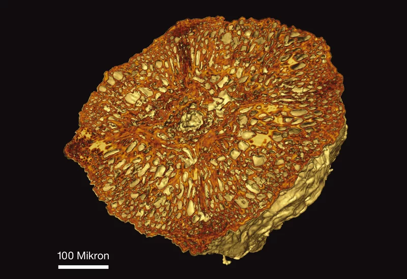
Using synchrotron light to produce 3D images
The properties of synchrotron light are also of benefit in synchrotron tomography. This technique provides an insight into the interior and three-dimensional structure of a very wide variety of objects, at a resolution of less than one micrometre (one thousandth of a millimetre). In this process, researchers benefit from the extremely intensive and highly parallel beam available. The technique also offers the opportunity of differentiating clearly between different materials in the tomogram and, for example, showing structures made from a particular substance on an individual basis. The objects investigated by synchrotron tomography range from engineering materials to rocks to biological tissue – and even to archaeological finds.
1 Adapted by permission from Macmillan Publishers Ltd: D. A. Pomeranz Krummel, C.Oubridge et al. Nature 458, 475-480(link is external), Copyright 2009