
First-time 3D imaging of internal magnetic patterns
Magnets are found in motors, in energy production and in data storage. A deeper understanding of the basic properties of magnetic materials could therefore impact our everyday technology. A study by Scientists at the Paul Scherrer Institute PSI in Switzerland, the ETH Zurich and the University of Glasgow has the potential to further this understanding. The researchers have for the first time made visible the directions of the magnetisation inside an object thicker than ever before in 3D and down to details ten thousand times smaller than a millimetre (100 nanometres). They were able to map the three dimensional arrangement of the magnetic moments. These can be thought of as tiny magnetic compass needles inside the material that collectively define its magnetic structure. The scientists achieved their visualisation inside a gadolinium-cobalt magnet using an experimental imaging technique called hard X-ray magnetic tomography which was developed at PSI. The result revealed intriguing intertwining patterns and, within them, so-called Bloch points. At a Bloch point, the magnetic needles abruptly change their direction. Bloch points were predicted theoretically in 1965 but have only now been observed directly with these new measurements. The researchers published their study in the renowned scientific journal Nature.

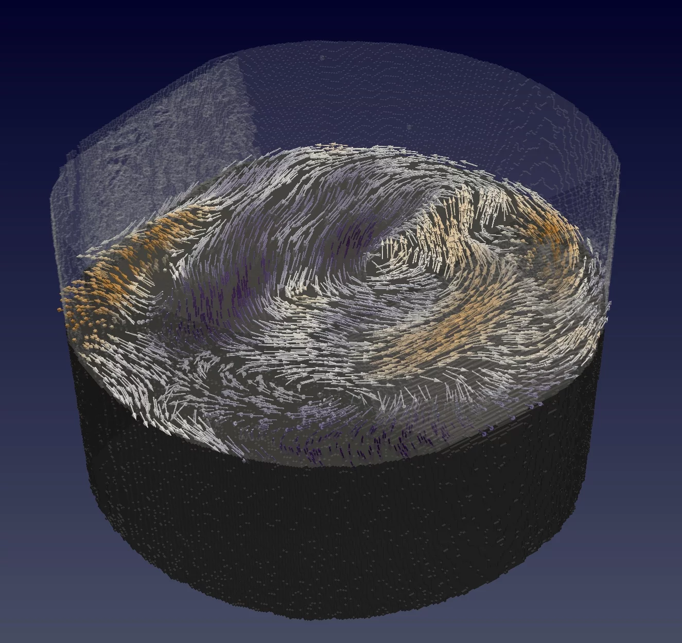
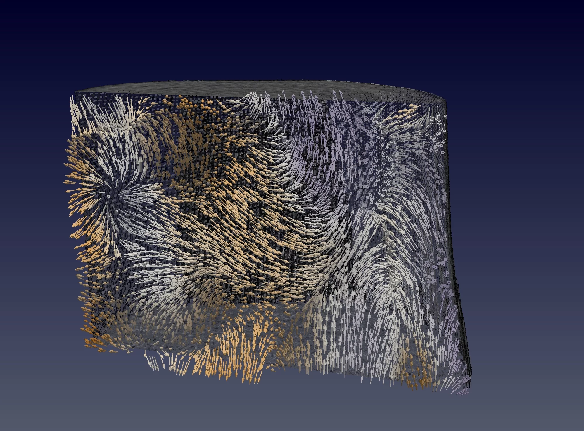
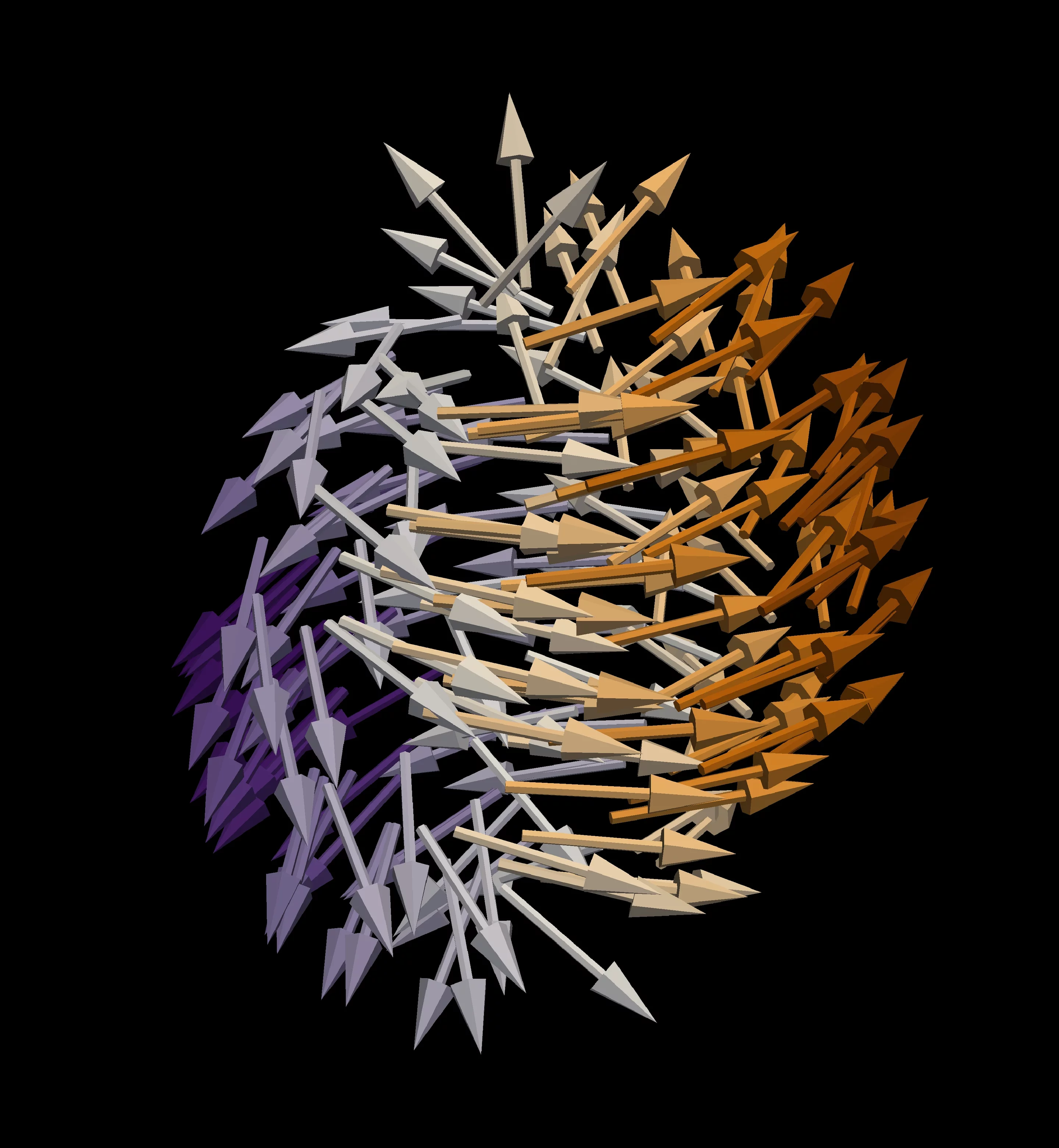
magnetic needles– visualised by arrows – to downward pointing ones. This singularity is surrounded by a swirling magnetisation pattern which is analogous to the structure of a tornado. (Graphics: Paul Scherrer Institute/Claire Donnelly)
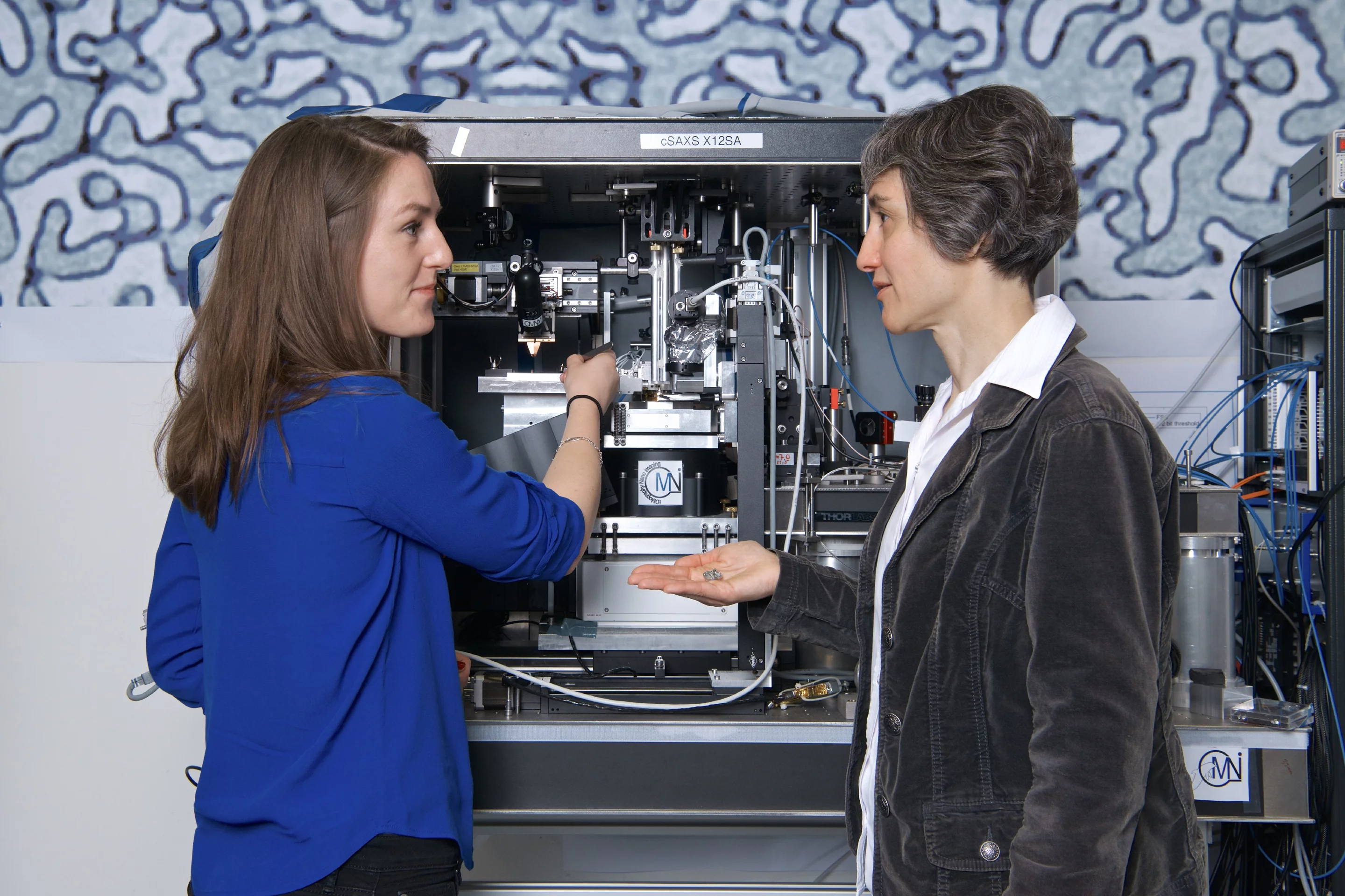
A team of scientists from the Paul Scherrer Institute PSI, the ETH Zurich and the University of Glasgow have for the first time been able to image the magnetic structure within a small 3D object on the nanometre scale. The magnetic structure is an arrangement of magnetic moments, each of which can be thought of as a tiny magnetic compass needle. The studied object was a micrometre-sized pillar (thousandth of a millimetre in diameter) made of the material gadolinium-cobalt, which acts like a ferromagnet. Within it, the scientists visualised the magnetic patterns that occur on a scale ten thousand times smaller than a millimetre – in other words, the smallest detail they could make visible in their 3D images was around 100 nanometres. The sophisticated imaging was achieved by a technique called hard X-ray magnetic tomography that was newly developed at PSI in the course of this proof-of-principle study.
Up to now, imaging magnetism and magnetic patterns at this small scale could only be done in thin films or on the surfaces of objects
, explains Laura Heyderman, principal investigator of the study, researcher at PSI and professor at ETH Zurich. We really feel like we are diving inside the magnetic material, seeing and understanding the 3D arrangement of the tiny magnetic compass needles.
These tiny needles ‘feel’ each other and hence are not oriented randomly, but instead form well-defined patterns throughout the magnetic object.
Basic magnetic structures and first-time visualisation of Bloch-Points
The scientists quickly realised that the magnetic patterns consisted of tangled fundamental magnetic structures: They recognised domains, in other words, regions of homogenous magnetisation, and domain walls, the boundaries separating two different domains. They also observed magnetic vortices, which have a structure analogous to that of tornadoes, and all of these structures intertwined to create a complex and unique pattern. Seeing these basic and well-known structures come together in a complex 3D network made sense and was very beautiful and rewarding
, says Claire Donnelly, first author of the study.
One specific kind of pattern stood out and gave additional significance to the scientists’ results: a pair of magnetic singularities, so-called Bloch points. Bloch points contain an infinitesimally small region within which the magnetic compass needles
abruptly change their direction. Singularities in general have fascinated scientists in a variety of research fields. Well known examples are black holes in space. In ferromagnets, the magnetisation can generally be considered continuous on the nanoscale. At these singularities, however, this description breaks down
, says Sebastian Gliga of the University of Glasgow and visiting scientist at PSI. Bloch points constitute monopoles of the magnetisation and although they were first predicted over 60 years ago, they have never been directly observed.
Magnetic X-ray tomography: 3D mapping with nanoscale resolution
The experimental technique of magnetic X-ray tomography employed in this study draws on a basic principle from computer tomography (CT). Similar to medical CT scans, many X-ray images of the sample are taken one after the other from many different directions with a small angle in-between adjacent images. The measurements were carried out at the cSAXS beamline of the synchrotron light source SLS at PSI using advanced instrumentation for X-ray nanotomography under the OMNY project and a recently developed imaging technique called ptychography. Employing computer calculations and a novel reconstruction algorithm developed at PSI, all of the data collected this way was combined to form the final 3D map of the magnetisation.
The scientists employed so-called ‘hard’ X-rays from the SLS at PSI. In comparison with ‘soft’ X-rays, hard X-rays have higher energy. Lower energy soft X-rays have already very successfully been used to achieve a similar map of the magnetic moments
, Claire Donnelly explains. But soft X-rays hardly penetrate such samples so you can only use them to see the magnetisation of a thin film or at the surface of a bulk object.
In order to really dive
inside their magnet, the PSI scientists chose hard X-rays of higher energy, at the price of obtaining a much weaker signal: Many people did not believe that we would be able to achieve this 3D magnetic imaging with hard X-rays
, Laura Heyderman recalls.
Tailoring the magnets of the future
The researchers see their achievement as a contribution to a deeper understanding of the basic properties of magnetic materials. Moreover, the ability to image inside magnets could be applied to many of today’s technological problems: Magnets are found in motors, in energy production and in data storage – creating better magnets thus has a huge potential of improving many every-day applications.
Text: Paul Scherrer Institute/Laura Hennemann
About PSI
The Paul Scherrer Institute PSI develops, builds and operates large, complex research facilities and makes them available to the national and international research community. The institute's own key research priorities are in the fields of matter and materials, energy and environment and human health. PSI is committed to the training of future generations. Therefore about one quarter of our staff are post-docs, post-graduates or apprentices. Altogether PSI employs 2100 people, thus being the largest research institute in Switzerland. The annual budget amounts to approximately CHF 380 million. PSI is part of the ETH Domain, with the other members being the two Swiss Federal Institutes of Technology, ETH Zurich and EPFL Lausanne, as well as Eawag (Swiss Federal Institute of Aquatic Science and Technology), Empa (Swiss Federal Laboratories for Materials Science and Technology) and WSL (Swiss Federal Institute for Forest, Snow and Landscape Research).
(Last updated in May 2017)
Additional information
- 3-D X-ray imaging makes the finest details of a computer chip visible (16. March 2017)
- 3D nanostructure of a bone made visible (19. November 2015)
- Nanometres in 3D (20. March 2015)
- Sixteen nanometres in 3D (11. June 2014)
Contact
Claire Donnelly
Laboratory for Multiscale materials experiments
Paul Scherrer Institute, 5232 Villigen PSI, Switzerland
Telephone: +41 56 310 30 94, e-mail: claire.donnelly@psi.ch [English]
Prof. Dr. Laura Heyderman
Head of Laboratory for Multiscale materials experiments
Paul Scherrer Institute, 5232 Villigen PSI, Switzerland
Telephone: +41 56 310 26 13, e-mail: laura.heyderman@psi.ch [German, English, French]
Original Publication
Three-dimensional magnetization structures revealed with X-ray vector nanotomography
Claire Donnelly, Manuel Guizar-Sicairos, Valerio Scagnoli, Sebastian Gliga, Mirko Holler, Jörg Raabe, Laura J. Heyderman
Nature 20 July 2017
DOI: 10.1038/nature23006