Experiments performed by British researchers at the Paul Scherrer Institute clarify an important step in how energy is gained from food
A central process in any living organism is that food reacts with oxygen from the air and, in the process, energy is released that is used for the production of a substance known as ATP. ATP can store this energy and distribute it throughout the whole organism. Until recently, an important detail was missing for a full understanding of this process – it was known that, during the process, protons are transported across a membrane within the cell, but, for a major part of the process, it was not known how. The explanation has now been provided by three researchers from the UK. Performing experiments at the Swiss Light Source (SLS) and the European Synchrotron Radiation Facility (ESRF) in Grenoble, they determined the structure of the responsible molecule and used this result to explain the relevant mechanism. They have shown that part of the molecule acts as a tiny piston, pushing protons through the membrane.
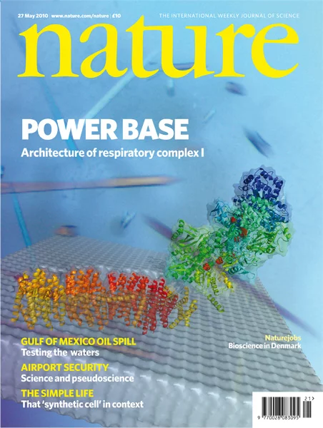
The SLS is one of the leading centres for this kind of investigation and is praised for the high quality of its X-ray beams and its state-of-the-art detectors. Three SLS beamlines are dedicated to the determination of the structures of large bio molecules, and each year attract many leading scientists from all over the world. The work of the British scientists has been published in the journal Nature, which in itself constitutes an important success in any scientist’s career. In addition to this, an image of the molecule investigated was printed on the cover of that issue – a fact considered by most scientists to be a particular distinction.
Reprinted by permission from Macmillan Publishers Ltd: Efremov, R. G., Baradaran, R. & Sazanov, L. A. Nature 465, 441–445 (2010)
Mitochondria are sometimes called cellular power plants, because, in these cell components, substances stemming from food react with oxygen from the air. In this reaction, energy is released that is used in the mitochondria to form the substance Adenosine triphosphate (ATP), which can store energy and distribute it throughout the whole organism. When studied in detail, this is a highly complicated process, with numerous bio molecules involved. Firstly, electrons are released by the reaction with oxygen and initiate the combination of molecules sitting in the inner membrane of the mitochondrion. Mitochondria have two membranes – an outer one forming its external boundary and an inner one separating its innermost region from the zone between the two membranes. The molecule combination in the inner membrane pumps protons out of the central part of the mitochondrion – against the potential gradient, i.e. in a sense “uphill”. These protons then return through the membrane by passing through another molecule that can produce ATP. As the protons travel with the potential gradient – “downhill” this time – energy is released that can be used for the synthesis of ATP.
Until recently, there was an important gap in the understanding of this process – it was unclear how the molecule combination known as respiratory complex I, which pumps the protons out of the innermost region of the mitochondrion, is built. Now, the researchers Rouslan G. Efremov, Rozbeh Baradaran and Leonid A. Sazanov from the Medical Research Council Mitochondrial Biology Unit in Cambridge, UK, have succeeded in determining the structure of the complex and deducing how it is able to push protons through the membrane. The complex has three proton channels passing through the membrane, and each of these channels is connected to an elongated part of the molecule that can act as a piston. When electrons arrive at a part of the molecule that can accept electrons, the whole complex becomes distorted, the piston moves, and can now push the protons through the channels by bending nearby parts of the molecule (see image). For their experiments, the researchers used molecules from bacteria that have a simpler structure than those present in the human organism, but still work according to the same principle.
An important part of the investigations has been performed at the Swiss Light Source (SLS) of the Paul Scherrer Institute, at the beamline X06SA. Here, a method known as X-ray crystallography is used in which scientists direct a beam of X-ray light at a crystalline substance and then observe the directions in which different parts of the beams are deflected. Using a complex mathematical procedure, they can then deduce the structure of the molecule under investigation from these patterns.
The SLS provides three beamlines for structure determination of biological molecules using synchrotron X-rays that are among the best in the world. The very intense and well-collimated X-ray beams provided at SLS, and the unique detectors that have been developed at PSI, allow an extraordinarily detailed determination of the molecules’ structures to be made.
The X-rays produced at the SLS are emitted by electrons moving at almost the speed of light along a circular path with a circumference of 282 metres. When compared with X-rays produced by a normal X-ray tube, synchrotron X-rays from the SLS are much more “brilliant”, which allows particularly sophisticated experiments to be performed. The SLS is open to scientific users from all over the world – as are PSI’s other large-scale facilities. A committee of scientists from different countries chooses the best proposals each year and allocates beam time to them.
About PSI
The Paul Scherrer Institute develops, builds and operates large, complex research facilities, and makes them available to the national and international research community. The Institute's own key research priorities are in the investigation of condensed matter and in the materials sciences, elementary particle physics, biology and medicine, and energy and the environment. PSI is Switzerland's largest research institution, with 1300 members of staff and an annual budget of approximately 260 million CHF.
About the MRC Mitochondrial Biology Unit
The Medical Research Council Mitochondrial Biology Unit (Cambridge UK) is set up to explore how mitochondria work, how they reproduce themselves inside our cells and to study their role in human diseases.
Contacts
Dr. Leo SazanovMRC Mitochondrial Biology Unit Cambridge, United Kingdom,
Phone: +44 1223 252910; E-mail: leo.sazanov@mrc-mbu.cam.ac.uk
Dr. Takashi Tomizaki,
Laboratory for Macromolecules and Bioimaging, Paul Scherrer Institute, Villigen PSI, Switzerland.
Phone: +41 56 310 51 29; E-mail: takashi.tomizaki@psi.ch
Dr. Clemens Schulze-Briese,
Head Laboratory for Macromolecules and Bioimaging, Paul Scherrer Institute, Villigen PSI, Switzerland.
Phone: +41 56 310 45 33; E-mail: clemens.schulze@psi.ch
Original publication
The architecture of respiratory complex IRouslan G. Efremov, Rozbeh Baradaran & Leonid A. Sazanov
Nature 465, 441–445 (2010)
DOI: 10.1038/nature09066


