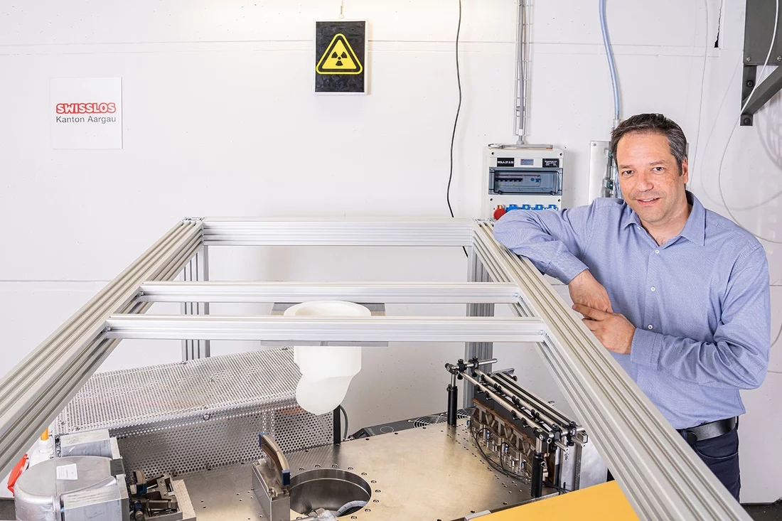
Marco Stampanoni, Zhentian Wang and their team have been nominated for this year’s European Inventor Award. The two scientists have been conducting research at PSI and ETH Zurich for more than ten years, in the pursuit of improved diagnostic methods for the early detection of breast cancer. The nomination for this prestigious award is in recognition of their pioneering work. The winners will be announced at the award ceremony on the 21 June 2022.
(Video: European Patent Office)
Since 2006, the European Patent Office (EPO) has presented an annual award to honour scientists whose ground-breaking inventions contribute to the technological and economic progress of our society and thereby, help to considerably improve our daily lives. The list includes five categories, with three nominees in each. Marco Stampanoni and Zhentian Wang are finalists in the category “non-EPO countries” – this covers inventions developed in countries that are not members of the EPO, or those developed in collaboration with non-EPO countries.
The two scientists have conceived an innovative diagnostic method that allows accurate and painless diagnosis of breast cancer. Their invention is based on a technique known as phase-contrast imaging. This X-ray method not only detects how much radiation is absorbed by the tissue, but also how the radiation changes direction slightly as it passes through different tissue densities. This additional information can be captured and processed into an image. “Our diagnostic method produces incredibly sharp images with excellent detail and contrast, making it easier than using traditional mammography to identify possible pre-stages of breast cancer, such as microcalcification, thickening or asymmetry,” Marco Stampanoni explains.
Drawback of the conventional method
Many countries operate screening programmes where women can undergo regular preventative checks for breast cancer once they reach a certain age. The most common method is mammography, which also uses X-rays. Here, the breast is pressed between two Plexiglas plates whilst X-rays are taken. The easiest way to envisage this, is like casting a shadow: most of the radiation passes straight through the breast – only a small amount is actually absorbed by the tissue. Any areas that are thicker or calcified, absorb more radiation than the surrounding healthy tissue. Any differences are displayed in a conventional X-ray image, where the suspicious areas appear lighter than the healthy tissue.
What sounds simple in theory, is not at all straightforward in practice. Any thickening of breast tissue is very difficult to detect, as the densities of the suspicious and healthy tissue are so similar, making distinction difficult. If there is suspicion of cancer, further diagnostic imaging is required such as an MRI scan or ultrasound. Ultimately, only a tissue biopsy delivers a definite diagnosis.
The difficulty of diagnosis produces many false-positive results, causing unnecessary emotional stress for the women affected. Around 8 out of 10 suspicious findings eventually turn out to be benign. This is one of the reasons why the effectiveness of regular mammogram screening is often questioned.
But how is it possible to produce high-resolution images without increasing the amount of radiation to a harmful level? Marco Stampanoni, Zhentian Wang and their team have found a solution.
Extracting every bit of information from light
The radiation produced by X-rays are nothing more than light – very high-energy light that is invisible to the human eye. When light waves hit a medium, for example, they can be fully or partly absorbed – a phenomenon that forms the basis of classical X-ray imaging. However, light can also be bent or broken by a medium, which forces a change of direction in the light wave as it leaves the medium. In addition, the light waves may slow down at different rates in a medium where the density distribution varies, which triggers phase shifting.
“In conventional X-ray imaging, all these phenomena are interpreted as noise, and filtered out. However, they actually contain important information about the density distribution within the medium under investigation,” says Zhentian Wang. When detected and analysed correctly, this information allows high-resolution X-ray images to be produced without having to increase the radiation dose.
To be able to use these phenomena to produce a more accurate diagnosis, the two scientists rely on grating-interferometry, a technique originally introduced at PSI by Christian David and Franz Pfeiffer for identifying the characteristics of synchrotron X-ray beams. The method is based on extremely fine gratings that converts the X-ray light into an interference signal. The absorption, phase, refraction and scattering properties of the object under investigation can then be calculated and an image produced from this.
This technique, first used in fundamental research, has been steadily refined and adapted. Countless hours worked by numerous scientists, together with numerous publications and patents, continue to push the research forward. Following close collaboration between Kantonsspital Baden, University Hospital Zurich and the company Philips, the first preclinical studies were performed on tissue samples and a prototype developed for two-dimensional imaging of patients. The very promising results led to the founding of the company GratXray AG in 2017, which is currently working on the development of a unique prototype for 3D imaging. The scientists want to use this to show how the procedure could work in everyday clinical practice. In 2017, GratXray AG won the Swiss Technology Award and now, the two researchers have the honour of being nominated for the European Inventor Award. “We’re very proud of this important international recognition. It shows how complex fundamental research combined with the inventive spirit and passion of an enthusiastic research team can produce a concrete application for the common good,” says Marco Stampanoni in acknowledging the significance of such a prestigious nomination.
The most common form of cancer there is
Breast cancer is the most common cancer in women – and since the end of 2021, it has been the most prevalent type of cancer worldwide, according to the World Health Organization. The risk of getting the disease also increases with age. Although women are mainly affected, men can develop breast cancer as well. As with any form of cancer, the earlier it is detected, the greater the chances of survival. Many countries therefore, rely on mass screening programmes where women undergo regular screening once they reach a certain age. The effectiveness of this preventative screening has still not been proven and its value has been repeatedly questioned. Phase-contrast imaging could help improve early detection and make screening programmes more attractive.
Text: Paul Scherrer Institute/Benjamin A. Senn
Profiles
Marco Stampanoni has worked at PSI since 2003. His career originally began at ETH Zurich, where he received the ETH silver medal for his outstanding PhD thesis on X-ray tomography. After switching to PSI he initially worked as a beamline scientist at the Swiss Light Source SLS before being appointed head of the X-Ray Tomography Group in 2005. In addition to his role as a scientist at PSI, he has been a professor at ETH Zurich since 2008, progressing from assistant to associate professor before eventually becoming Full Professor for X-Ray Imaging in 2017. Since 2018 Marco Stampanoni has also been President of the Research Commission of the Paul Scherrer Institute.
Zhentian Wang began his career at the Department of Engineering Physics at Tsinghua University in Beijing. He joined PSI in 2010, where he worked as a postdoc in Marco Stampanoni’s research group. Together, they refined the phase-contrast method and are working on developing it into clinical applications. Zhentian Wang is co-founder of the PSI spin-off GratXray AG, where he works as Technical Director. Since August 2021, he has been teaching and researching as an associate professor at Tsinghua University, specialising in the area of X-ray imaging.
Popular Prize
In addition to their nominations in the respective categories, all finalists are also nominated for the “Popular Prize”, whose winners are chosen by the general public. Everyone has a vote, and can cast a vote for their favourite nominee here: Popular Prize
The award ceremony at 12 noon on 21 June will be streamed live via the following link: https://inventoraward.epo.org/index
Further information
- PSI spin-off GratXray wins Swiss Technology Award 2017 – article, 17 November 2017
- Phase contrast improves mammography – media release, 15 May 2014
- A promising new method for the diagnosis of breast cancer – media release, 24 October 2013
- Investigation of a new method for the diagnosis of cancer in breast tissue – media release, 2 August 2011
More information on the topic of breast cancer
- World Health Organization: https://www.who.int/news-room/fact-sheets/detail/breast-cancer
- Swiss Cancer League (German only): https://www.krebsliga.ch/ueber-krebs/krebsarten/brustkrebs
Contact
Prof. Dr. Marco Stampanoni
Institute for Biomedical Engineering at ETH Zurich
Laboratory for Macromolecules and Imaging
Paul Scherrer Institute, Forschungsstrasse 111, 5232 Villigen PSI, Switzerland
Telephone: +41 56 310 47 24, e-mail: marco.stampanoni@psi.ch [German, English, Italian, French]
Assoc. Prof. Dr. Wang Zhentian
Department of Engineering Physics
Tsinghua University, Haidian District, 100084, Beijing, China
Telephone: +86 13716413267, e-mail: wangzhentian@tsinghua.edu.cn [English, Chinese]
Copyright
PSI provides image and/or video material free of charge for media coverage of the content of the above text. Use of this material for other purposes is not permitted. This also includes the transfer of the image and video material into databases as well as sale by third parties.


