
The Paul Scherrer Institute PSI is involved in a project conducted by several research institutes (spearheaded by EPFL) to devise an X-ray machine especially for developing countries. The device should be able to cope with tropical climes and be easy and cheap to repair. PSI researchers are focusing on producing a cost-effective detector that is necessary for the imaging. The detector registers the X-ray light much like a chip in a digital camera.

Robust X-ray machine for modern diagnostics in developing countries Two thirds of the global population don’t have access to X-ray machines, even though X-ray images are basic diagnostic tools in modern medicine. Although developing countries keep receiving old X-ray units from industrial countries, the relief they provide is short-lived as they are not adapted to the tropical conditions on the ground. They can’t withstand the heat, humidity, dust or irregular electricity supplies. The WHO estimates that 70 per cent of all medical equipment donated is never actually used in the recipient country.
Up to tough demands
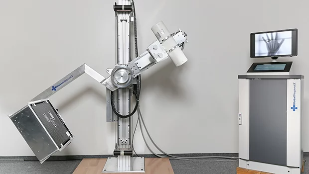
Together with GlobalDiagnostiX, a consortium headed by ETH Lausanne EPFL1 has developed the first digital X-ray machine especially with developing countries in mind. PSI was also involved. Postdoc David Haberthür explains: Calculated over ten years, including the purchase, operation and supplies, modern film X-ray units cost around 500,000. The aim of the project is to reduce these overall costs to a tenth. At the same time, the device is supposed to withstand temperatures of up to 40 degrees Celsius and tolerate humidity levels of up to 95 per cent, not to mention exposure to dust, which is also a common problem.
Bridging powercuts
The new development also tolerates instable power supplies. The prototype is able to function for up to five hours without any electricity whatsoever. The sudden high power consumption of X-ray machines also poses problems for hospitals. An X-ray unit requires a very high amount of electricity in a short space of time for the X-ray tube. Thanks to a specially developed module, GlobalDiagnostiX is able to supply this without constantly overloading the hospital’s electricity supply.
Moreover, the device is easy to operate, which is important as it is frequently not possible to train the often extremely busy members of staff sufficiently in how to use complicated equipment.
PSI’s job was to come up with a concept for a suitable X-ray detector and build it as a prototype in conjunction with its partners. It’s supposed to be as cost-effective as possible but still meet the latest medical standards in terms of image quality and low radiation exposure,
explains Haberthür. The digital detector replaces in the X-ray unit the X-ray film, which is still used frequently in developing countries. The detector converts the X-ray beams into an image, much like a digital camera.
6,000 images a year
The device is designed to take more than 6,000 images per year. Its main task will be to perform the most common examinations. It isn’t a high-end device for high-performance medicine. But it can do 99 per cent of what’s needed in everyday hospital life in a planned environment,
continues Haberthür. In order to provide the best possible image quality, Haberthür combined the detectors necessary for the imaging with different lenses and a scintillator. The latter converts X-ray light into visible light – as only this can be recorded by the image detectors.
In the prototype, twelve structurally identical modules made of these three components are arranged in such a way that they can be used to X-ray an area measuring 43 by 43 centimetres. Like a series of individual cameras, they work much like an insect’s compound eye. Each one only forms a small part of the overall picture. Software puts together the complete picture from the individual images, like the insect’s brain. The challenge was to determine the best combination of components. There were two criteria: on the one hand, all the components had to be available on the market; on the other hand, it should also be possible for the entire device to be repaired by the local technicians on the ground.
Dozens of combinations tested
We worked out different variations and sometimes conducted simulations to select the ideal component combination for the predefined requirements,
recalls Haberthür. The components deemed suitable then needed to be tested in all combinations imaginable to filter out the best ones. Finally, Haberthür teamed up with the student Ivan Kasanzew to build and test 124 different combinations in the lab.
A design still needed to be found that would also withstand rougher treatment without becoming damaged. This took place in collaboration with the EPFL and the Institute of Industrial Automation at the University of Applied Science Western Switzerland (HES-SO) in Yverdon-les-Bains. As Haberthür explains: Our detector is incorporated into a stable metal frame a lot more sturdily than usual and can also withstand traumas. If you drop a conventional device, the detector’s had it.
Besides the solid construction, the modular, compound-eye-like structure also boosts the device’s robustness.
If one of the detector’s twelve modules breaks, the whole system is not immediately paralysed. The patient might have to place his or her hand a bit differently. But you can still carry on taking images until the spare part arrives.
The prototype of the complete X-ray device is already fully functional. The unit now needs to be put through its paces at a test hospital in Cameroon. Only if it proves itself under challenging conditions on the ground will it then find its market.
1 The EssentialTech Team from the Cooperation & Development Centre at EPFL was in charge of the coordination. A complete list of the institutions involved is available on the website: http://www.globaldiagnostix.org/
Text: Paul Scherrer Institute/Alexandra von Ascheraden
Additional information
Website of GlobaldiagnostiXContact
Dr. David HaberthürPaul Scherrer Institute
5232 Villigen
Switzerland
Telephone: +41 56 310 31 80
E-mail: david.haberthuer@psi.ch