The interior of fossils and artefacts that are hundreds, thousands and yes, sometimes millions of years old can be examined at two PSI research facilities. A conversation with Federica Marone and Eberhard Lehmann, who are opening a new view into the past with their methods.
Ms. Marone, Mr. Lehmann - palaeontologists and archaeologists regularly come to you to look inside fossils and ancient objects with your non-destructive analysis methods. How do they know that their questions can be answered at PSI?
Marone: In our research group at the Swiss Light Source SLS, this collaboration began more than ten years ago. An article by a professor in England, in which fossil embryos of a wormlike creature were presented, came to the attention of my colleague Marco Stampanoni. The researchers had analysed these fossils only on the basis of their external structure – at our beamline they would also have been able to look at the interior of this small, roughly one-millimetre thick spherule. So Stampanoni contacted these people and invited them to PSI. They knew nothing about synchrotrons and tomography but, thanks to us, realised that in this way they could gain completely new insights. And after the first joint article was published, other palaeontologists also got in touch with us.
Lehmann: It was similar with us: Originally no historian knew what our method is capable of, but after the first examples were published, word got around. Our first customers
came from the Vindonissa Museum in Brugg, just a few kilometres away from PSI. In the 1990s, they brought us a Roman sword, a so-called gladius, that had just recently been discovered. We examined it and found out that the wooden scabbard was still intact. Over the centuries this had corroded together with the blade to such an extent that it could no longer be discerned with the naked eye, but with the neutrons we were able to make the wood grain very clearly visible.
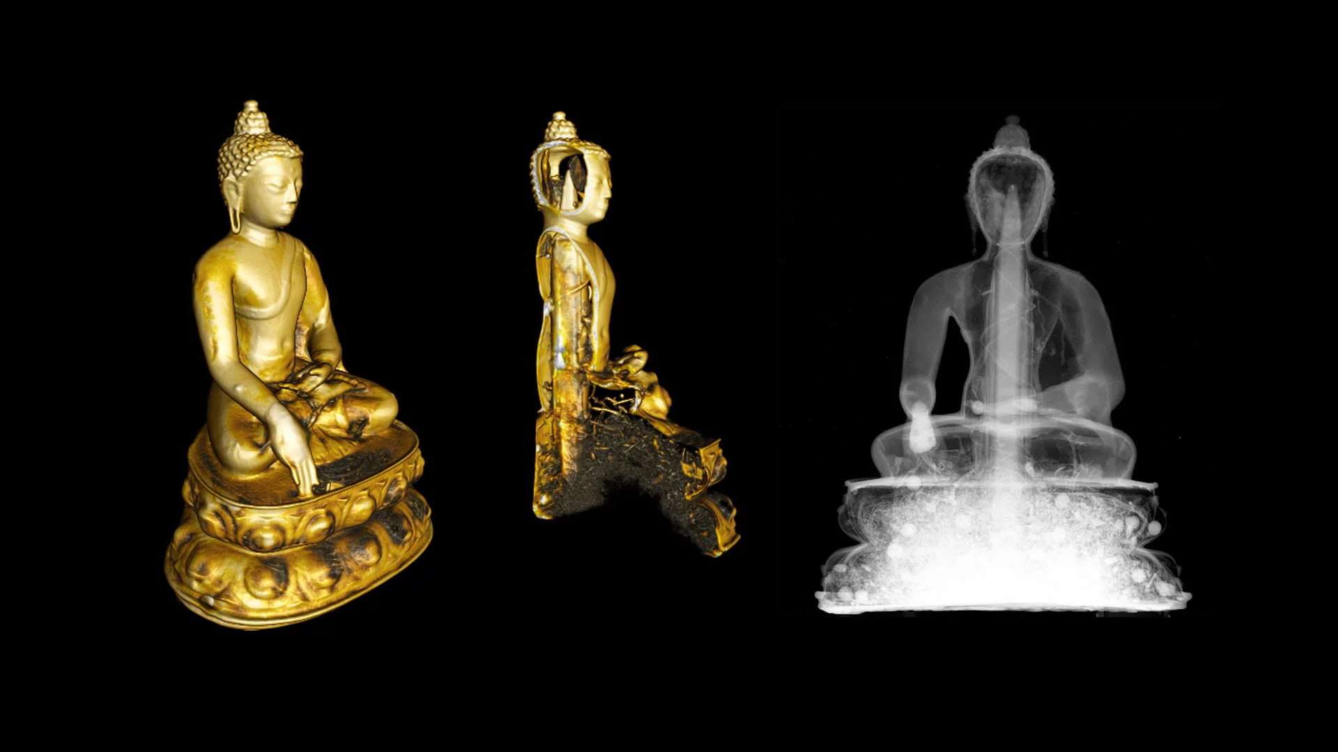
Buddha Shakyamuni, 14th or 15th century CE
What else can be discovered about ancient objects or palaeontological samples through the measurement methods used at PSI?
Marone: With X-ray tomography, we can examine the interior of objects and materials without destroying them. This is important for the archaeologists and palaeontologists alike, since the objects are often unique. Sometimes, though, it can't be avoided and the palaeontologists plan to cut one of their fossils. Then they bring it to us beforehand to decide, with the help of our imaging, which they should cut and exactly where to do it, so that what's inside will be damaged as little as possible.
On the other hand, these researchers can already find out an unbelievable amount about the development of plants and animals just by looking at our images. For example, we made studies of fossilised jawbones of a prehistoric fish species, and there the tissues, the cells, and all of the growth lines could be very precisely seen. From that, our research partners were able to draw conclusions about the evolutionary development of the jaw and determine: Teeth came into being at the same time as or shortly after the jaw, several hundred million years ago.
Lehmann: The X-ray tomography group has its pool
of partners, and we in neutron beam analysis have a somewhat different one - we have considerably more historians than palaeontologists. That is due to the methodology: While X-ray tomography at PSI focuses mainly on the microscopic structures and can go into great detail, we work on a coarser scale. And: With the neutrons, completely different materials are transparent
than with X-rays.
Imaging with X-rays and neutrons at PSI
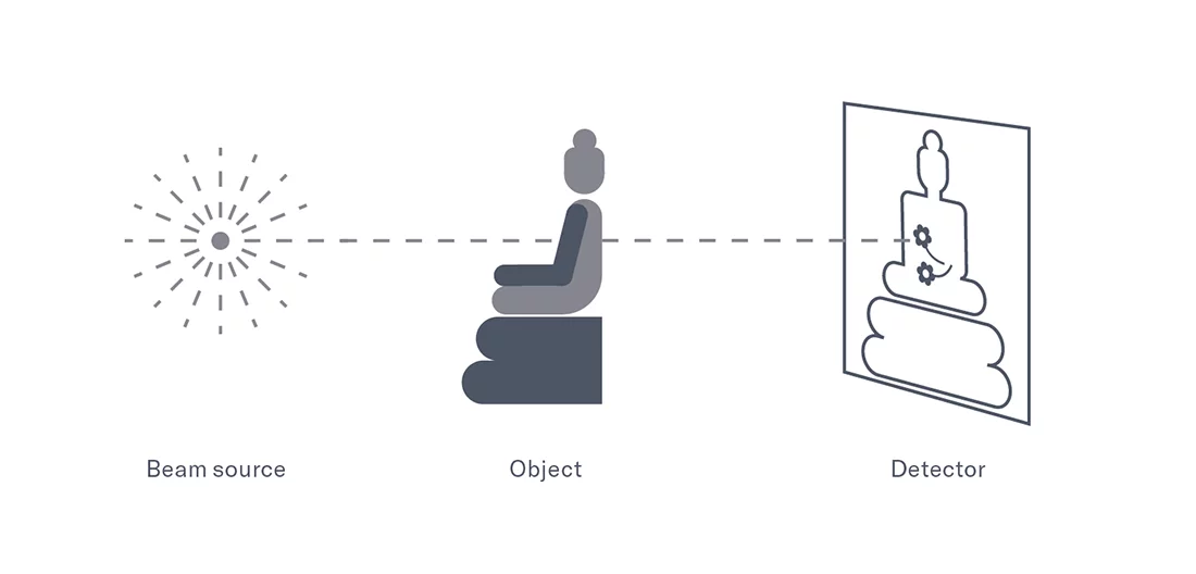
In addition to such radiographic images, both types of beams can also generate a three-dimensional, virtual reconstruction of the object. For that, it is first illuminated repeatedly from different directions; in the end, all of the data gathered are merged in the computer to form a so-called tomogram.
At PSI, extremely intense X-rays are available at the Swiss Light Source SLS, enabling imaging with high resolution. Meanwhile, the spallation neutron source SINQ allows the relatively rare imaging with neutrons.
What is the biggest surprise that you have experienced in working with historical objects?
Lehmann: That was definitely the examination of Buddhist statues. These can't be imaged well with X-rays, because they are made of metal. With neutrons, however, you can see: The statue is hollow, and inside it are wood, textile, and also plant materials such as flowers. We do this in collaboration with Michael Henss, who is the expert for this topic. It was with him that we investigated many of these Buddhas - and that, for me, was an aha
experience, what can be in there. And then we were naturally curious and looked further. Since then we have studied more than 60 such statues. In some of them there were written scrolls, in others gemstones - completely different kinds of things.
Marone: One surprising insight that came to light just recently was that fungi have been around much longer than was previously assumed. Up to now it had been thought that they are around 400 million years old. But now some of our partners have found traces of fungi in rock layers that could be up to 2.4 million years old. Their filaments, branching out and tangled together in the cracks, have been preserved as fossils. The palaeontologists are comparing them to present-day fungi and thus are able to carry out a biological classification.
Lehmann: The age can be determined according to which rock layer the sample came from?
Marone: Exactly. The rock layer was dated radiometrically, that is, with the help of small amounts of lead and uranium that occur in it naturally. I am always fascinated by the kinds of knowledge these palaeontologists have. They know the rock just as perfectly as the biology and evolution, and they bring it all together.
Lehmann: It's true, you meet very interesting people in this work – people who have achieved such a high level of education are often real characters. They surely must find us a bit quirky as well.
When one comes into the halls of neutron analysis and X-ray tomography, everything is very big and technical – am I right in assuming that this is rather unusual for your cooperation partners?
Marone: Yes, that's true. We always try to arrange everything as simply as possible and to provide assistance so that our external partners can find their bearings when they're with us.
Lehmann: Partners who have already been with us a couple of times are by now doing their own analyses. Some have entire projects running, even doctoral research studies, and come back regularly. There are also others who simply bring us a sample that we then examine, because we don't want to let any complete technical laypersons near our machines.
Marone: That works for us most of the time. Especially in the beginning, we help the external researchers find the right settings and explain how the machine is used.
Lehmann: Some of our users come only once, with a single sample ...
Marone: Oh, they sometimes come to us with a hundred samples at once. I never handle the samples myself because such age-old fossils are, as I said, unique, and some belong to museums. I do not want to be responsible if something happens to one of them. At our beamline, the researchers mount their samples themselves.
What do you have to do in order to find a common language with such a different scientific culture?
Lehmann: Learn from one another. The archaeologists have to learn how our tomography image can be interpreted, and we need to learn what kinds of materials and methods existed in a particular time. Sometimes we decide that we are not at all the right partner – for example, when something is too delicate. Then we send them along to SLS. That's why the dialogue here within PSI is also very important.
Marone: With us, too, the dialogue functions pretty well – our partners are for the most part positively interested, and by now we also know what is realistic and what doesn't work.
One last question: Is there one experience that especially sticks in your memory?
Marone: Now that is a somewhat special story. The palaeontologists, when they study something here, are always discovering new species, which they then are free to name. And they named one plant species Tomcatia, after our beamline ...
Lehmann: Not after you?
Marone: Of course, one species was also named after me, the Lignierispermum maroneae, and now the Lobospermum stampanoni has been named after my colleague Marco Stampanoni. That really was something very special for us.
Lehmann: In general, I am inspired again and again by the good collaboration with the archaeologists. Recently, for example, the people at the Roman Museum in Avenches came directly to us with their metal finds after a new dig. That shows me how much our method and the contribution that we are able to make with it in the archaeology scene have taken root there.
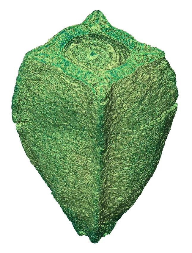
Lignierispermum maroneae, 110 million years old
About the scientists
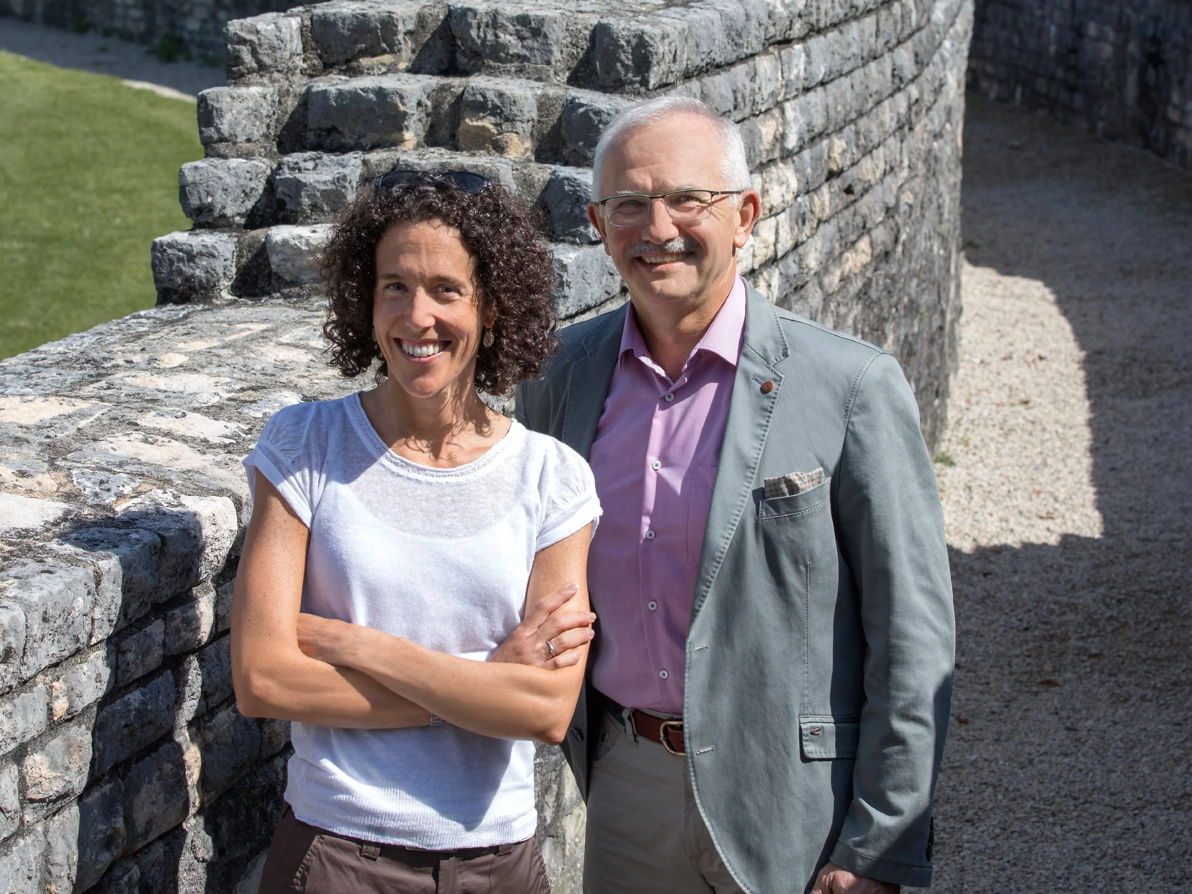
Eberhard Lehmann was born in 1952 in Saxony, where he studied and did his doctoral work in physics. In 1991 he came to PSI where, at the spallation neutron source SINQ, he refined the techniques for imaging with neutrons and led his own research group until his retirement in 2017. At the SINQ beamlines, neutron imaging illuminates ancient objects as well as automobile components, plants, roots, and many other things.
Interview: Luise Loges