
Call for expressions of interest: Beamline partners at the SLS for PX II and PX III
We invite companies and institutions to secure access to the beamlines X10SA/PX II and X06DA/PX III through a long term contract.
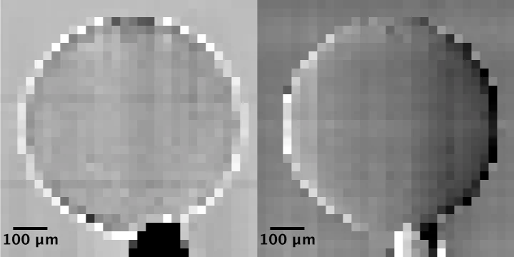
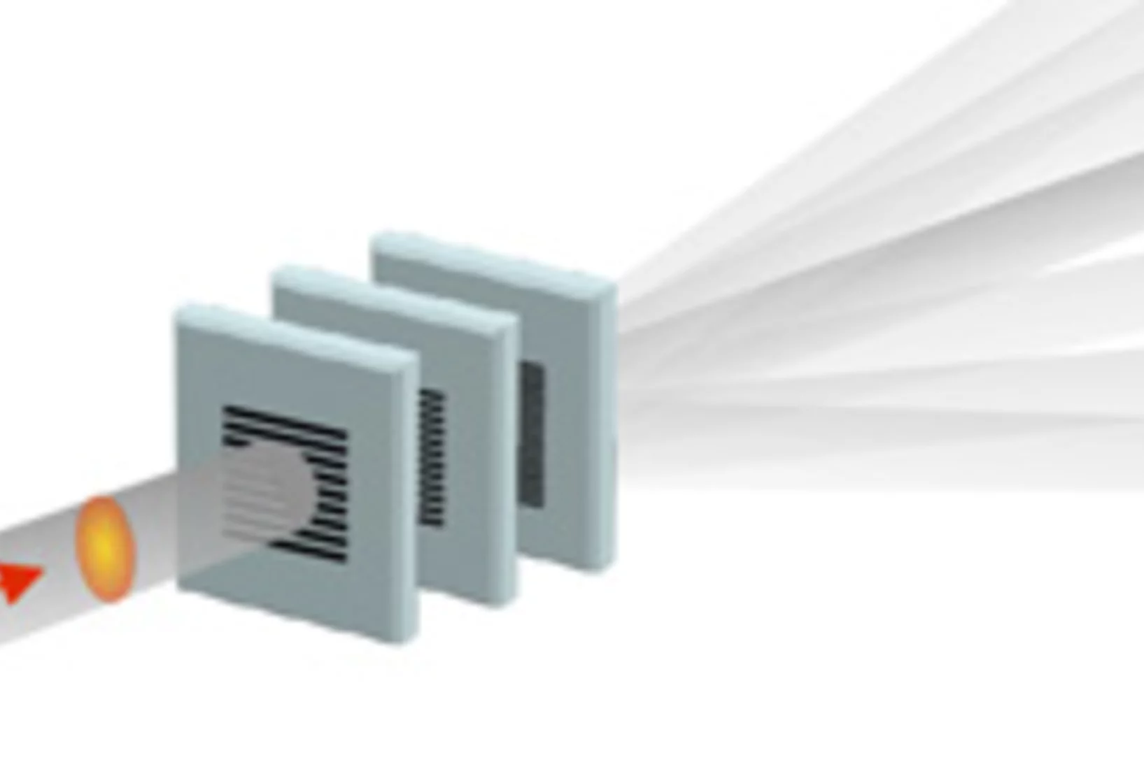
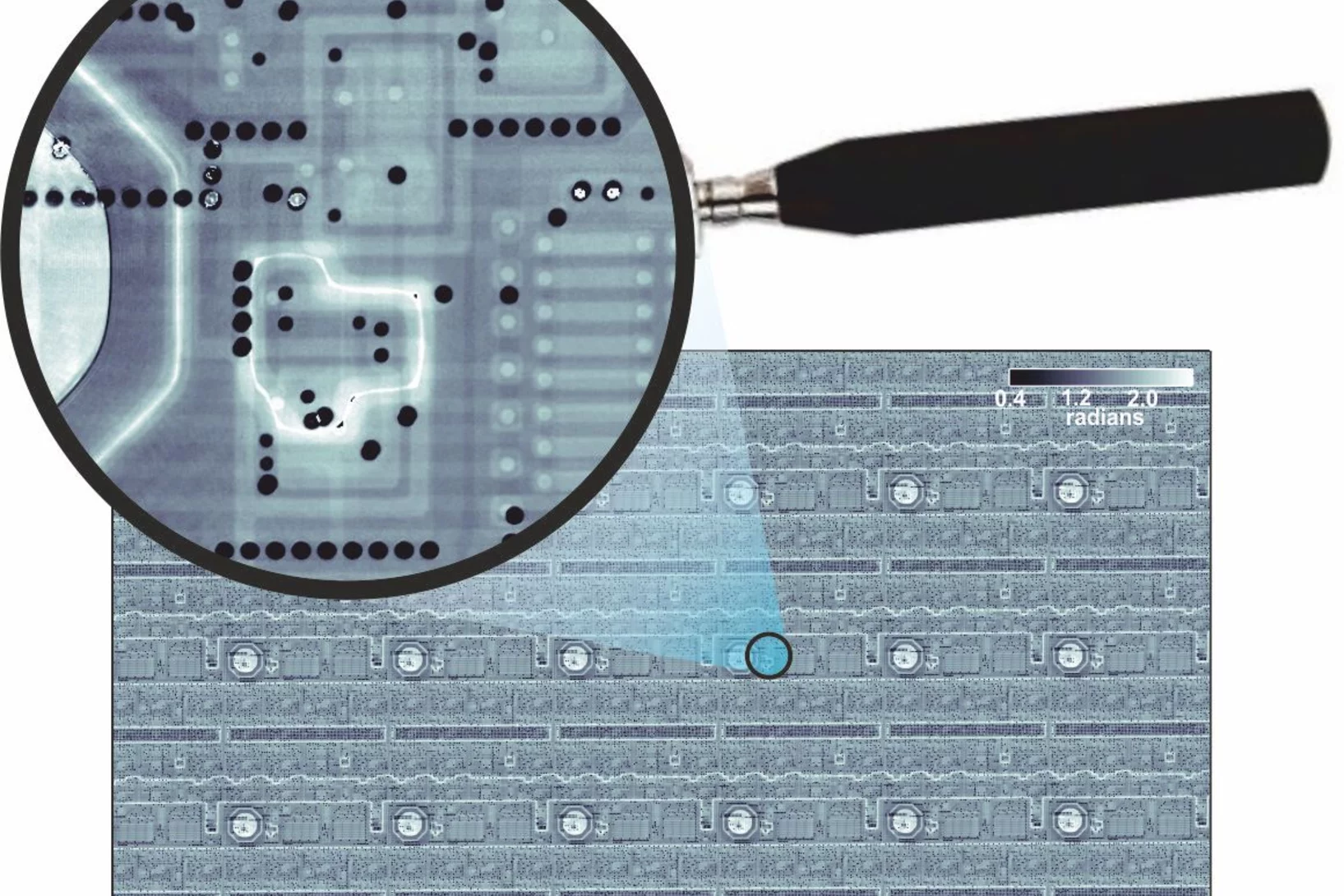
Single shot grating interferometry demonstrated using direct conversion detection
Researchers at the Paul Scherrer Institute's Swiss Light Source in Villigen, Switzerland, have developed an X-ray grating interferometry setup which does not require an analyzer grating, by directly detecting the fringes generated by the phase grating with a high resolution detector. The 25um pitch GOTTHARD microstrip detector utilizes a direct conversion sensor in which the charge generated from a single absorbed photon is collected by more than one channel. Therefore it is possible to interpolate to achieve a position resolution finer than the strip pitch.
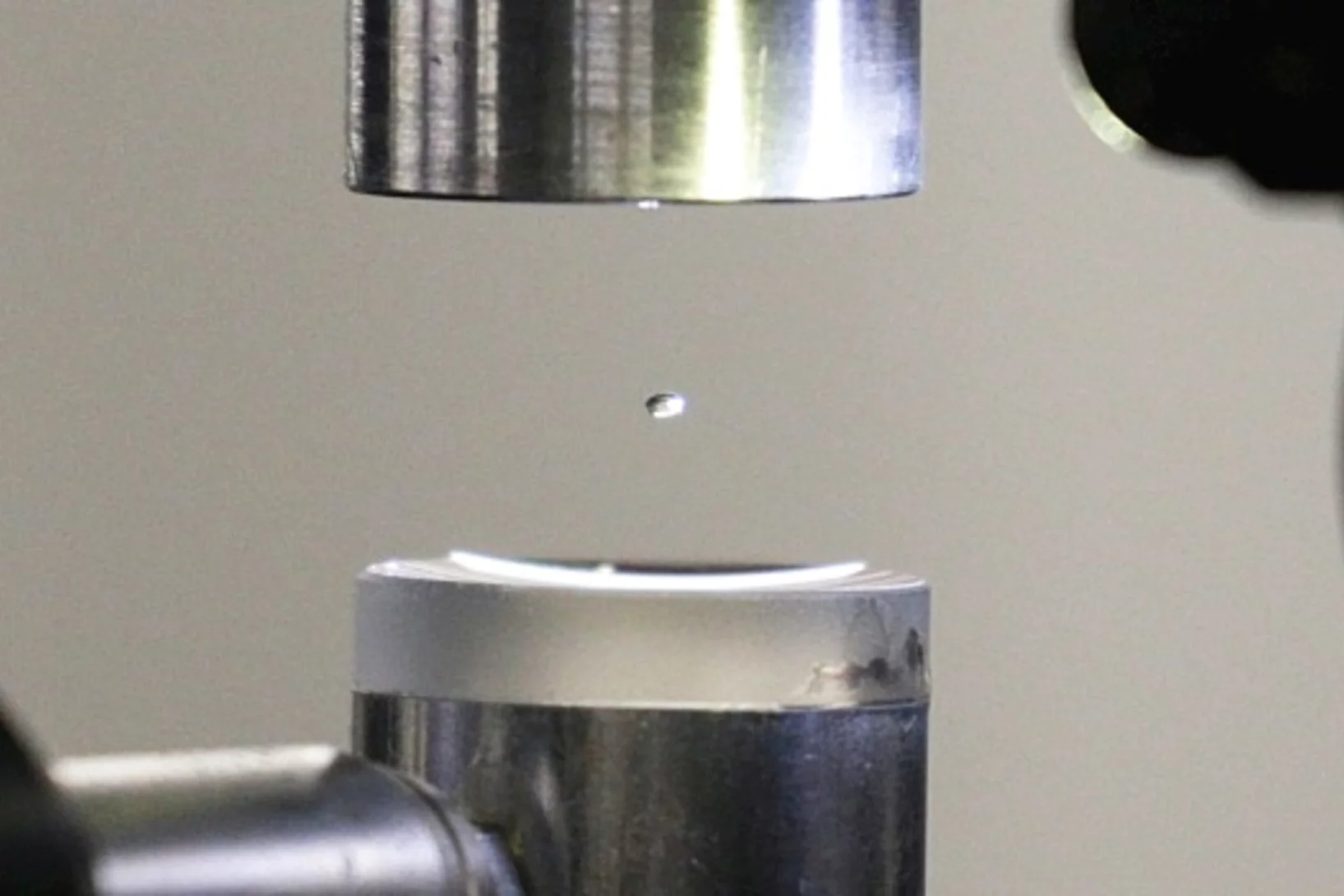
Experiment in a hovering droplet
At the PSI, the exact structure of proteins is deciphered in the standard way, with X-rays. Now two PSI researchers have used a clever trick to advance this method further: Instead of pinning down the proteins, they are studying them within a levitating drop of liquid.
How does food look like on the nanoscale?
The answer to this question could save food industry a lot of money and reduce food waste caused by faulty production. Researchers from the University of Copenhagen and the Paul Scherrer Institut have obtained a 3D image of food on the nanoscale using ptychographic X-ray computed tomography. This work paves the way towards a more detailed knowledge of the structure of complex food systems.

Watching lithium move in battery materials
In order to understand limitations in current battery materials and systematically engineer better ones, it is helpful to be able to directly visualize the lithium dynamics in materials during battery charge and discharge. Researchers at ETH Zurich and Paul Scherrer Institute have demonstrated a way to do this.
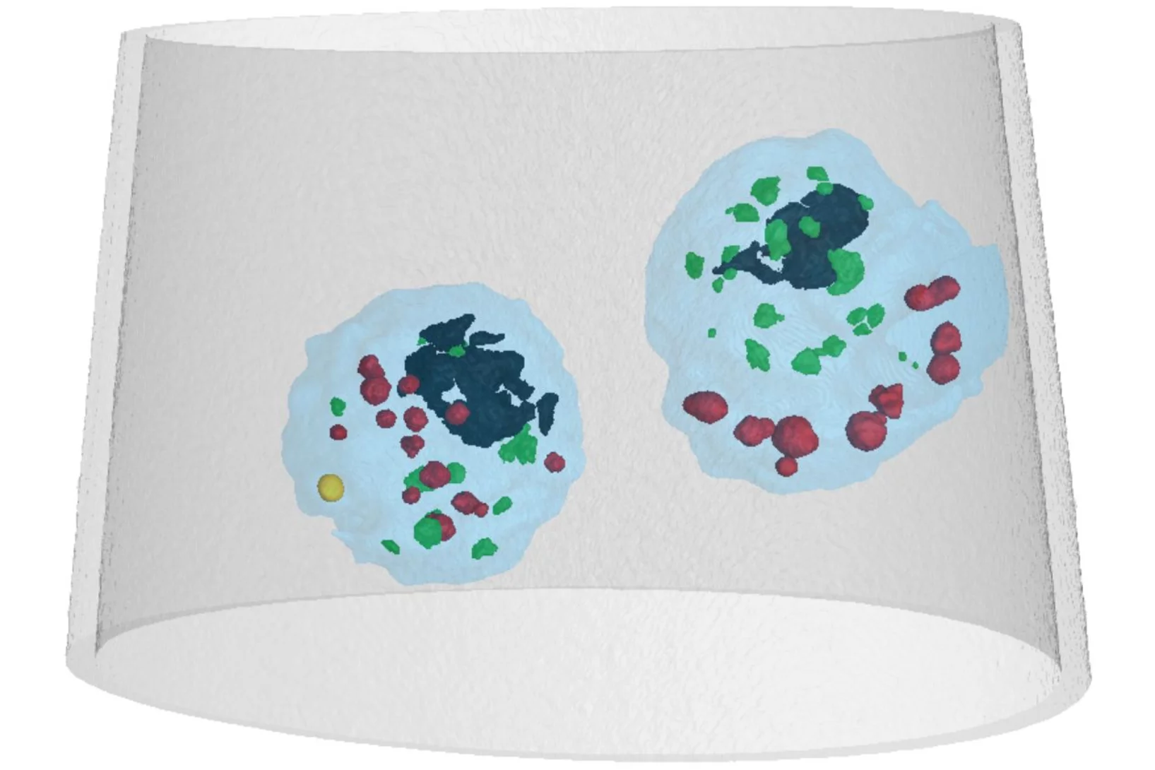
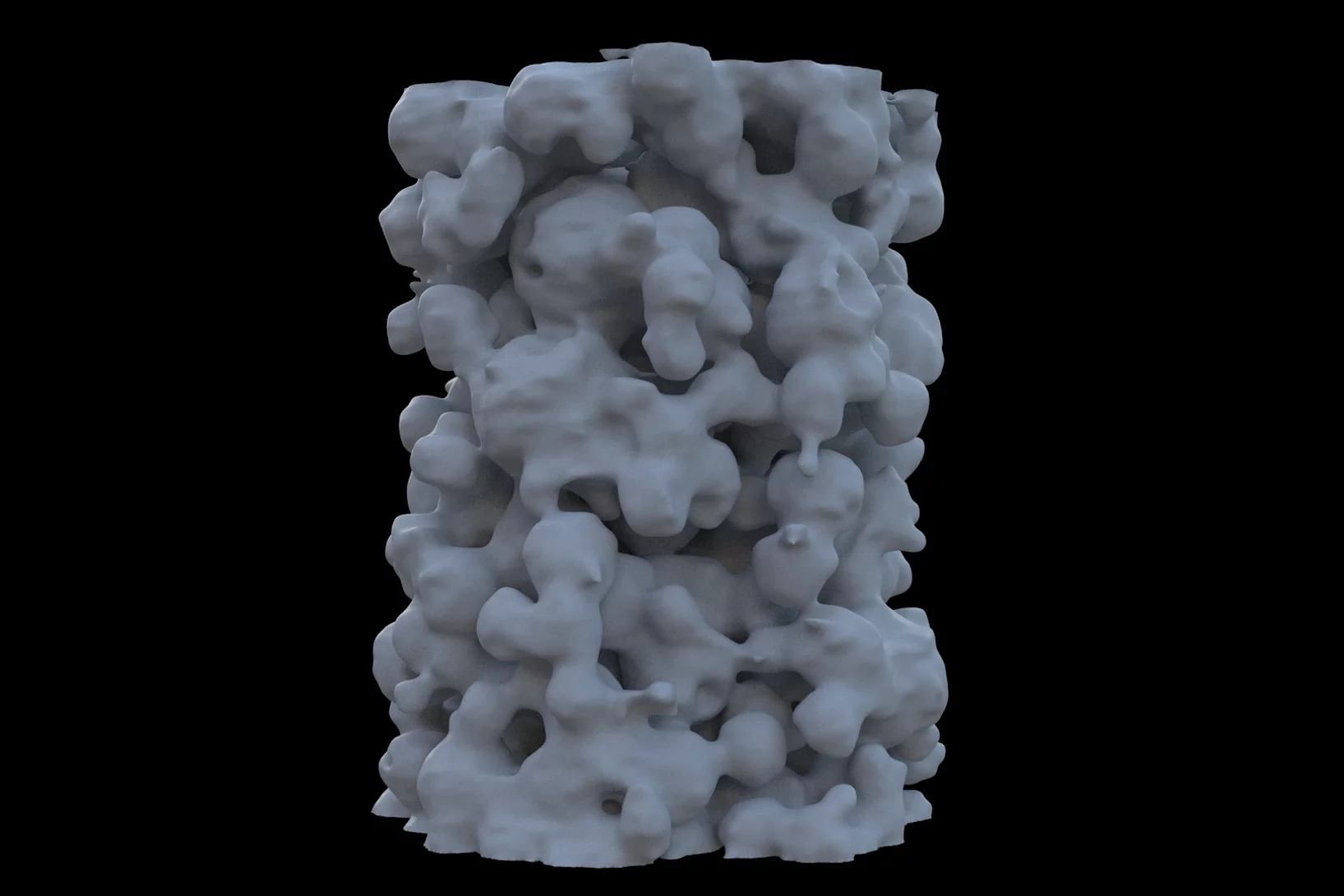
Mass density distribution of intact cell ultrastructure
The determination of the mass density of cellular compartments is one of the many analytical tools that biologists need to unravel the extremely complex structure of biological systems. Cryo X-ray nanotomography reveals absolute mass density maps of frozen hydrated cells in three dimensions.
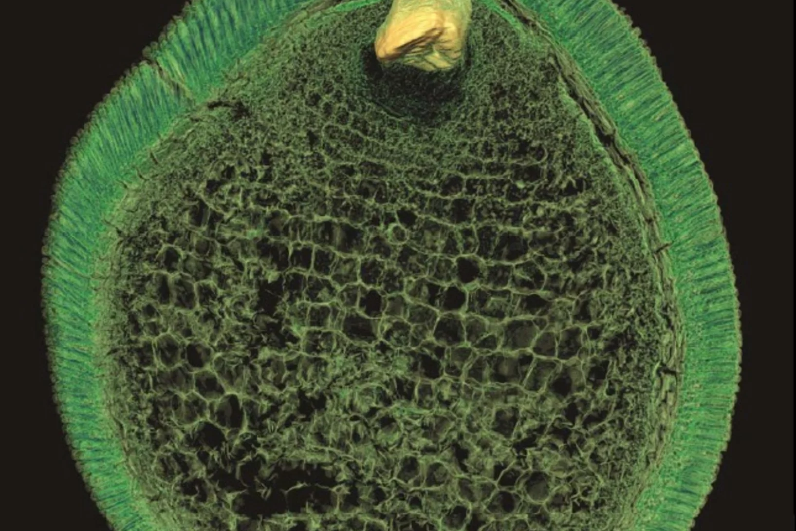
Preserved Embryos Illustrate Seed Dormancy in Early Angiosperms
The discovery of exceptionally well-preserved, tiny fossil seeds dating back to the Early Cretaceous corroborates that flowering plants were small opportunistic colonizers at that time, according to a new Yale-led study.
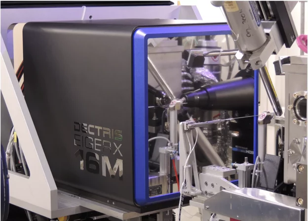
First EIGER X 16M in operation at the Swiss Light Source
The macromolecular crystallography beamline X06SA at the Swiss Light Source, a synchrotron operated by Paul Scherrer Institute, is the first one in the world to upgrade its detector to an EIGER X 16M.
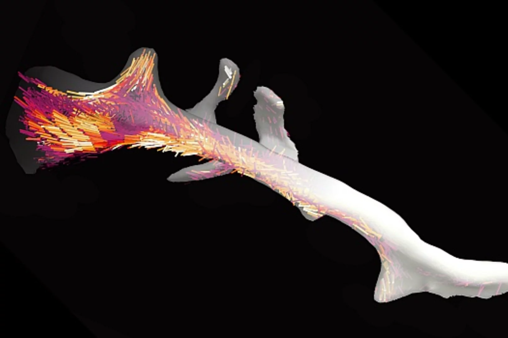
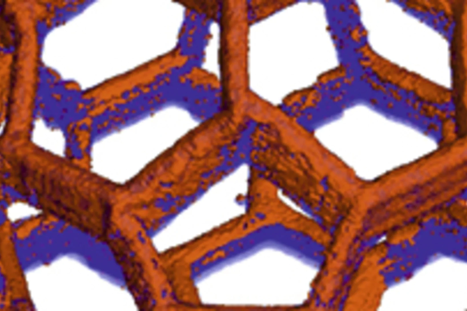
3D nanostructure of a bone made visible
Bones are made up of tiny fibres that are roughly a thousand times finer than a human hair. Researchers at the Paul Scherrer Institute PSI have developed a new computer-based algorithm with which they were able to visualize the localised order and alignment of these nanostructures inside an entire piece of bone for the first time.
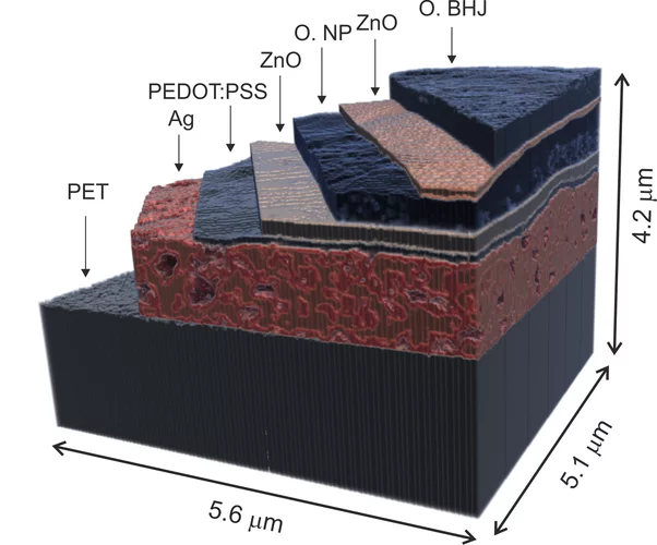
X-ray nanotomography aids the production of eco-friendly solar cells
Polymer solar cells are in the spotlight for sustainable energy production of the future. Characterization of these devices by X-ray nanotomography helps to improve their production using environmentally friendly materials.
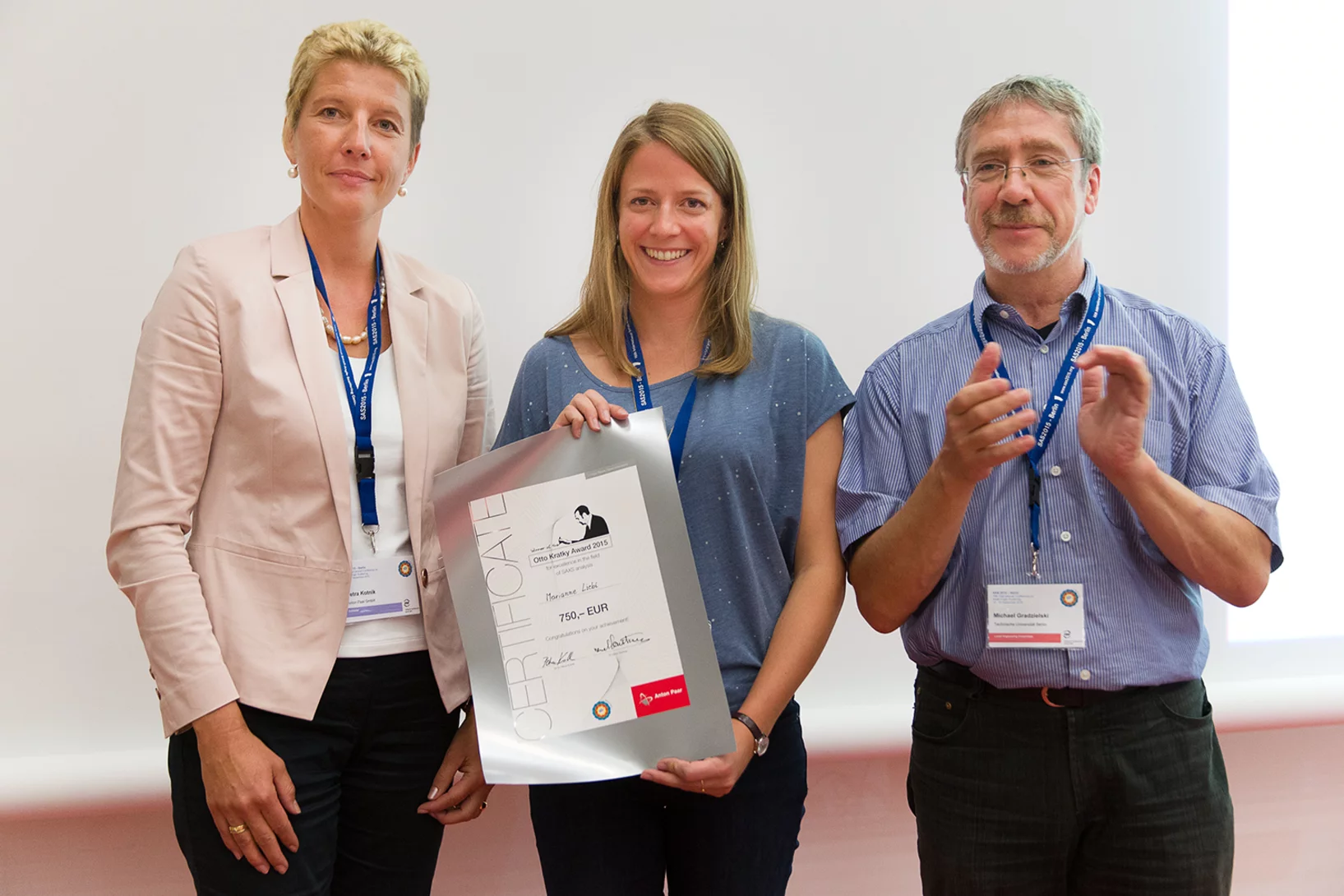
2015 Otto Kratky award
Marianne Liebi was awarded the 2015 Otto Kratky award by the Helmholtz-Centre Berlin for excellence in the field of small-angle X-ray scattering (SAXS) analysis. The award was bestowed in the last SAS2015 conference in Berlin. Marianne is a postdoctoral fellow in the coherent X-ray scattering group (CXS) in PSI, carrying out research in scanning SAXS measurement and analysis in 2D and 3D. Image credit ©HZB/Michael Setzpfandt
In Situ Serial Crystallography Workshop at the SLS
The Macromolecular Crystallography group at SLS is organizing a three days workshop on in situ serial crystallography (http://indico.psi.ch/event/issx) between November 17 and 19, 2015. It will be dedicated in the presentation of a novel method facilitating the structure determination of membrane proteins, which are highly important pharmaceutical targets but are difficult to handle using 'classical' crystallographic tools. Designed for 20 Ph.D. students, postdocs and young scientists from both academia and industry, the workshop will consist of introductory lectures, followed by hands-on practicals on in meso or lipidic cubic phase (LCP) crystallization, on in situ serial crystallography data collection using a micro-sized beam and on data processing.
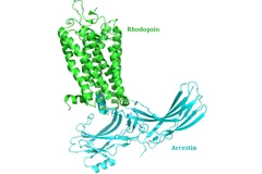
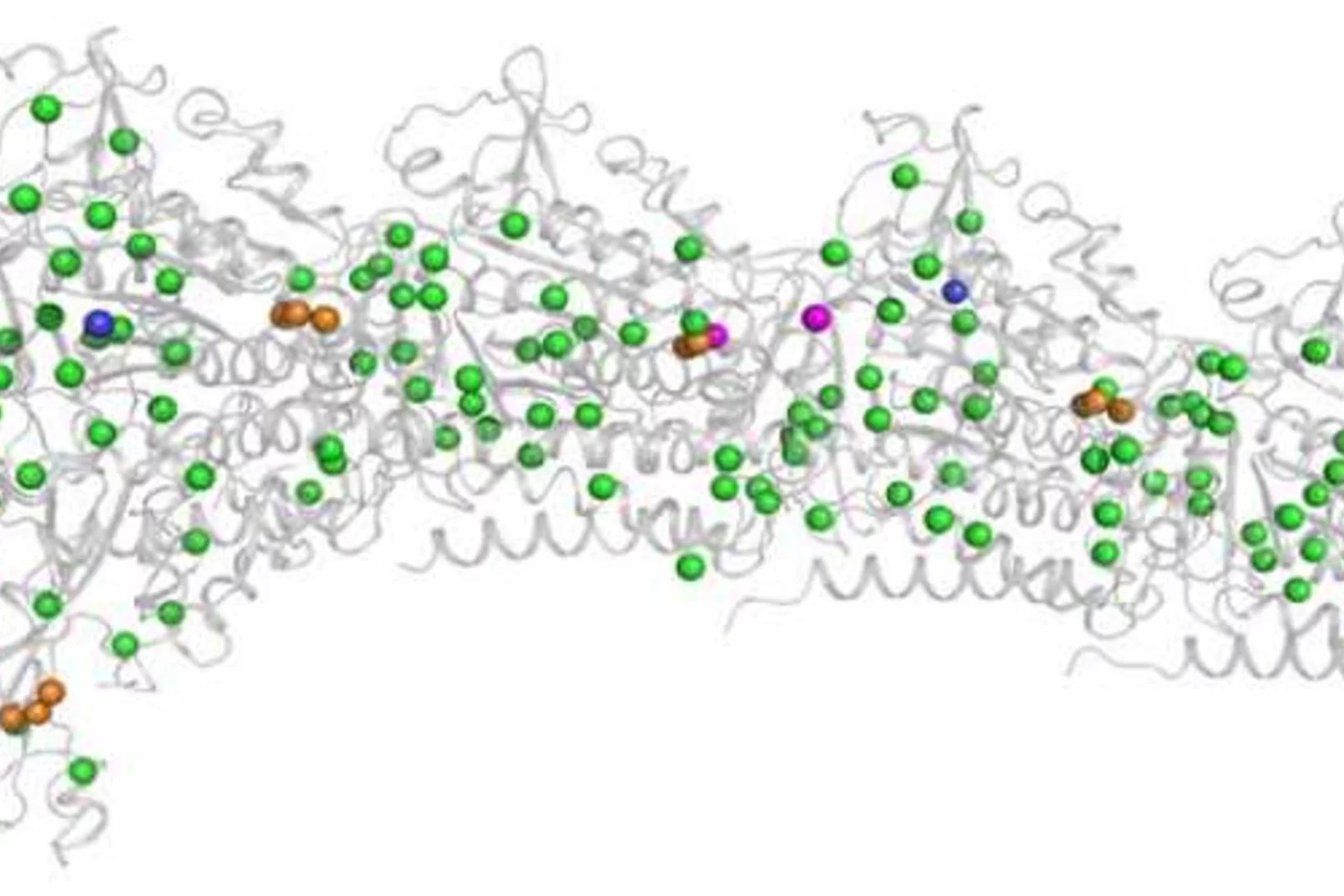
New insight into receptor signalling
A team of 72 investigators across 25 institutions including researchers from the Paul Scherrer Institut obtained the X-ray structure of a rhodopsinàarrestin complex, which represents a major milestone in the area of G-protein-coupled-receptor (GPCR), a protein family recognized in the award of the 2012 Nobel Prize in Chemistry.
Element-Specific X-Ray Phase Tomography of 3D Structures at the Nanoscale
Recent advances in fabrication techniques to create mesoscopic 3D structures have led to significant developments in a variety of fields including biology, photonics, and magnetism. Further progress in these areas benefits from their full quantitative and structural characterization.
Together, not alone
Decoding biomolecules at SwissFEL and SLSProteins are a coveted but stubborn research object. A method developed for x-ray free-electron lasers and PSI’s future SwissFEL should now help researchers to make good headway in this field. It involves x-raying many small, identical protein samples consecutively at short intervals, thereby avoiding the main problem that protein research has faced thus far: producing samples in a sufficient size.
From inside an eggshell
Tiny cavities inside eggshells supply the materials that stimulate and control the shell’s growth. Using a novel imaging technique, researchers from the Paul Scherrer Institute (PSI), ETH Zurich and the Dutch FOM Institute AMOLF have succeeded in depicting these voids in 3D for the first time. In doing so, they lift an old limitation of tomographic images and hope that one day medicine will also benefit from their method.
Multiresolution X-ray tomography, getting a clear view of the interior
Researchers at PSI have developed a technique that combines tomography measurements at different resolution levels to allow quantitative interpretation for nanoscale tomography on an interior region of interest of the sample. In collaboration with researchers of the institute AMOLF in the Netherlands and ETH Zurich in Switzerland they showcase their technique by studying the porous structure within a section of an avian eggshell. The detailed measurements of the interior of the sample allowed the researchers to quantify the ordering and distribution of an intricate network of pores within the shell.
Split x-ray flash shows rapid processes
SwissFEL, PSI’s x-ray laser, is to render the individual steps of very rapid processes visible. A new method will facilitate especially precise experiments: the individual x-ray flashes are split into several parts that arrive at the object under examination one by one. The principle of the method harks back to the ideas of the earliest high-speed photography.
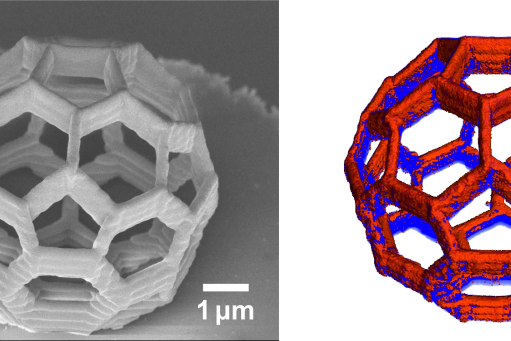
Nanometres in 3D
Scientists at the Paul Scherrer Institute and ETH Zurich have created 3D images of tiny objects showing details down to 25 nanometres. In addition to the shape, the scientists determined how particular chemical elements were distributed in their sample and whether these elements were in a chemical compound or in their pure state.
Shortcut to protein portraits
All living organisms, from bacteria to humans, rely on proteins to perform their vital functions. How these proteins accomplish their tasks depends on their structure. Researchers from the Paul Scherrer Institute have now devised a novel method to determine the crystal structure of proteins using X-ray light, which could also hasten the development of new drugs in future. The study will be published in the journal Nature Methods on 15 December.
Innovation Award on Synchrotron Radiation 2014 for high-resolution 3D hard X-ray microscopy
The 2014 Innovation Award on Synchrotron Radiation was bestowed to researchers Ana Diaz, Manuel Guizar-Sicairos, Mirko Holler, and Jörg Raabe from the Paul Scherrer Institut, Switzerland, for their contributions to method and instrumentation development, which have set new standards in high-resolution 3D hard X-ray microscopy.
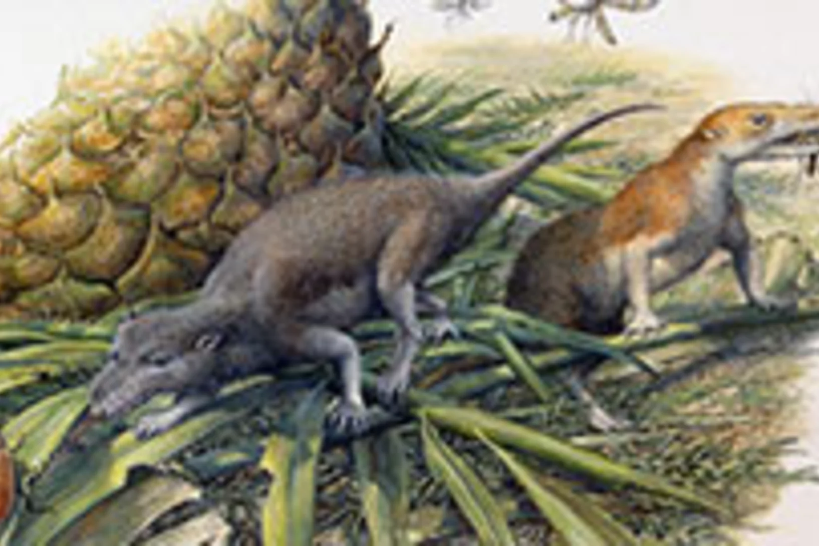
Jurassic Welsh mammals were picky eaters, study finds
New analyses of tiny fossil mammals from South Wales are shedding light on the function and diets of our earliest ancestors, a team led by researchers from the Universities of Bristol and Leicester report in the journal Nature. The team used CT scanning with synchrotron X-rays at PSI’s Swiss Light Source to reveal in unprecedented detail the internal anatomy of the mammals’ tiny jaws.
Fast scanning coherent X-ray imaging using Eiger
The smaller pixel size, high frame rate, and high dynamic range of next-generation photon counting pixel detectors expedites measurements based on coherent diffractive imaging (CDI). The latter comprises methods that exploit the coherence of X-ray synchrotron sources to replace imaging optics by reconstruction algorithms. Researchers from the Paul Scherrer Institut have recently demonstrated fast CDI image acquisition above 25,000 resolution elements per second using an in-house developed Eiger detector. This rate is state of the art for diffractive imaging and even on a par with the fastest scanning X-ray transmission instruments. High image throughput is of crucial importance for both materials and biological sciences for studies with representative population sampling.

Phase contrast improves mammography
Phase contrast X-ray imaging has enabled researchers at ETH Zurich, the Paul Scherrer Institute (PSI) and the Kantonsspital Baden to perform mammographic imaging that allows greater precision in the assessment of breast cancer and its precursors. The technique could improve biopsy diagnostics and follow-up.
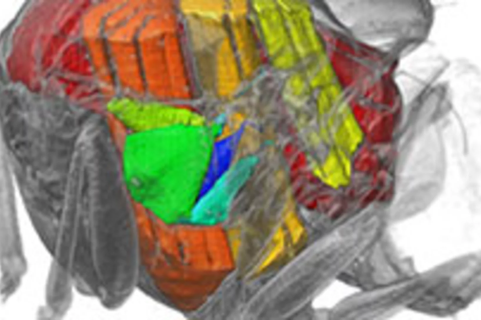
X-rays film inside live flying insects – in 3D
Scientists have used a particle accelerator to obtain high-speed 3D X-ray visualizations of the flight muscles of flies. The team from Oxford University, Imperial College, and the Paul Scherrer Institute (PSI) developed a groundbreaking new CT scanning technique at the PSI’s Swiss Light Source to allow them to film inside live flying insects. The movies offer a glimpse into the inner workings of one of nature’s most complex mechanisms, showing that structural deformations are the key to understanding how a fly controls its wingbeat.
X-ray tomography reaches 16 nm isotropic 3D resolution
Researchers at PSI reported a demonstration of X-ray tomography with an unmatched isotropic 3D resolution of 16 nm in Scientific Reports. The measurement was performed at the cSAXS beamline at the Swiss Light Source using a prototype instrument of the OMNY (tOMography Nano crYo) project. Whereas this prototype measures at room temperature and atmospheric pressure, the OMNY system, to be commissioned later this year, will provide a cryogenic sample environment in ultra-high vacuum without compromising imaging capabilities. The researchers believe that such a combination of advanced imaging with state-of-the-art instrumentation is a promising path to fill the resolution gap between electron microscopy and X-ray imaging, also in case of radiation-sensitive materials such as polymer structures and biological systems.
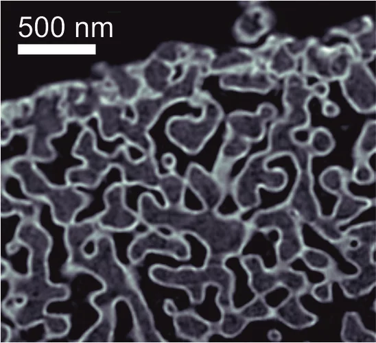
Unique insight into carbon fibers on the nanoscale
The investigation of the mechanical properties of carbon fibers benefits from highly resolved three-dimensional density maps within representative volumes, but such images are not easily obtained with standard methods. Scientists from the Paul Scherrer Institut in collaboration with Honda R&D in Germany have recently visualized density distributions on the sub-micrometer scale within entire carbon fiber sections, revealing surprising graphite distributions within the fibers. This capability will prove useful for the systematic characterization of fibers, contributing to the development of light and robust materials at lower costs.
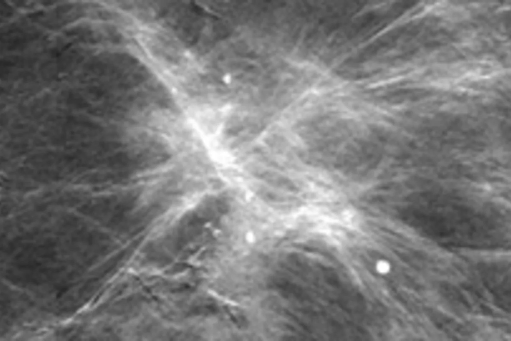
A promising new method for the diagnosis of breast cancer
A new mammography procedure that could generate substantial added value for the diagnosis of breast cancer in medical practice has just been published in the scientific journal Investigative Radiology. The method was developed at PSI in cooperation with the Certified Breast Centre at the Kantonsspital (cantonal hospital in) Baden and Philips as an industrial partner and is making the tiniest tissue changes visible. This has the potential to improve the early detection of breast cancer. Further studies in women suffering from breast cancer are to prove in a definitive manner the added value of the method.
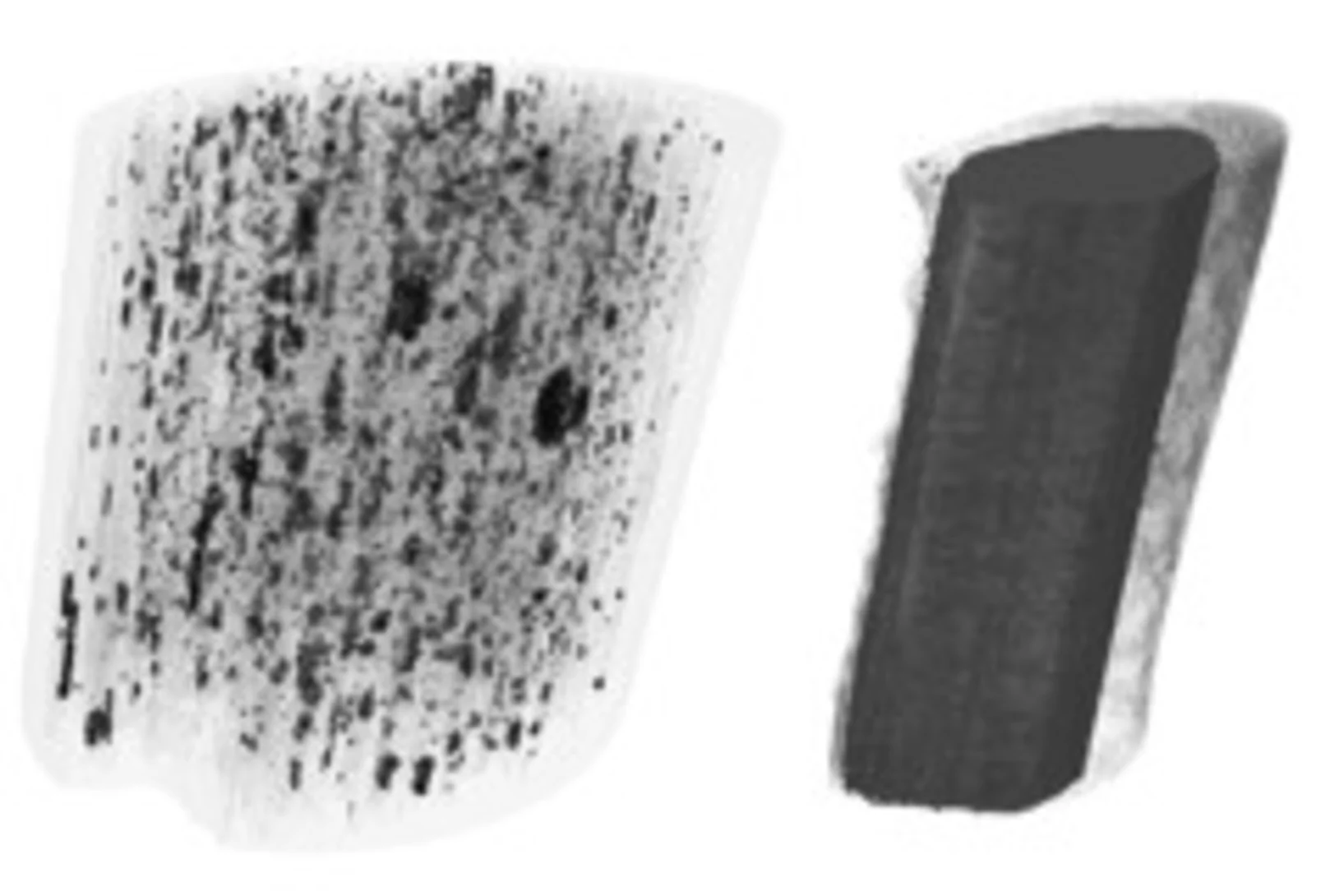
Why lithium-ion-batteries fail
Materials in lithium ion battery electrodes expand and contract during charge and discharge. These volume changes drive particle fracture, which shortens battery lifetime. A group of ETH and PSI scientists have quantified this effect for the first time using high-resolution 3D movies recorded using x-ray tomography at the Swiss Light Source.
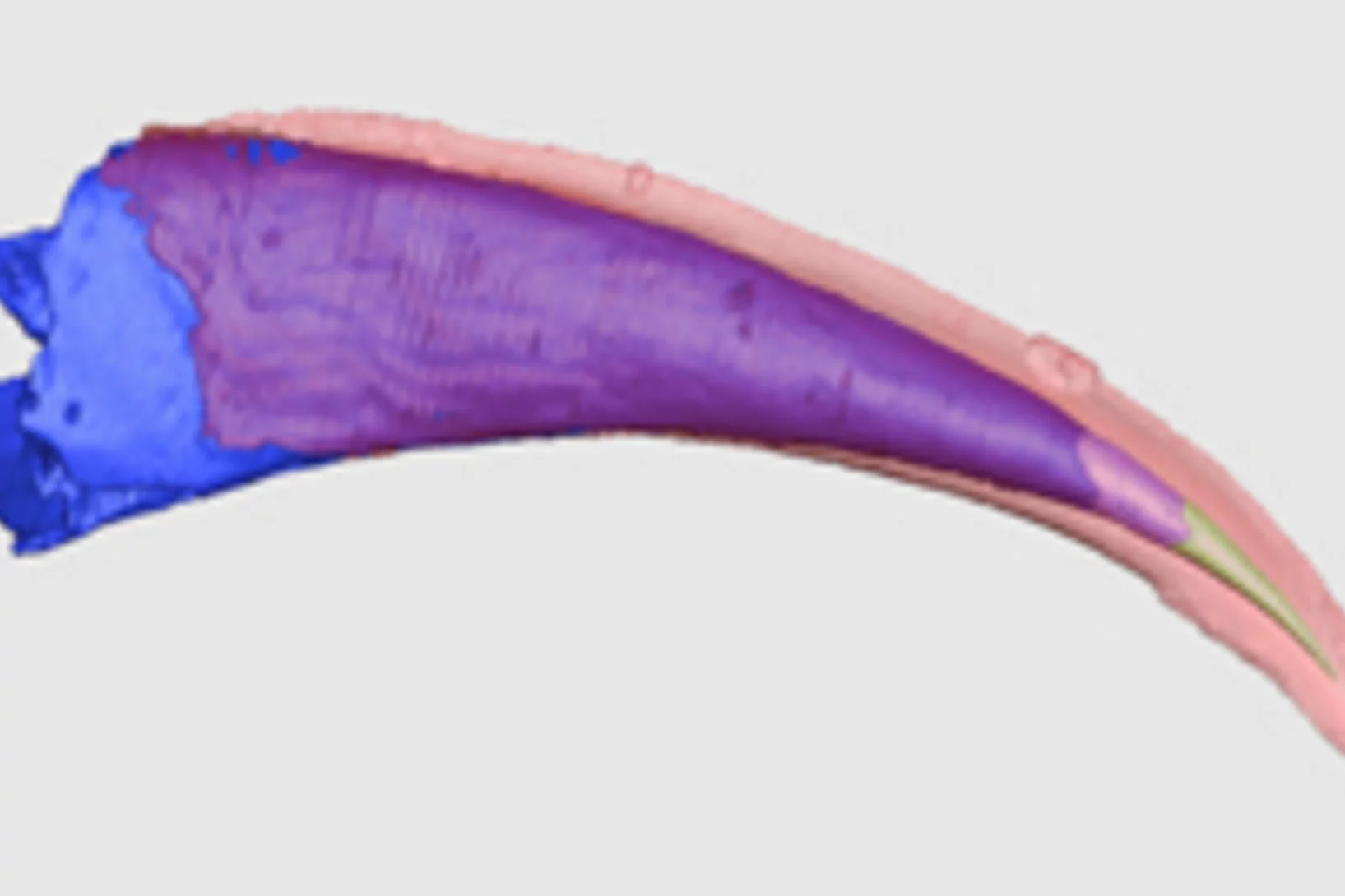
Die Ursprünge der ersten Fische mit Zähnen
Mit Hilfe von Röntgenlicht aus der Synchrotron Lichtquelle Schweiz des PSI ist es Paläontologen der Universität Bristol gelungen, ein Rätsel um den Ursprung der ersten Wirbeltiere mit harten Körperteilen zu lösen. Sie haben gezeigt, dass die Zähne altertümlicher Fische (der sogenannten Conodonten) unabhängig von den Zähnen und Kiefern heutiger Wirbeltiere entstanden sind. Die Zähne dieser Wirbeltiere haben sich vielmehr aus einem Panzer entwickelt, der dem Schutz vor den Conodonten, den ersten Raubtieren, diente.