Super-Resolution X-ray Microscopy unveils the buried secrets of the nanoworld
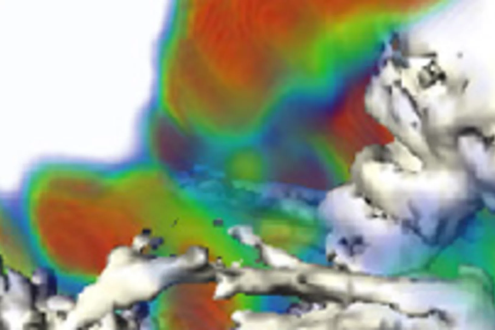
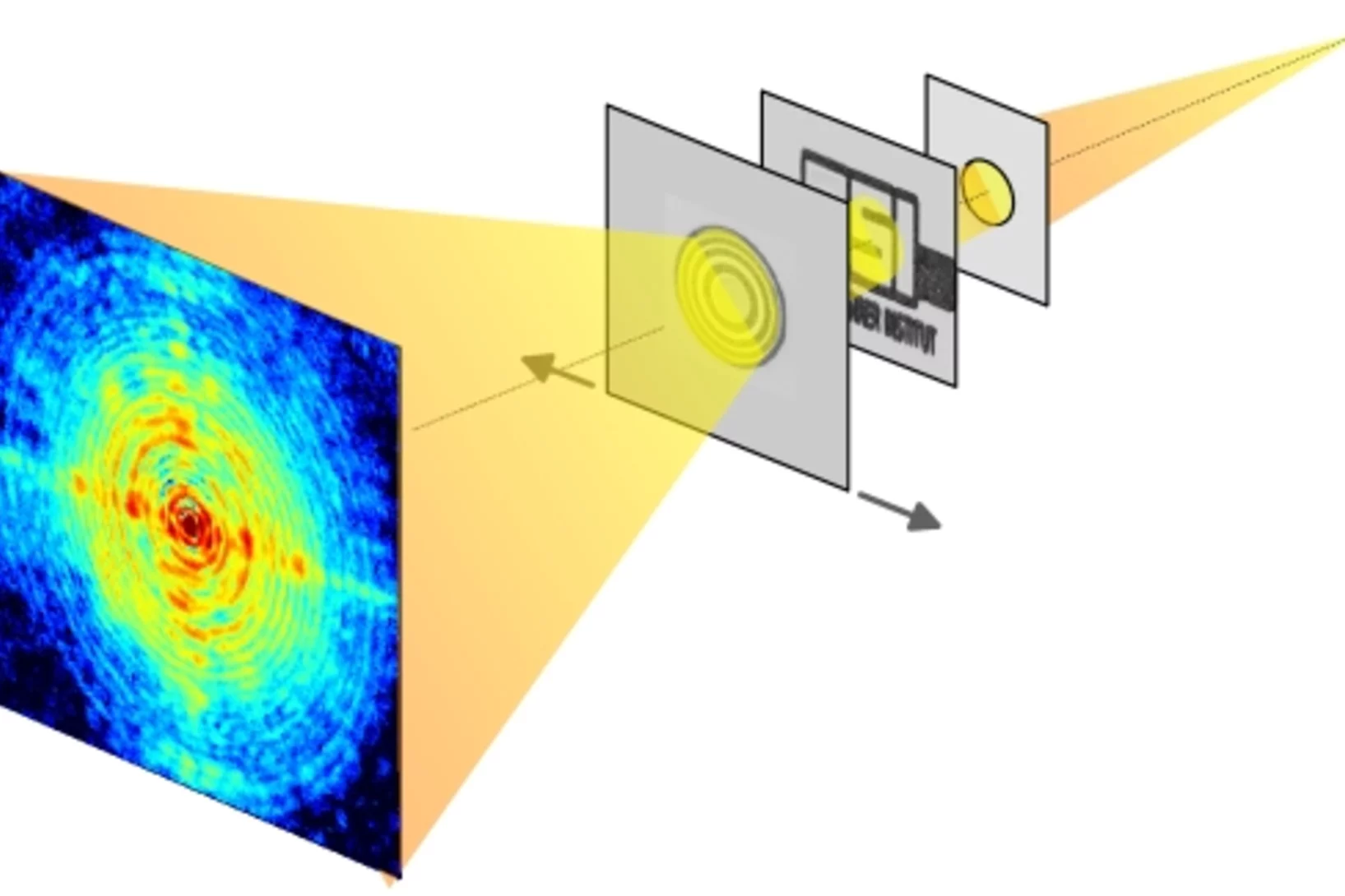
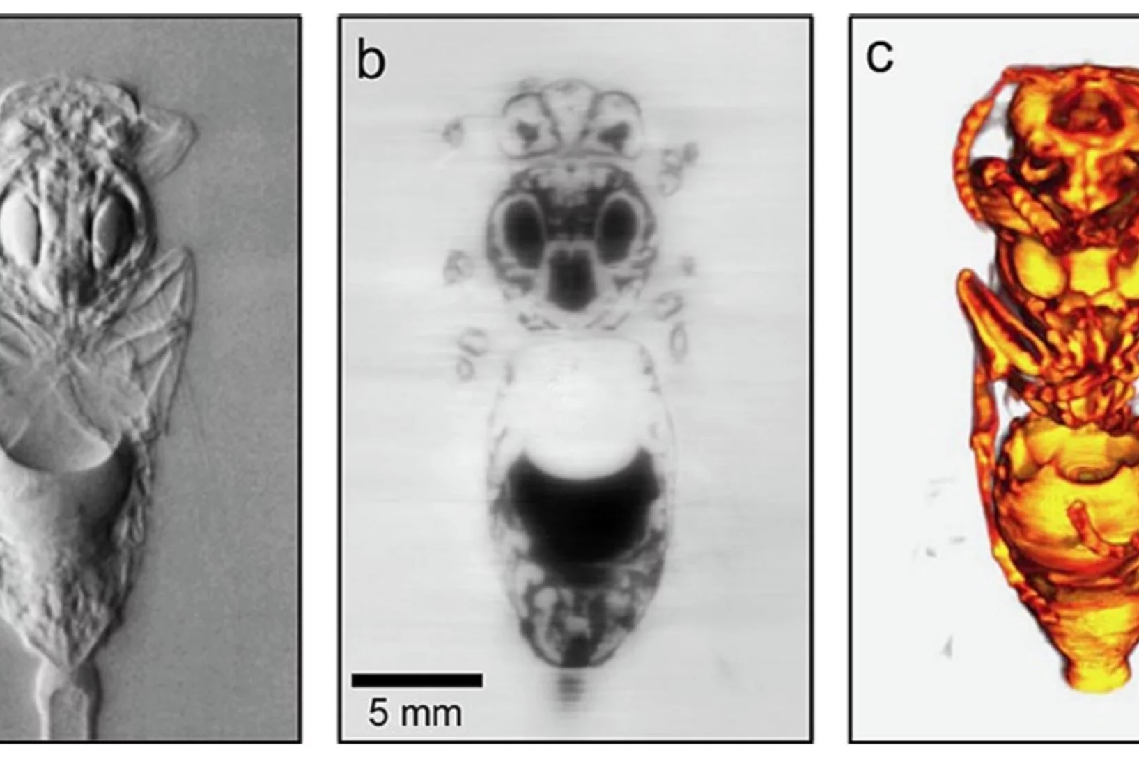
A novel super-resolution X-ray microscope developed by a team of researchers from the Paul Scherrer Institut (PSI) and EPFL in Switzerland combines the high penetration power of x-rays with high spatial resolution, making it possible for the first time to shed light on the detailed interior composition of semiconductor devices and cellular structures.
The first super-resolution images from this novel microscope will be published online July 18, 2008 in the journal Science.
Vitamin B12 ist das Trojanische Pferd der Krebsforscher am schweizerischen Zentrum für radiopharmazeutische Wissenschaft
Erste Klinische Studie soll Mitte Juni beginnen.This news release is only available in German.
Paul Scherrer Institut-Technologie an Bord eines neuen Navigationssatelliten
Erste Ergebnisse werden schon im Juni erwartet. Am vergangenen 27. April 2008 wurde GIOVE-B, der zweite Testsatellit des europäischen Navigationssystems GALILEO, auf seinen Orbit gebracht. Durch den Van-Allen-Gürtel und rund 23 200 km über der Erdoberfläche zieht GIOVE-B seine Kreise durch eine Region der irdischen Magnetosphäre, die permanent unter Beschuss durch einen intensiven Schauer hochenergetischer Strahlung steht. Ein am Paul Scherrer Institut (PSI) entwickeltes und getestetes System zur Strahlungsüberwachung sammelt wertvolle Daten über das Weltraumwetter und sorgt zugleich für den sicheren Betrieb der Bordinstrumente.This news release is only available in German.
Die Belenos Clean Power AG und das Paul Scherrer Institut beschliessen die Schweizer Brennstoffzelle
Die Vision geht in die operative Phase. Ein nachhaltiger, sauberer Energieverbrauch und eine individuelle Mobilität mit sauberer, CO2-freier Energie, das ist die Vision der Belenos Clean Power AG. Um dieses Ziel zu erreichen, müssen Anstrengungen unternommen werden, die die ganze Kette von der nachhaltigen Primärenergie à der Sonne à über die saubere Energie für Haushalte, Fabriken u. ä., bis zum effizienten, emissionsfreien Auto-Antrieb umfassen.This news release is only available in French and German.
Europa zu Gast am Paul Scherrer Institut
Botschafter der Europäischen Union und der Beitrittskandidaten der EU informierten sich über Forschung mit Grossgeräten. Die Kombination von Grossforschungsgeräten, von denen jedes Einzelne an sich schon einzigartig ist, wie die Spallations-Neutronenquelle (SINQ), die Myonenanlage (MueSR) und die Synchrotron Lichtquelle Schweiz (SLS) ermöglicht es Forschenden aus der ganzen Welt, hier Experimente durchzuführen, wie z. B. die Untersuchung von neuen Supraleitern oder Brennstoffzellen, die in dieser Komposition woanders nicht möglich sind. Das PSI setzt etwa 70% seines Budgets für die Aufgaben des Benutzerlabors ein.This news release is only available in German.
Jahresmedienkonferenz vom 6. Mai 2008
Das PSI feiert dieses Jahr seinen 20. Geburtstag. Seit seiner Gründung 1988 haben sich die Forschungsschwerpunkte des PSI deutlich verändert.Aus den beiden Vorgängerinstituten EIR und SIN, welche primär auf Kernenergieforschung und Grundlagenforschung in Kern- und Teilchenphysik ausgerichtet waren, ist ein Forschungszentrum entstanden, welches sich mit wissenschaftlichen Fragestellungen in Physik, Materialwissenschaften, Chemie, Biologie und Medizin, sowie ganzheitlich mit zukünftiger Energietechnik und ihren Umweltauswirkungen beschäftigt.This news release is only available in German. %
Nanoscale depth resolution of lattice dynamics
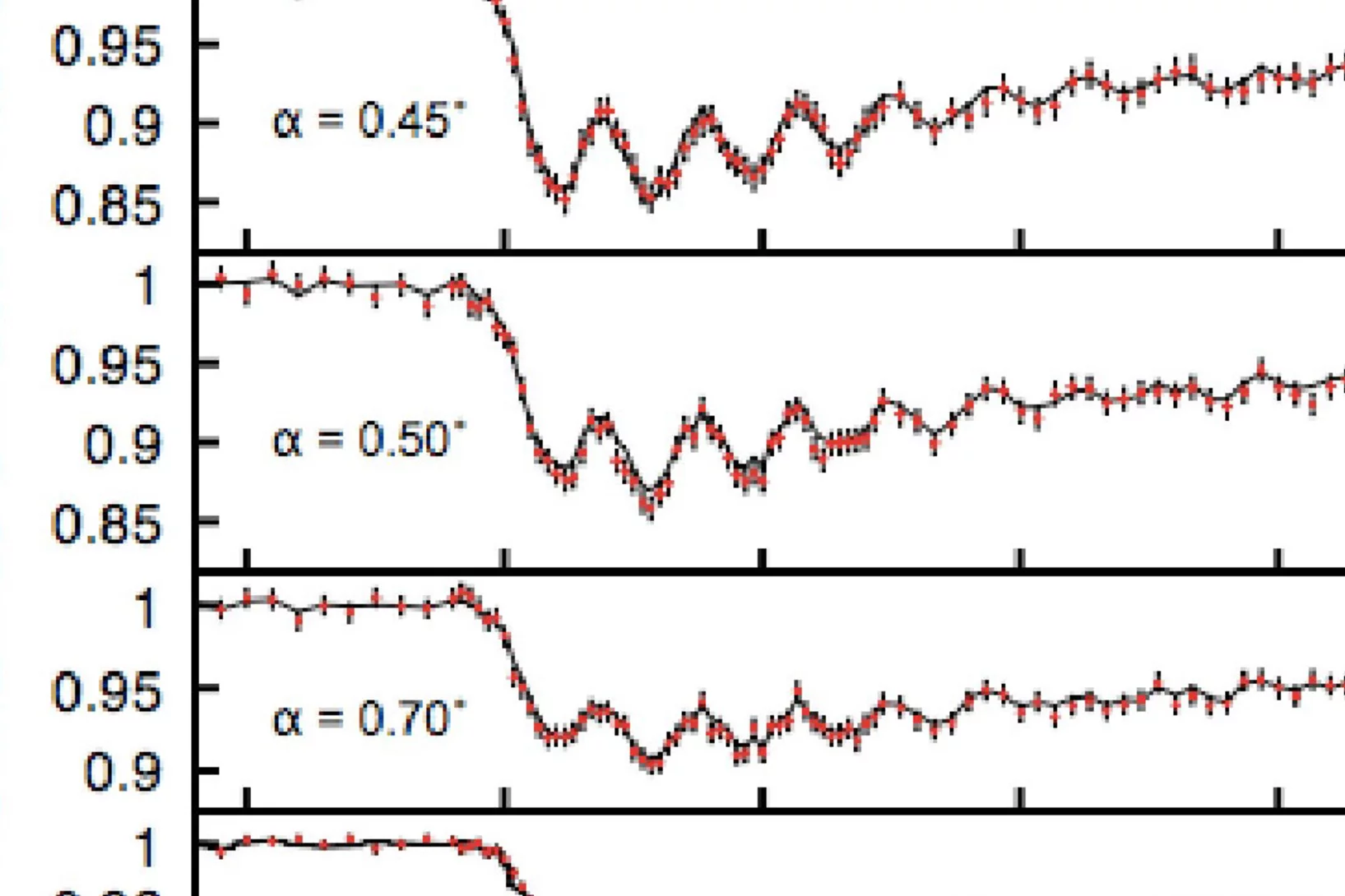
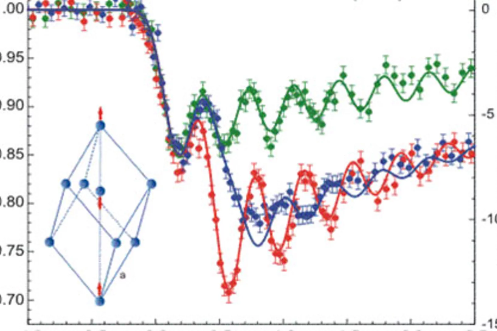
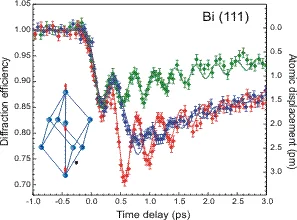
We employ grazing-incidence femtosecond x-ray diffraction to characterize the coherent, femtosecond laser-induced lattice motion of a bismuth crystal as a function of depth from the surface with a temporal resolution of ~200 fs. The data show direct consequences on the lattice motion from carrier diffusion and electron-hole interaction, allowing us to estimate the effective diffusion rate for the highly excited carriers and the electron-hole interaction time.
Coherent Diffraction Imaging Using Phase Front Modifications
We introduce a coherent diffractive imaging technique that utilizes multiple exposures with modifications to the phase profile of the transmitted wave front to compensate for the missing phase information.
Das Paul Scherrer Institut öffnete am 13. April seine Pforten und lud zum KLIMAsonntag ein
Das Paul Scherrer Institut öffnete am 13. April seine Pforten und lud zum KLIMAsonntag ein. Den strahlendem Sonnenschein nutzten mehr als 1300 Besucher die Gelegenheit sich am PSI in Villigen über das Thema Klimawandel und die Folgen zu informieren. Anreiz dazu gaben vier Erlebniswelten.This news release is only available in German.
Neue Verantwortliche für Kommunikation am PSI
Die Stelle der Kommunikationsleitung am Paul Scherrer Institut (PSI) ist seit Anfang des Monats wieder besetzt. Dagmar Baroke hat die Nachfolge von Beat Gerber angetreten.This news release is only available in German.
Eröffnung Schülerlabor iLab am Paul Scherrer Institut
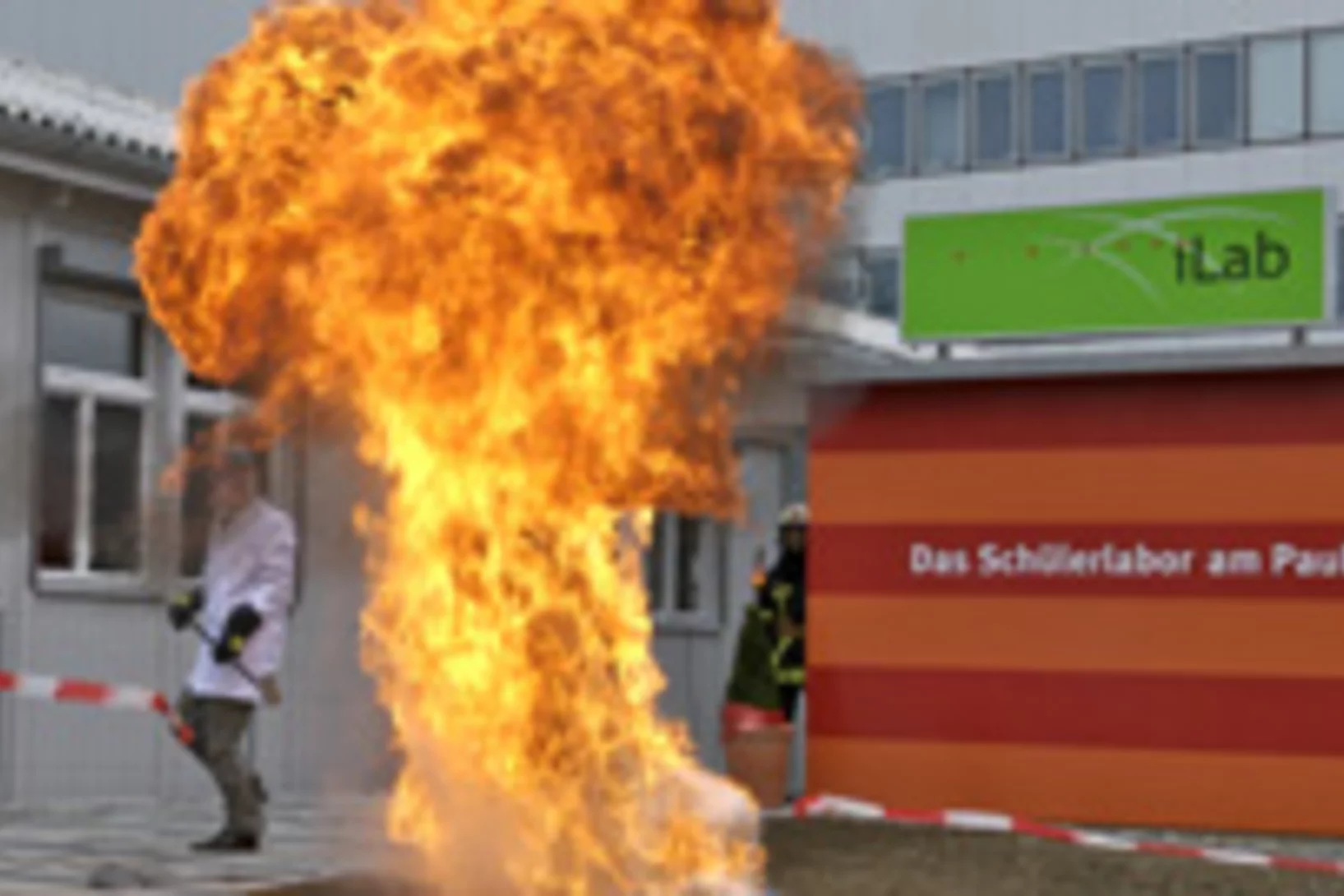
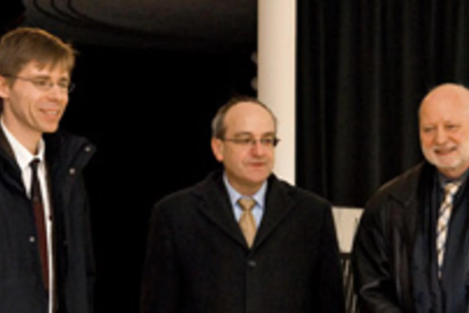
Eröffnung Schülerlabor iLab am Paul Scherrer Institut. Ab heute hat die Schweiz ein einzigartiges Schülerlabor für Naturwissenschaften und Technik. Regierungsrat Rainer Huber vom Bildungsdepartement des Kantons Aargau und PSI-Direktor a.i., Martin Jermann, eröffneten das iLab vor geladenen Gästen.Bei der Nachwuchsförderung in Naturwissenschaften und Technik geht das Paul Scherrer Institut (PSI) neue Wege. Zum Auftakt seines 20-Jahre-Jubiläums eröffnete das PSI in Villigen heute ein in der Schweiz einzigartiges Schülerlabor. Überall fehlt es an Nachwuchs in Natur- und Ingenieurwissenschaften. Das PSI will in seinem Jubiläumsjahr ein Zeichen setzen und die junge Generation für eine berufliche Karriere als Ingenieurin oder Naturwissenschaftler begeistern.This news release is only available in German.
President of ETH Board visits PSI
Glarus attorney Fritz Schiesser (54) has served as President of the ETH Board since the beginning of January. Today on Monday, 4 February, Fritz Schiesser paid his first visit to PSI since assuming office.
Scharfe Röntgenbilder im Krankenhaus und am Flughafen
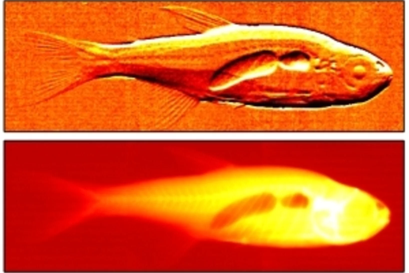
Publication in Nature. Researchers at the Paul Scherrer Institute (PSI) and the EPFL in Switzerland have developed a novel method for producing dark-field X-ray images at wavelengths used in typical medical and industrial imaging equipment. Dark-field images provide more detail than ordinary X-ray radiographs and could be used to diagnose the onset of osteoporosis, breast cancer or Alzheimer's disease, to identify explosives in hand luggage, or to pinpoint hairline cracks or corrosion in functional structures.
X-ray dark-field imaging using a grating interferometer
A type of X-ray imaging that shows detail otherwise lost, and which is compatible with conventional radiography instrumentation is now feasible, reports a study published online in Nature Materials. This technique offers unprecedented resolution for several applications, including medical imaging, security screening and industrial non-destructive testing.
New discovery in superconductor research
Publication in ScienceSuperconductors take advantage of electron pairing to transport electrical current without resistance. They are therefore of central significance in energy research. An international team of scientists has published the latest research results in this field in today's edition of Science magazine
Stable Source of Femtosecond X-Ray Pulses at SLS – Pushing atoms on a swing
The typical time scale of atomic motion during fundamental physical processes such as phase transitions in solids or molecular dynamics in chemical reactions ranges from ten to hundreds of femtoseconds. The direct observation of these processes on an atomic length scale therefore requires utrashort light pulses at wavelengths capable of resolving the underlying atomic structures.
Pushing atoms on a swing
The typical time scale of atomic motion during fundamental physical processes such as phase transitions in solids or molecular dynamics in chemical reactions ranges from ten to hundreds of femtoseconds. The direct observation of these processes on an atomic length scale therefore requires utrashort light pulses at wavelengths capable of resolving the underlying atomic structures.
Vibrational spectra of adsorbates from DFT
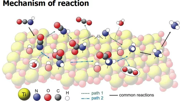
The hydrolysis of isocyanic acid was studied experimentally and theoretically and a reaction mechanism on different catalysts was established. The decreasing NOx emission limits for diesel vehicles impel the further development of the existing NOx deactivation technologies, particularly the selective catalytic reduction (SCR) of nitrogen oxides with urea. In the urea-SCR process, urea is injected into the hot exhaust gas, where it thermally decomposes into isocyanic acid (HNCO) and ammonia.
Vibrational Spectra of Adsorbates from DFT
The hydrolysis of isocyanic acid was studied experimentally and theoretically and a reaction mechanism on different catalysts was established. The decreasing NOx emission limits for diesel vehicles impel the further development of the existing NOx deactivation technologies, particularly the selective catalytic reduction (SCR) of nitrogen oxides with urea. In the urea-SCR process, urea is injected into the hot exhaust gas, where it thermally decomposes into isocyanic acid (HNCO) and ammonia.
The conducting meat in the insulating sandwich
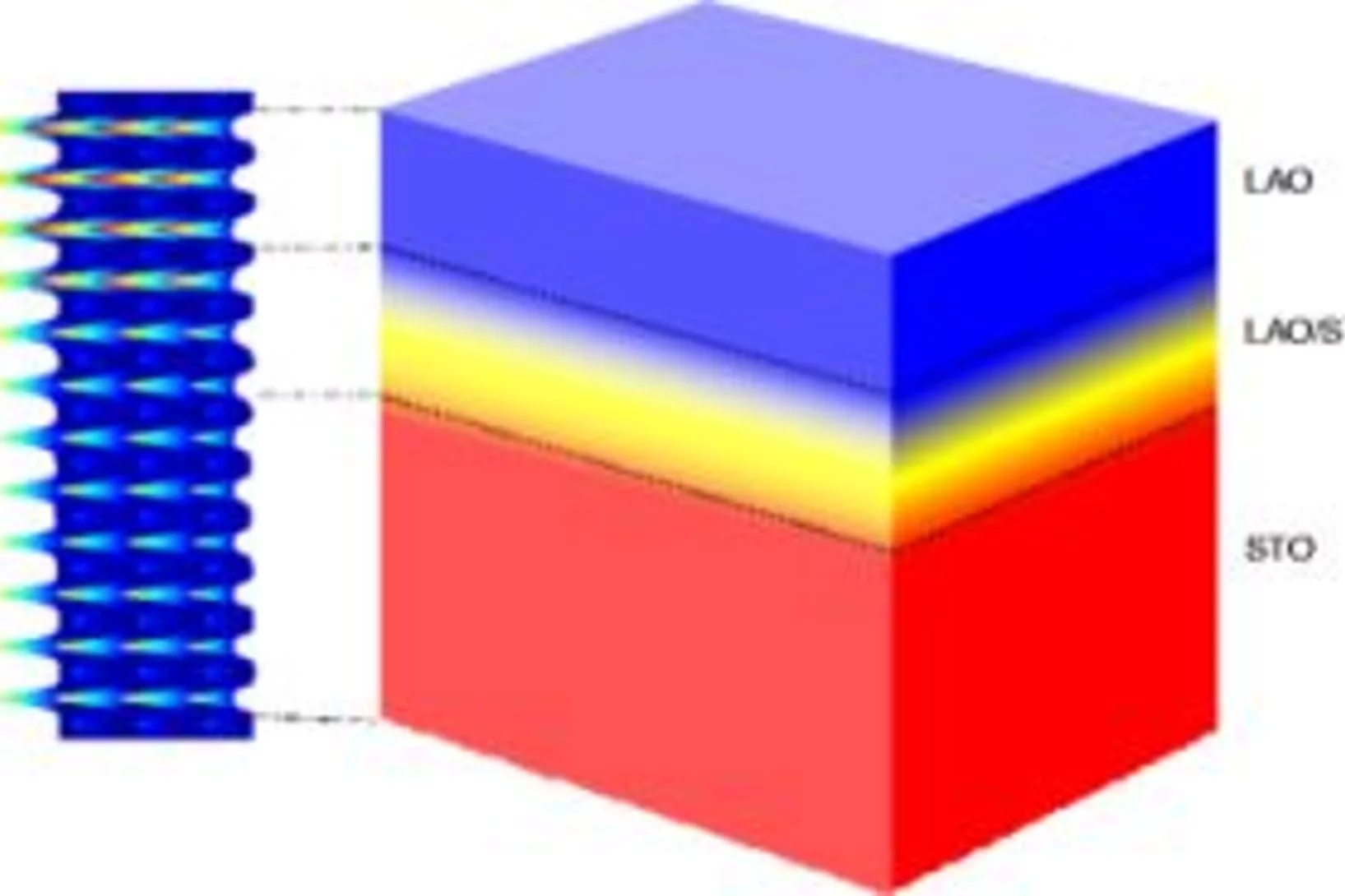
In 2004, it was discovered that when a layer of LaAlO3 (LAO) is in contact with a layer of SrTiO3 (STO), an ultrathin layer of highly conducting material is formed where they contact one another, despite the fact that both LAO and STO are insulators. The underlying physics responsible for this phenomenon is still much disputed, despite a worldwide concerted research drive since then to explain it.
The exciting story of TiSe2
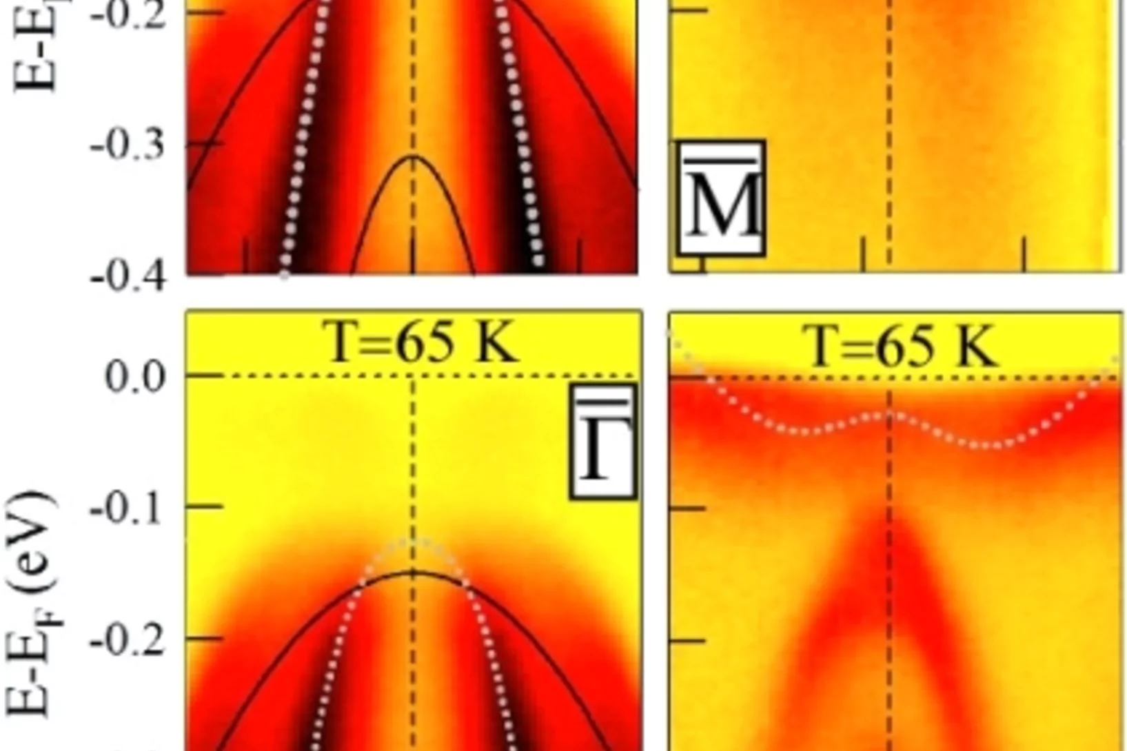
In this story of TiSe2, experiment and theory meet to provide an explanation for a long-standing enigma. In this system, the electrons rearrange themselves spontaneously at low temperature, resulting in a new periodicity from that of the original lattice. This phase change is driven by a decrease in the total energy of the system. However, the nature of this transition has been a matter of controversy for a long time.
Is Smaller Stronger?
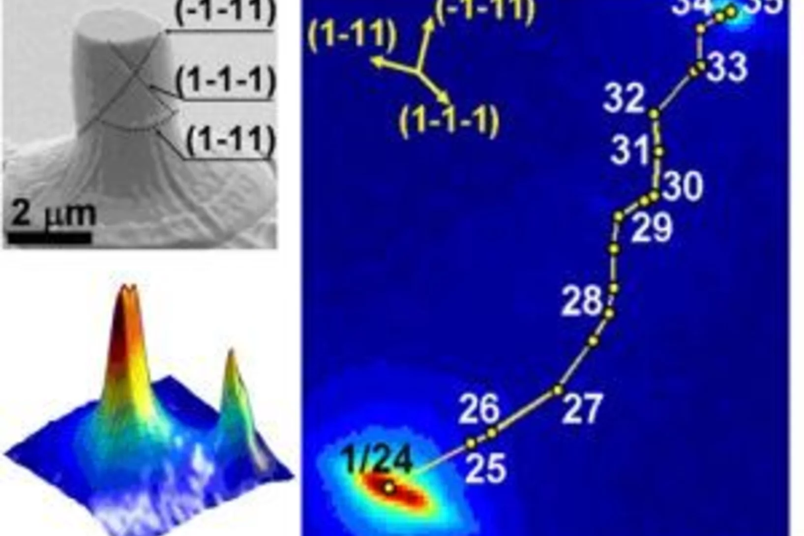
In 2004 researcher discovered that a single crystalline metal is stronger when the sample volume is reduced to the micron or even submicron range. In an ongoing debate on the origin of this phenomenon classical deformation theories are questioned. The suspicion that structural defects, i.e. deviations from perfect crystalline structure would play an important role in the smaller is stronger effect, could not be verified because of the lack of an appropriate measuring technique.
Making the invisible visible
Using x-rays scientists have learned to make the invisible visible. Since almost 100 years doctors use the difference in x-ray absorption between bones and tissue to diagonose their patients.
A virus in a nutshell
Nature has found remarkable ways to protect sensitive objects. One example is the seed of a nut which is protected by its shell. Another are viruses from the cypovirus family. They are hidden inside tiny natural crystals where they can survive harsh conditions until they meet their target, the gut of the silkworm. Here the virus is released from the crystal causing a virus infection of the worm. Researchers have now unraveled the structure of these natural protein crystals.
A microscope without a lens
It is known since a long time that x-rays, which are nothing but light of very short wave length, can be used for microscopy. This is particularly attractive because due to the small wave length x-rays allow studying objects which are invisibly small in an optical microscope.
Looking inside fossilised embryos
Although only recently discovered, the fossil record of embryonic development has already begun to challenge cherished hypotheses on the origin of major animal groups. Synchrotron-based X-ray Tomographic Microscopy has provided unparalleled insight into the anatomy and preservation of these fossil remains and this has allowed us to test competing hypotheses on their nature.
Phase Imaging with Neutrons
Neutrons are usually considered as small massive particles with a size of about 10^-15 meters. Due to the wave-particle duality of quantum mechanics, however, they can equivalently be considered as matter wave packets whose spatial extent may be large enough to show interference effects similar to what can be observed with visible laser light.
How to avoid atomic sandpaper
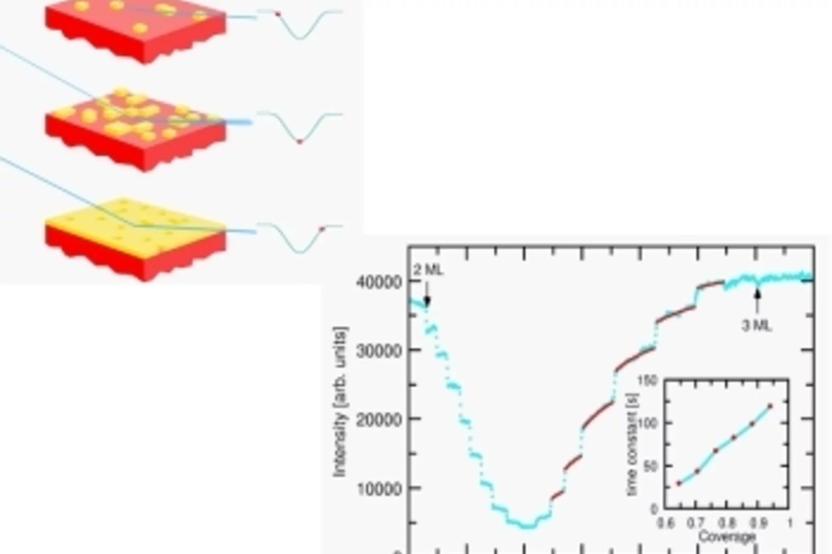
When growing thin films of novel materials, smooth surfaces are a must. How else could one stack them layer by layer, as needed in optical coatings, sensors or conductors? One method known to produce atomically smooth films is Pulsed Laser Deposition (PLD).
Seeing ever finer Details with X-Rays
X-ray radiographic absorption imaging is an invaluable tool in medical diagnostics and materials science. For biological tissue samples, polymers, or fiber composites, however, the use of conventional X-ray radiography is limited due to their weak absorption. This is resolved at highly brilliant X-ray synchrotron or micro-focus sources by using phase-sensitive imaging methods to improve contrast.
Shining light on superconductors
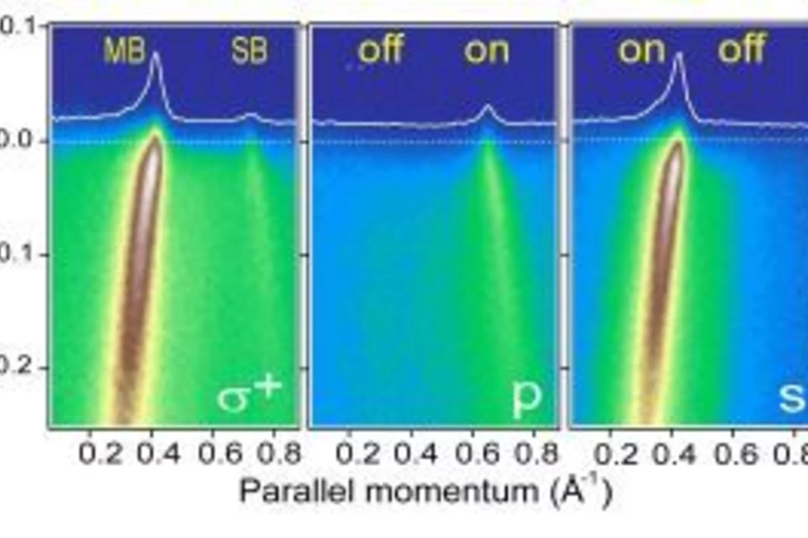
More than 20 years ago researchers in Switzerland discovered that certain materials transport electrical current without any loss. For this they need to be cooled, but because they do so at relatively high temperatures (up to -150°C) they are called high temperature superconductors. How exactly the electrons transport current in such materials is still a mystery. But the electrons can be studied using the photoelectric effect where light of high energy knocks an electron out of the material.


