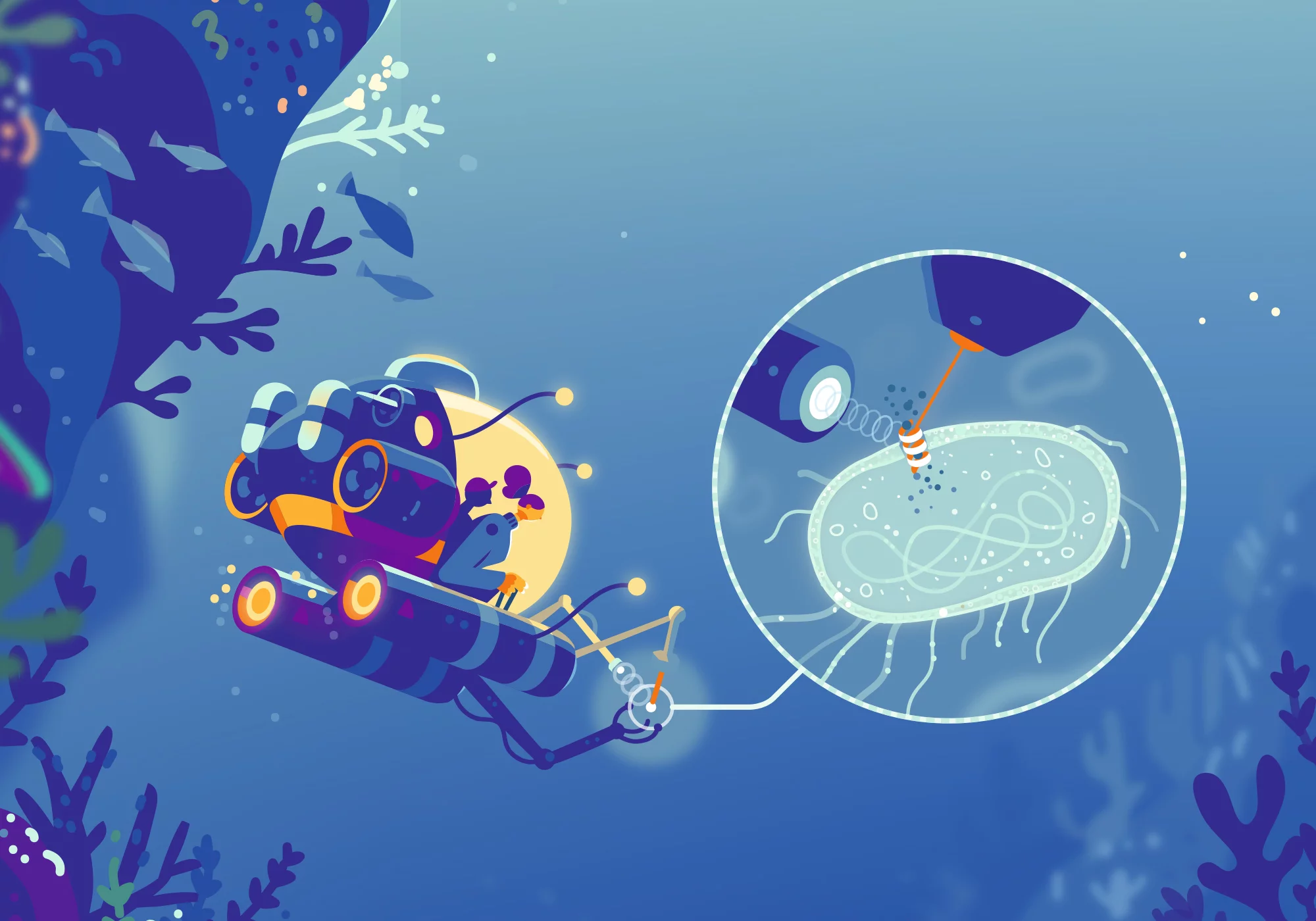
The world of microbes and viruses is extremely old and exceedingly diverse. With the help of the large research facilities at PSI, researchers are peering deep inside this alien cosmos and investigating, above all, the proteins of these exotic beings.
Since they emerged as the first life on our planet around 3.5 billion years ago, they have shaped the Earth in ways no other form of life has: microorganisms. In this motley crew there are such diverse representatives as bacteria, archaea, algae, yeasts, amoebas, and parasites including the malaria pathogen. But as diverse as microorganisms may be, there's one biological form of existence not included in the group: viruses. That's because these are a borderline case between the animate and the inanimate. They do not have their own metabolism and therefore always need a host in order to awaken to life and reproduce. The vast majority of microorganisms and viruses are harmless or even very useful for humans, for example for digestion or to produce food, to purify wastewater, or to form humus. A few, such as the pathogens that cause dangerous diseases, are harmful to humans and animals.
So it's no wonder that researchers at PSI are also studying microorganisms and viruses. Since these are so tiny, sometimes only a hundredth or even a thousandth of the thickness of a human hair in size, they and their components can only be studied thoroughly under extremely high magnification. Conventional light microscopes are by no means sufficient for this. Therefore, scientists like Biology and Chemistry Division Head Gebhard Schertler and his team rely on the large research facilities at PSI. At the Swiss Light Source SLS or the X-ray free-electron laser SwissFEL, the proteins and biomolecules of microorganisms can be examined precisely with the help of X-ray or laser light down to the individual atoms, and the properties of their structures can be deciphered.
Schertler, a chemist, has a passion for this microcosm. Tiny beings such as Escherichia coli, archaea, baculoviruses, and others kept him busy in various laboratories during his 35-year research career. "Microbes are the workhorses of biotechnology," he says, speaking enthusiastically about their great importance for humans: "They can consist of just a single cell or can exist as dense cell layers of microorganisms, so-called biofilms, or even as tiny particles." In biotechnology and medicine, they can be used as miniature chemical factories to manufacture and study products such as amino acids, drugs, or enzymes.
Single-celled organism with a solar collector
In the PSI laboratories, researchers work with proteins from a wide variety of microorganisms and viruses. Among them are harmless fragments of one of the most powerful poisons of all, botulinum toxin, which under the name Botox helps in treating some neurological diseases – or, in the beauty industry, smoothes wrinkles. Using the X-ray light from SLS, biophysicist Roger Benoit and his team determined the structure of a protein complex that reveals exactly how the toxin binds to a nerve cell and then blocks its activity. These findings could be useful for the development of improved botox drugs that are less likely to result in an overdose.
With the aid of PSI's facilities, researchers can not only elucidate the rigid structure of molecules, but also can record their movements. Thus, PSI researchers are investigating complex light-driven ion pumps, for example from so-called extremophile archaea, which are capable of surviving in the most inhospitable places on Earth. The pumps serve as a model for the researchers to study light-driven metabolic processes and structural changes in proteins, for example at SwissFEL.
Movie time at SwissFEL
Just recently, Jörg Standfuss and his team at SwissFEL shed new light on how a retinal-controlled protein from a microorganism that usually lives in the world's oceans works. In its middle, the protein contains a form of vitamin A, the retinal molecule, which serves as a light receptor. When this molecule is hit by light, it absorbs a small part of it and changes its shape. This process sets a pump in motion, which transports sodium out of the cell. The researchers have succeeded in filming this marine bacterial sodium pump in action. The PSI researchers hope that the knowledge about how such light-driven pumps work can be used in many ways. "Since the activity of nerve cells in multicellular organisms is regulated by sodium pumps in their membranes, these light-driven bacterial sodium pumps can likewise control the activity of nerve cells, and they can be put to use in so-called optogenetics," explains Standfuss. "If you insert them into nerve cells, using molecular genetic methods, you can control them specifically using light signals and thus investigate the functioning of specific brain regions." With the knowledge gained, new advances in neurobiology could be achieved.
Taking aim at coronaviruses
The possibilities for using SwissFEL and SLS for research on proteins from microbes and viruses are extremely diverse. Because of its excellent infrastructure, PSI is therefore a sought-after partner for other researchers and industry, and it supports research collaborations from all over the world. PSI has actively participated in the fight against the SARS-CoV-2 virus as well, with a large number of initiatives. In March 2020, the Institute invited external scientists to take advantage of the opportunity at SLS to employ the most modern techniques to elucidate the structure and functionality of SARS-CoV-2 in a timely manner, in order to develop active agents and diagnostics. In one collaboration, with researchers from the Goethe University in Frankfurt, the first results were available just a few weeks later. At one of the three SLS beamlines for macromolecular crystallography, the researchers examined the enzyme protease PLpro, which SARS-CoV-2 needs to assemble new virus particles in human cells. Experiments have shown how a possible inhibitor against PLpro can block the spread of the virus and increase antiviral immunity in human epithelial cells, the main entry point for the pathogen. These findings open up opportunities for future active agents.
The international team benefited from the fact that the SLS group for macromolecular crystallography, headed by Meitian Wang, has extensive experience in the characterisation of viral structures. PSI researcher Justyna Wojdyla and a Chinese research group have already elucidated the protein structures of various viruses that pose a danger to humans. For her analyses, she directed the strong X-ray light from SLS onto protein crystals from coronaviruses such as MERS-CoV and HKU1, as well as the Alongshan virus. While the two coronaviruses affect the airways and lungs in particular, the tick-borne Alongshan virus causes persistent headaches, tiredness, and nausea. As with SARS-CoV-2, all of the viruses studied have in common that neither a vaccine nor an effective antiviral therapy has been available to date. "Our experiments on the SLS beamline have helped us to better understand the structure and function of viruses," says Wojdyla. For the investigation of the coronavirus HKU1, the X-ray crystallography specialist used a special technique on the MX beamline of SLS: single-wavelength anomalous dispersion. In this procedure, the protein crystal is only exposed to the strong X-ray light for a very short time, reducing possible radiation damage to the molecule and thus the risk of errors during data acquisition.
Microbes in motion
In addition to viral and bacterial protein crystals, PSI researchers are also deciphering other structures of tiny single-celled organisms, such as the two flagella of the green alga Chlamydomonas. This freshwater-living microbe could help us understand the mechanism by which the novel coronavirus enters into the body. Chlamydomonas’ flagella are fine cell extensions made of proteins, which are found in a similar form, as cilia, in the human respiratory tract. When the cilia move there, they undulate like a field of seaweed in the ocean. There are also cilia in the fluid-filled chambers of the brain, on embryos, and in the uterine tubes. For example, they convey the egg cell and sperm and, if there are genetic defects, can cause infertility. "The cilia play a major role in a great many transport functions in the body," explains Takashi Ishikawa, who has worked at PSI for ten years and studies green algae as a model system. "If they fail, important protective mechanisms are disabled." In the respiratory tract, they move mucus and bacteria to the outside – and are a target for coronaviruses. In the early infection phase, SARS-CoV-1, the causative agent of the first SARS epidemic, and SARS-CoV-2 penetrate the airways through the cilia.
So Ishikawa wants to understand exactly how the movement of the flagella comes about and what inhibits it. With the help of cryo-X-ray tomography at SLS and cryo-electron microscopy, he examines the cell extensions, just a few micrometres long, inside which rests a delicate framework of protein tubes: the so-called microtubules. On them sit tens of thousands of tiny molecular motors, which actively move the flagella. Ishikawa's test object, Chlamydomonas, is particularly ingenious: Either its two flagella beat in time, as if the alga were swimming the breaststroke, or they make undulating bends to the side. The amount of calcium ions determines which swimming program the alga follows when it is under way. The Japanese scientist now wants to find out, with the help of X-ray tomography on the cSAXS beamline at SLS, what effect this has on the complex interaction of the molecular motors in the cell.
New active agents are the goal
There is a completely different microorganism, less harmless than green algae, that researchers also want to find out about, likewise with help from SLS: the malaria pathogen Plasmodium falciparum. Unfortunately, the physiology of these tiny parasites is very similar to that of humans – at least on a cellular level. It is therefore difficult to develop active substances against Plasmodium that do not produce severe side effects. Therefore, using synchrotron light from SLS, PSI researchers are looking for small structural differences in the supporting apparatus, the so-called cell skeleton, between the parasite and humans. These small differences could help in developing drugs that interfere with the growth of parasite cells but not human cells.
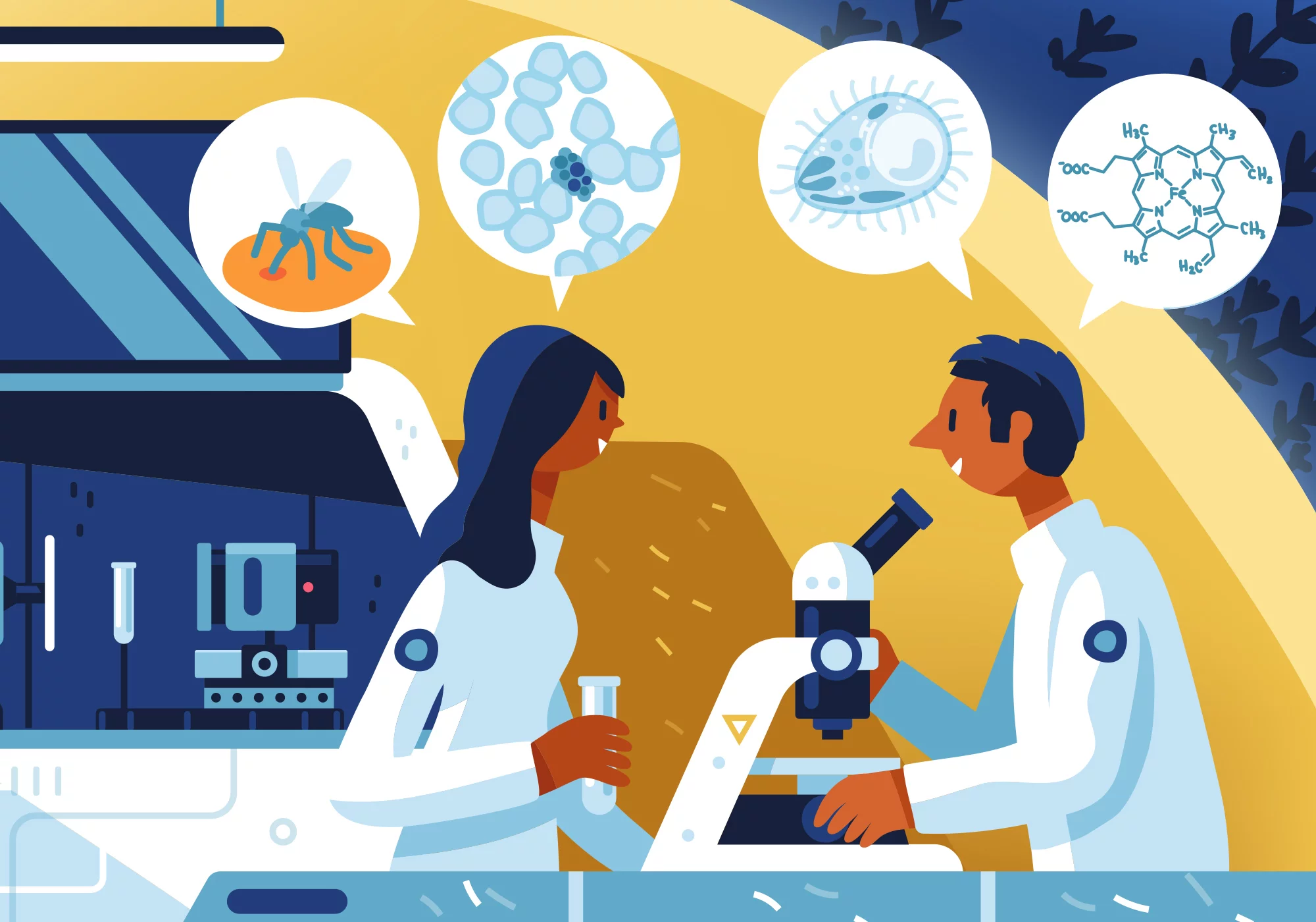
In an international research project involving close collaboration with Sergey Kapishnikov from the Weizmann Institute in Israel, PSI researcher Daniel Grolimund has demonstrated another possibility, which could switch off the malaria pathogen in the future. The trick is to undermine its survival strategy. When Plasmodium reproduces in its host's red blood cells, after the host has been bitten by the Anopheles mosquito, it digests the blood pigment haemoglobin there. A toxic iron complex, the haem, is released from the haemoglobin. This is harmful to Plasmodium, which is why the parasite converts it into an insoluble crystal package. The researchers wanted to understand exactly how this process takes place. "Using X-ray fluorescence microscopy at SLS, we measured how much haem was distributed in the parasite and then calculated how quickly the parasite converts it," says Grolimund. The results show how much effort Plasmodium has to make to enclose haem in a crystal package. Among other things, it uses an auxiliary protein as a tool, the PV5 protein. The researchers hope that if this tool could be disabled with a suitable active ingredient, the malaria pathogen would lose its protection against haem and die. But first, many more experiments are needed.
"Research into basic biological processes in microorganisms and the analysis of biomolecules of pathogens is a success story for PSI," summarises Gebhard Schertler. On the one hand, it shows the importance of basic research for the reputation of PSI, and on the other hand, it allows the Institute to respond quickly to new challenges such as the Covid-19 pandemic.
Text: Sabine Goldhahn
Copyright
PSI provides image and/or video material free of charge for media coverage of the content of the above text. Use of this material for other purposes is not permitted. This also includes the transfer of the image and video material into databases as well as sale by third parties.


