
Forty years of research on cell growth
PSI researcher Kurt Ballmer-Hofer is concerned with the question of how tumours could be starved by preventing the development of blood vessels. After 40 years of research that yielded many fundamental insights about the formation of blood vessels, one of the key molecules has been found; further research is expected to enable clinical applications. Ballmer-Hofer will be following these future advances from the perspective of retirement.
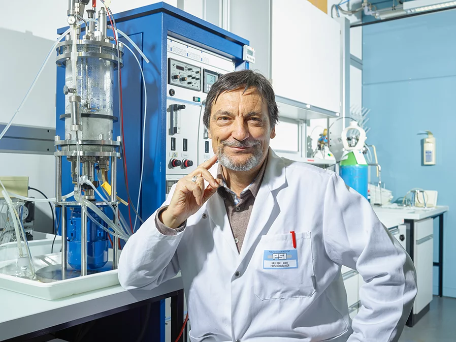
For forty years, Kurt Ballmer-Hofer has been delving into the question of how cells grow – an important basis for research on cancer cells because, in such malignant diseases, undesirable cells grow uncontrollably.
When I entered into research in the mid-1970s, we were just beginning to understand some of the fundamental mechanisms regulating cell growth. For example, the way a virus makes cells grow and, at the same time, produce new viruses,
recalls the chemist and long-time group leader in the Laboratory for Biomolecular Research at the Paul Scherrer Institute. Why cancer cells proliferate uncontrolled was equally unclear. Normally, cells halt their growth after each cell division, until they are prompted to further growth and division by intracellular "messenger" molecules. In cancer this stop signal is suppressed – but how?
Helpful yeast
When Ballmer-Hofer came to the PSI in 1997, his mission was to concentrate on the messenger molecule VEGF (see box). It had been observed that tumours trigger the formation of VEGF. In turn the released VEGF stimulates the formation of new blood vessels, which the tumour needs for nourishment. So for more than thirty years the idea has been circulating to block the formation of blood vessels in the tumour in order to cut it off from nutients, to "starve" it.
Twenty-five doctoral candidates, ten postdocs, and twenty of his own PSI research years later, Ballmer-Hofer knows enough about the form and functionality of VEGF to see that new possibilities for treatment are within reach.
The goal is to block the function of the receptor activated by VEGF and thereby prevent the formation of blood vessels.
Producing VEGF is a delicate operation. Many research groups have found it a tough nut to crack. One of the doctoral candidates of a collaborator of Ballmer-Hofer’s at PSI, too, tried many methods without success. In the end,
Ballmer-Hofer says, he recalled a lecture about Pichia pastoris, a yeast that thrives on rotting wood.
Bull's eye! Within a few weeks, he was able to produce VEGF in large quantities in yeast. The yield was further optimised in a specially adapted fermenter. Today this fermenter, Ballmer-Hofer notes, is the best-amortised piece of equipment in his laboratory. Thanks to his group's know-how, they have been able to supply various other research groups with VEGF and thus enter into a number of interesting scientific collaborations.
Now researchers could get down to the work of deciphering the structure of the VEGF molecule and its receptors. In biochemical terms, VEGF is a protein. Proteins are a bit like a wadded-up string of pearls. They consist of a sequence of different amino acids. Some of these amino acids – the pearls – repel each other, while others are mutually attracted. Thus the chain assumes one precisely defined folded form. And only when the folding is correct can VEGF bind to the cell's receptor and transmit the command to form new blood vessels. With knowledge of the exact structure or shape of the molecules involved, researchers can develop counteragents that prevent the function of the receptor.
It was tough going at the start. Through biochemical and microscopic analyses, researchers determined how individual amino acids in the receptor influence its behaviour and studied how cells respond to VEGF.
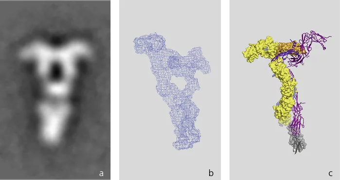
Meanwhile, the Swiss Light Source SLS was established at PSI. The SLS is like a gigantic microscope in which, by means of X-ray light, the tiniest structures – including VEGF and its receptors – can be deciphered.
The three-dimensional image of such structures can only be generated, however, when countless molecules can be crystallized – that is, rigidly embedded in a regular crystal lattice. For a long time, in the case of VEGF, this could not be achieved. Therefore researchers made do with a lower-resolution method, electron microscopy. We made thousands of raw images in the electron microscope and averaged them to form pictures,
Ballmer-Hofer says.
Unexpected result
Then came the big surprise: VEGF does not only directly bind to the receptor, but forms a figure 8
with two additional binding sites between the attached receptors. Ballmer-Hofer still finds this exciting: That makes science interesting – when something other than the expected result shows up. When nature contradicts you.
If these further binding sites are blocked, the blood vessel cells don't do what the cancer cell demands. It is like giving someone the wrong key to prevent him from getting past a locked door.
A doctoral candidate was able to produce crystals when he combined VEGF with another receptor. With the help of these crystals and using the latest tricks, a postdoctoral researcher was able to determine the structure of the complex between VEGF and the receptor at the SLS.
The next step now is to find a molecule that interferes with the function of the receptor itself. In cell cultures, we have already managed this
, Ballmer-Hofer says. Now we want to show that our concept also works in animals.
This will bring potential clinical applications closer to realisation. Ballmer-Hofer himself plans to follow these further advances from the perspective of a retiree.
Text: Alexandra von Ascheraden
VEGF (vascular endothelial growth factor) is an intercellular signaling molecule that is produced when an organ needs new blood or lymph vessels. It binds to a special receptor on the outer surface of endothelial cells, the cells that line blood vessels. When VEGF docks onto this receptor, it changes its structure and triggers a chemical reaction inside the cell. This induces the cell to divide and contribute to the growth of new vessels.
Contact
Prof. Dr. Kurt Ballmer, Laboratory of Biomolecular Research, Paul Scherrer InstituteTelephone: +41 56 310 41 65, E-mail: kurt.ballmer@psi.ch
Additional information
Attacking the lifeline of tumour cells: http://psi.ch/u5U5Original Publication
Thermodynamic and structural description of allosterically regulated VEGF receptor 2 dimerization. Brozzo, M.S. et al.Blood 119, 1781-1788 (2012)
DOI: 10.1182/blood-2011-11-390922
Structure of a VEGF-VEGF receptor complex determined by electron microscopy.
Ruch, C., Skiniotis, G., Steinmetz, M.O., Walz, T., & Ballmer-Hofer, K.
Nat. Struct. Mol. Biol. 14, 249-250 (2007);
DOI: 10.1038/nsmb1202