

Scanning Probe Microscopy has become an invaluable tool in the years since its discovery. A pool of two newly acquired instruments, a Bruker Nanoscope Multimode and a Bruker Dimension Icon are currently operated at PSI in order to support internal projects from LMN and all of PSI’s research divisions as well as external projects.
Scanning Probe Microscopy is capable of mapping a variety of surface properties involving electric, magnetic fields in non-contact geometry as well as attractive and repulsive forces in proximity of the substrate. Beyond imaging, local experiments with individual atoms and molecules can be performed and local materials properties can be determined and modified. A wide range of research topics ranging from life sciences to material science, chemistry and physics as well as surface and interface science and the engineering of surfaces and nanostructures make beneficial use of one or the other SPM technique.
Surfaces and Interfaces in air, in liquids and at a variety of other conditions can be imaged and experimented with. For experiments on delicate, atomically clean systems in the ultra high vacuum the facility of the Surface Science Lab is recommended which also provides the access to surface science techniques using photons, also at the Swiss Light Source.
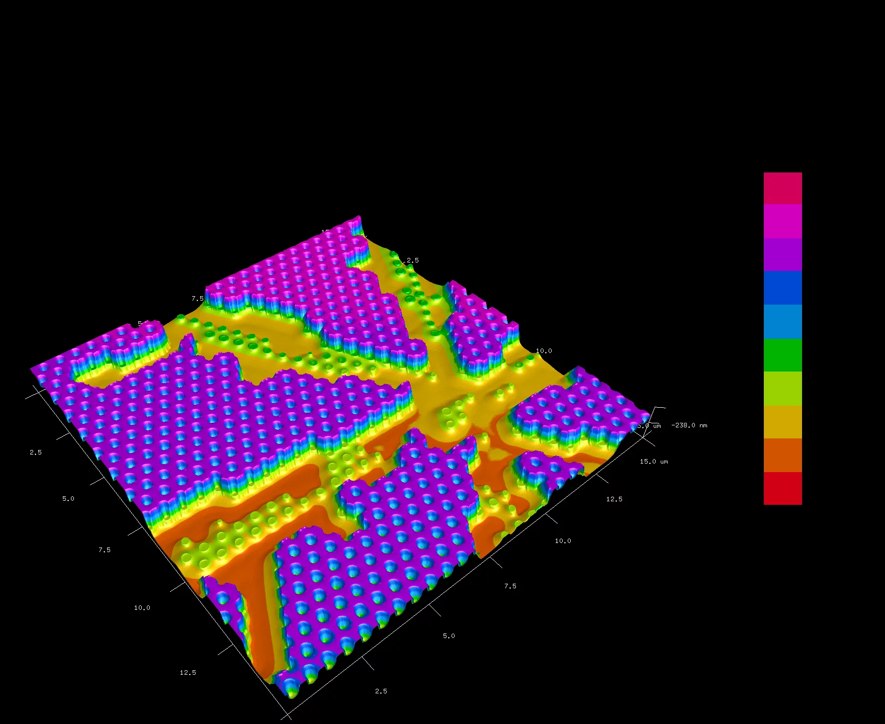
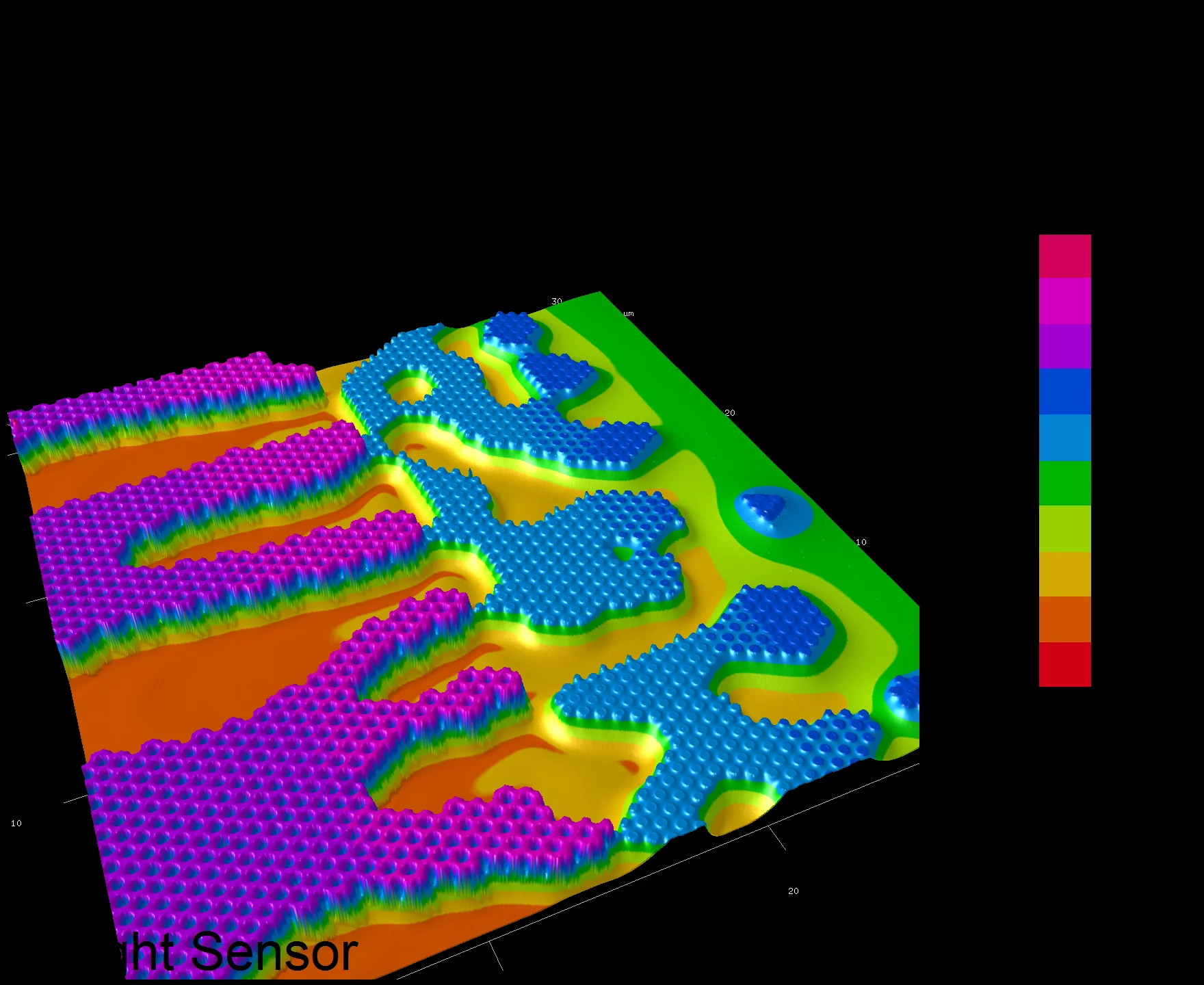
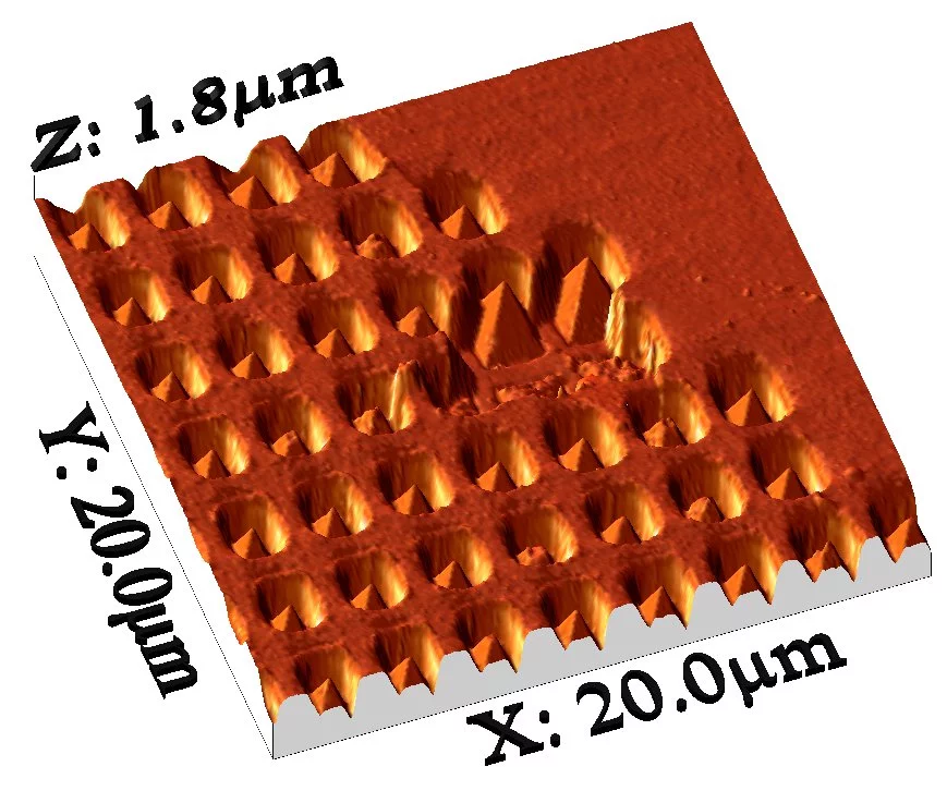
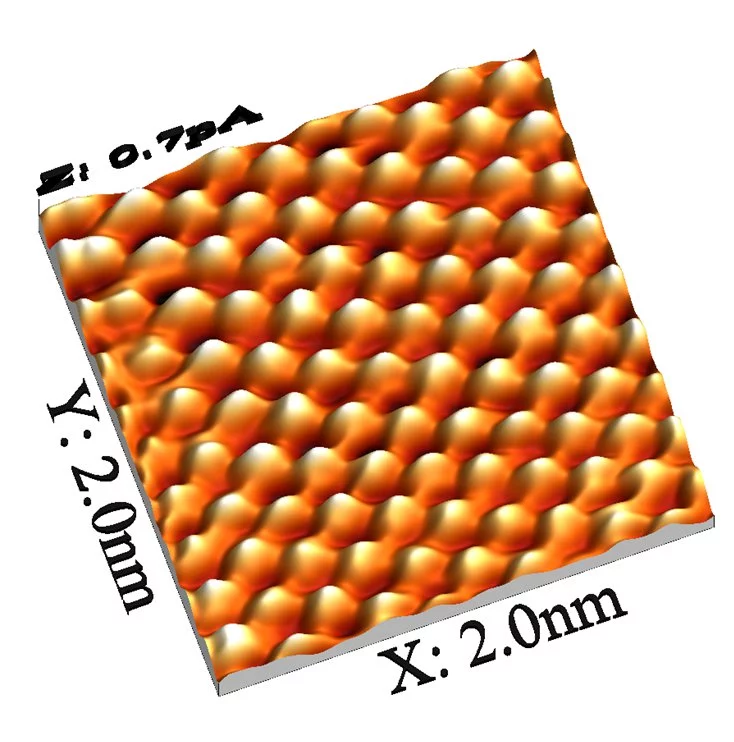
Examples of specimens probed with AFM
Instruments
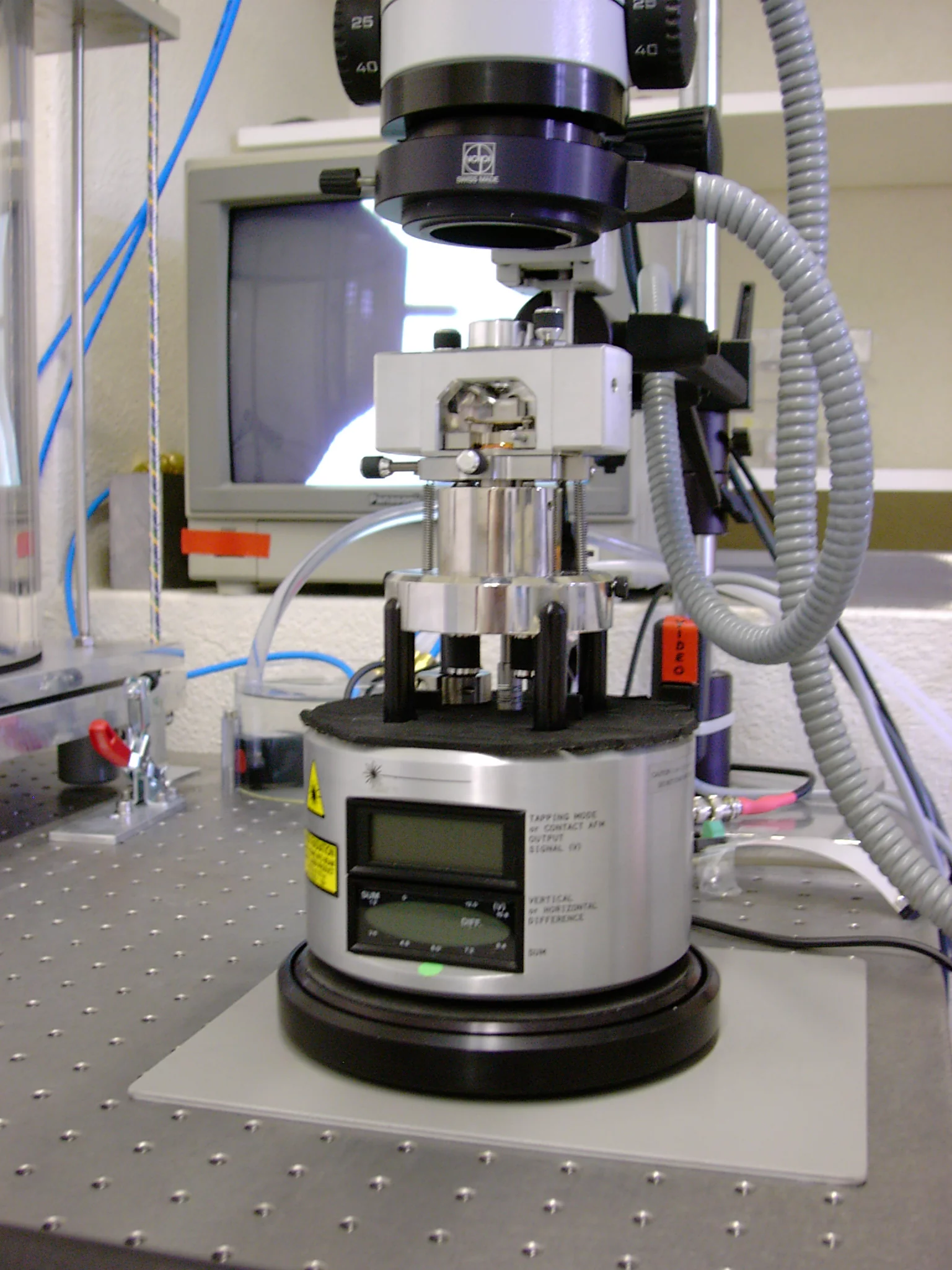
Bruker Nanoscope Multimode, Scanhead mounted below an Optical Microscope
- Maximum scan-size: 130µm,
- Maximum sample size: 12x12mm.
- AFM: Tapping Mode and Phase Imaging
- AFM: Contact Mode
- MFM: Magnetic Force Microscopy
- LFM: Lateral Force Microscopy
- CAFM: Conductive AFM
- STM: Scanning Tunneling Microscopy
- PFM: Peak Force Mode
- TRM: Torsional Resonance Mode
Bruker Dimension Icon Scanning Station mounted on a vibration and sound isolation table
In interaction with project partners we have successfully designed and implemented apparatus for very complex experiments. Do not hesitate to make an appointment for a discussion if you can imagine some benefit from nano-scopic images and experiments of your samples.
All users of the SPM Userlab at PSI are requested to acknowledge the use of the PSI SPM Userlab. Consequent to extended interaction periods and discussions leading to novel conclusions a specific personal acknowledgement and / or coauthorship is appropriate.
- Maximum scan-size: 90µm
- Maximum sample size: up to 6" Wafer
- AFM: Tapping Mode, Phase Imaging
- AFM: Contact Mode
- LFM: Lateral Force Microscopy
- MFM: Magnetic Force Microscopy
- PFM: Peak Force Mode
- TRM: Torsional Resonance Mode
- PF-QNM Quantitative Nanomechanical Property Mapping
- PF-Tuna:Peak Force Tunneling
- TR-Tuna: Torsional Resonance Tunneling
- SSRM: Scanning Spreading Resistance Microscopy
In interaction with project partners we have successfully designed and implemented apparatus for very complex experiments. Do not hesitate to make an appointment for a discussion if you can imagine some benefit from nano-scopic images and experiments of your samples.
All users of the SPM Userlab at PSI are requested to acknowledge the use of the PSI SPM Userlab. Consequent to extended interaction periods and discussions leading to novel conclusions a specific personal acknowledgement and / or coauthorship is appropriate.