Detectors

We develop specialized detectors for applications at synchrotrons and XFELs, while also advancing detector research

The Detector Group of the PSI Center for Photon Science has a long-standing history in developing single-photon counting X-ray hybrid detectors for synchrotron facilities. Our work began with MYTHEN and PILATUS, continued with EIGER, and is now focused on the next generation of detectors for the SLS 2.0 upgrade of the Swiss Light Source (SLS).

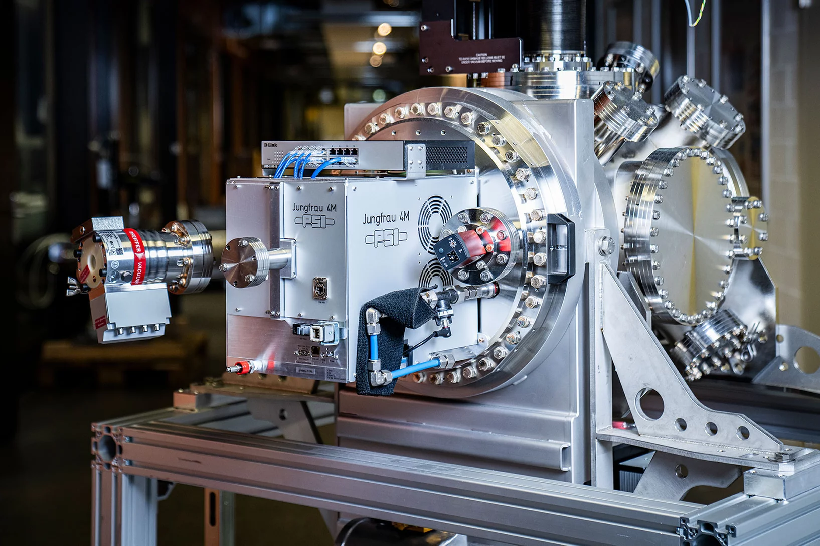
In addition, we are involved in the development of charge-integrating X-ray pixel detectors for XFELs. We are part of the AGIPD consortium, contributing to the AGIPD detector for the European XFEL, and are developing the GOTTHARD microstrip detector as well as JUNGFRAU, a high-performance pixel detector for SwissFEL.

Further details about these detectors can be found on our project pages.

Our core research interests include optimizing spatial resolution by reducing pixel size and utilizing charge sharing to extract maximum information about the photon absorption position. Much of our work has centered around studying charge sharing, particularly with microstrip detectors, and we are now developing the MOENCH pixel detector, featuring a 25 µm pitch and capable of 2D interpolation. We are also exploring new sensor materials to enhance detection efficiency at higher energies, using high-Z materials like CdTe and thick silicon sensors. Recently, we have spearheaded the development of sensors with intrinsic signal amplification to facilitate soft X-ray detection with hybrid pixel detector technology.

Additional information on our research can be found on the research pages.


teaser

Hindering the magnetic dead layer in manganites

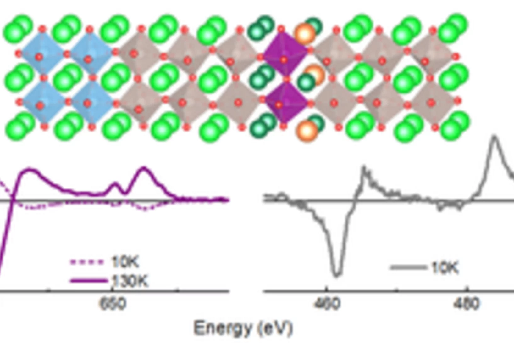
The authors demonstrate the stability of ferromagnetic order of one unit cell thick optimally doped manganite (La0.7Ba0.3MnO3, LBMO) epitaxially grown between two layers of SrRuO3 (SRO). LBMO shows ferromagnetism even above SRO Tc. Density Functional Theory calculations help understand the reasons behind this interesting result.

Cover Materials Today

Operando X-ray diffraction during laser 3D printing

Ultra-fast operando X-ray diffraction experiments reveal the temporal evolution of low and high temperature phases and the formation of residual stresses during laser 3D printing of a Ti-6Al-4V alloy. The profound influence of the length of the laser-scanning vector  on the evolving microstructure is revealed and elucidated.  

laminography chip

3D imaging for planar samples with zooming

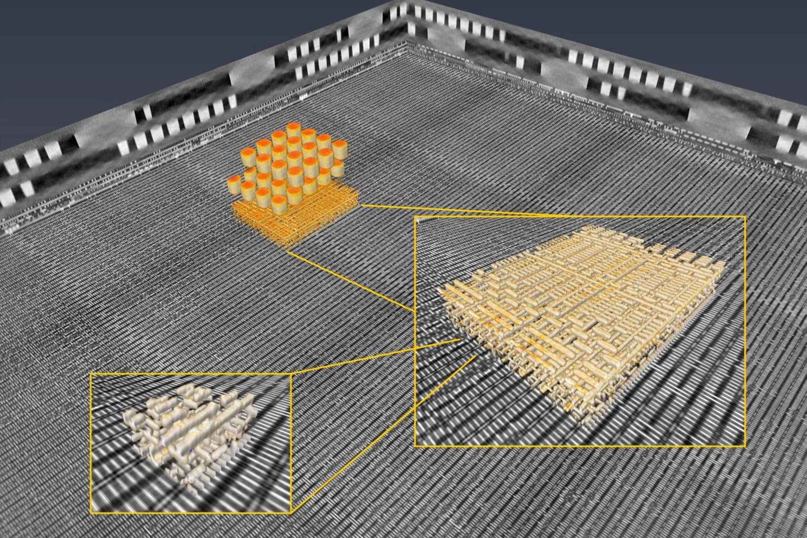
Researchers of the Paul Scherrer Institut have previously generated 3-D images of a commercially available computer chip. This was achieved using a high-resolution tomography method. Now they extended their imaging approach to a so-called laminography geometry to remove the requirement of preparing isolated samples, also enabling imaging at various magnification. For ptychographic X-ray laminography (PyXL) a new instrument was developed and built, and new data reconstruction algorithms were implemented to align the projections and reconstruct a 3D dataset. The new capabilities were demonstrated by imaging a 16 nm FinFET integrated circuit at 18.9 nm 3D resolution at the Swiss Light Source. The results are reported in the latest edition of the journal Nature Electronics. The imaging technique is not limited to integrated circuits, but can be used for high-resolution 3D imaging of flat extended samples. Thus the researchers start now to exploit other areas of science ranging from biology to magnetism.