

Self-amplified spontaneous emission (SASE) is currently the most common method for the generation of short radiation pulses at x-ray free-electron laser (XFEL) facilities. Due to its stochastic nature, the pulses produced fluctuate both in intensity and in spectral composition. These fluctuations can affect the measurements performed at XFEL facilities. To mitigate the effect on experimental results, it is crucial to develop a diagnostic tool capable of providing well-resolved spectral measurements for each individual pulse. In the hard x-ray energy range it is difficult to conceive spectrometers that does not compromise the quality of the beam delivered to the experiment. We developed two different spectrometer setups that use non-invasive, radiation hard and highly transmissive nanostructured diffraction gratings made of diamond. These gratings act as beam splitters by diffracting away a small portion of the main beam (~1%) which will be used for monitoring purposes. The remaining beam will be transmitted through the grating, and are available for the experiment.
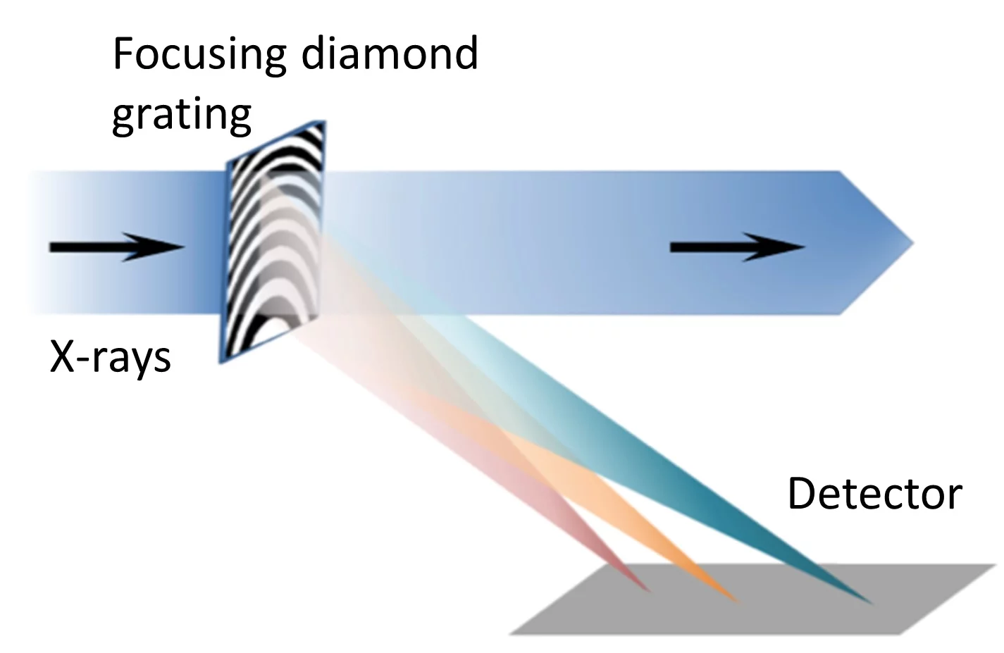
Figure 1: Schematic drawing of the experimental setup using an off-axis Fresnel zone plate. The diamond focusing grating diffracts and focuses a small portion of beam onto a fast-frame camera for spectral monitoring purposes.
One spectrometer type is based on an off-axis focusing Fresnel zone plate and a fast-frame recording device used to monitor the diffracted beam (Figure 1). The device was tested in the full beam of the XFEL at the Stanford Linear Accelerator, US [1].
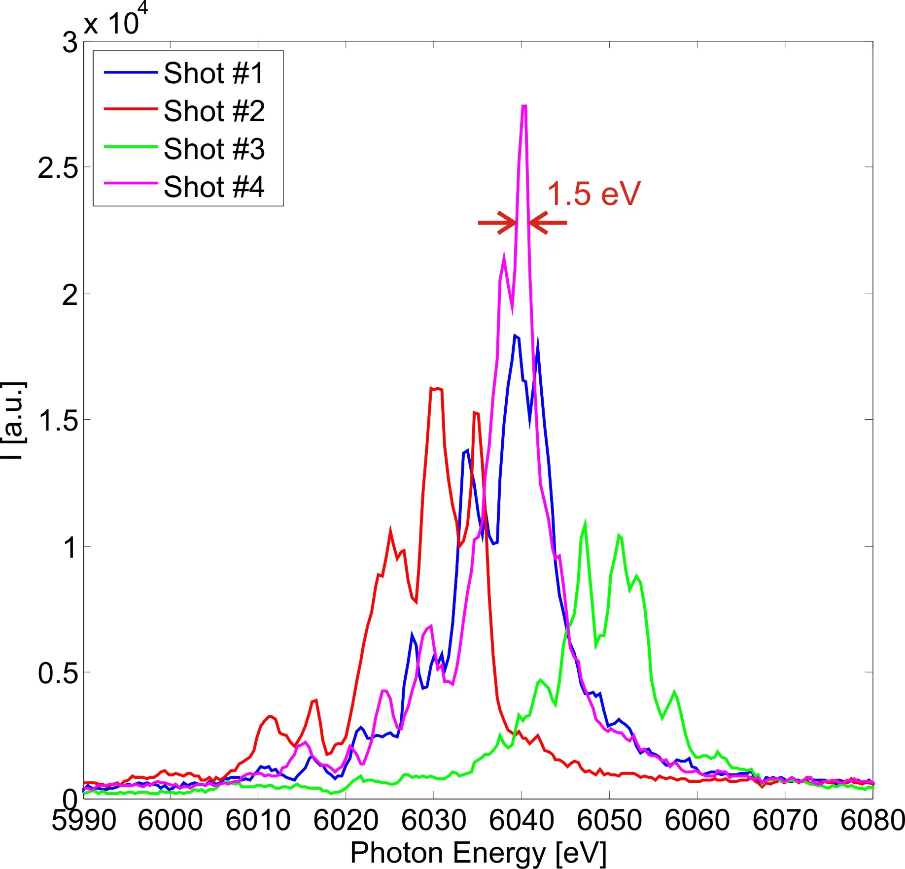
Figure 2: Single shot spectra recorded at the LCLS XFEL at 6 keV photon energy. A spectral resolution of 1.5 eV was obtained.
It proved to be radiation hard under full radiation load, and was capable of recording single shot spectra with ~1.5 eV resolution (Figure 2).
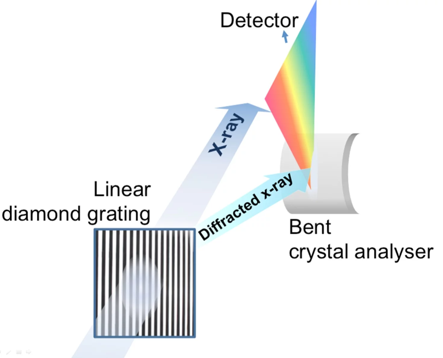
Figure 3: Schematic drawing of the setup using a linear diffraction grating. A small portion of the beam is diffracted onto a high resolution bent crystal spectrometer, and the spectra are then recorded by a fast-frame camera.
We have further developed the concept of beam-splitting to obtain better resolution by combining a nanostructured linear grating with a high-resolution diffraction crystal spectrometer (Figure 3) developed by a group at Stanford Linear Accelerator (SLAC), USA. The capabilities of this setup were demonstrated at the Linac Coherent Light Source at SLAC [2], using hard x-ray SASE pulses at a repetition rate of 120 Hz.
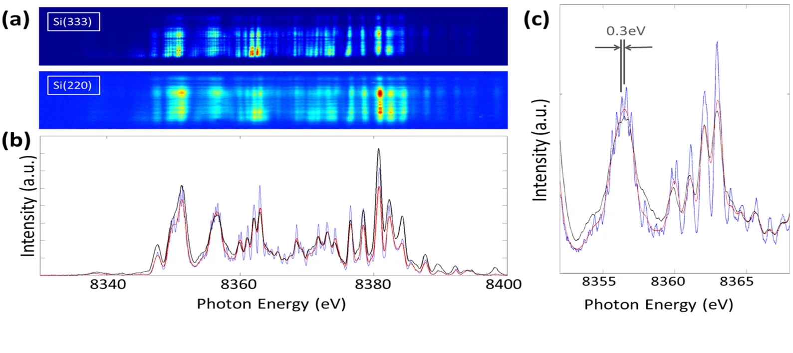
Figure 4: Comparison of single-shot spectra from the diffracted beam and the direct beam. (a) 2D images as recorded on the detectors, from the Si(333) (diffracted beam), and the Si(220) (direct beam) spectra. (b) Projections of the same shot from the Si(333) spectrometer (blue), the Si(333) spectrum after smoothening (red), and from the Si(220) spectrometer (black). The spectrum from the Si(220) reflection has similar overall features and intensity distribution. (c) Detail of the same spectra. SASE spikes separated by ∼0.3 eV were clearly resolved by the Si(333) spectrometer.
The recorded single-shot spectra had a resolving power of 3×10⁴ (Figure 4) [2]. The grating showed no degradation after days of exposure under such intense x-ray beam, satisfying the robustness required for experiment use, and no replacement or realignment of the grating was required during the measurements. The shot-to-shot spectral information can be used for the normalization of data obtained in scientific experiments. This type of spectrometer setup will be implemented at the two latest XFEL facilities: the SwissFEL [4] and the European XFEL [5].
Publications
- P. Karvinen, S. Rutishauser, A. Mozzanica, D. Greiffenberg, P.N. Juranić, A. Menzel, A. Lutman, J. Krzywinski, D.M. Fritz, H.T. Lemke, M. Cammarata, and C. David, Single-shot analysis of hard X-ray laser radiation using a non-invasive grating spectrometer, Optics Letters 37 (2012) p. 5073, DOI: 10.1364/OL.37.005073
- M. Makita, P. Karvinen, D. Zhu, P. Juranic, J. Grünert, S. Cartier, J. H. Jungmann-Smith, H.T. Lemke, A. Mozzanica, S. Nelson, L. Patthey, M. Sikorski, S. Song, Y. Feng, and C. David, High Resolution Single Shot Spectral Monitoring of Hard X-ray Free Electron Laser Radiation, Optica 2 (2015) p. 912, DOI: 10.1364/OPTICA.2.000912
- M. Makita, P. Karvinen, V.A. Guzenko, P. Vagovic, and C. David, Diamond diffraction gratings for experiments with intense hard x-rays, Microelectronic Engineering 176 (2017) p. 75, DOI: 10.1016/j.mee.2017.02.002
- J. Rehanek, M. Makita, P. Wiegand, P. Heimgartner, G. Seniutinas, U. Flechsig, V. Thominet, C. Schneider, A. Rodriguez Fernandez, C. David, L. Patthey and P. Juranić, The hard X-ray Photon Single-Shot Spectrometer of SwissFEL – initial characterization, Journal of Instrumentation 12 (2017) P05024, DOI:10.1088/1748-0221/12/05/P05024
- N. Kujala, J. Grünert, J. Liu, M. Makita, A. Zozulya, M. Sprung and C. David
Characterizing transmissive diamond gratings as beam splitter for hard X-ray single-shot spectrometer of European XFEL
Journal of Synchrotron Radiation 26 (2019) p. 708 – 713, DOI: 10.1107/S1600577519003382