Paul Scherrer Institute PSI
Forschungsstrasse 111
5232 Villigen PSI, Switzerland
+41 56 310 21 11
Paul Scherrer Institute PSI
Forschungsstrasse 111
5232 Villigen PSI, Switzerland
+41 56 310 21 11
Paul Scherrer Institute PSI
Forschungsstrasse 111
5232 Villigen PSI, Switzerland
+41 56 310 21 11
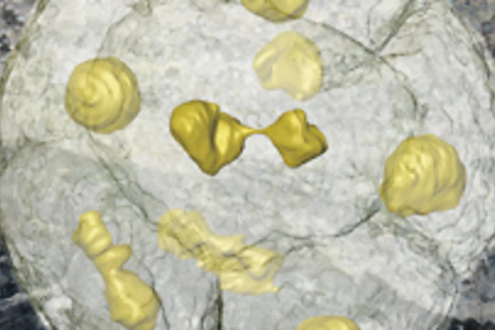
Einzellige Organismen, die vor über einer halben Milliarde Jahre gelebt haben und deren Fossilien in China gefunden wurden, sind wohl die unmittelbaren Vorläufer der frühesten Tiere. Die amöbenartigen Einzeller haben sich in einer Weise in zwei, vier, acht usw. Zellen geteilt, wie es heute tierische (und menschliche) Embryonen tun. Die Forscher glauben, dass diese Organismen einem der ersten Schritte vom Einzeller zum Vielzeller in der Entwicklung richtiger Tiere entsprechen.This news release is only available in German.
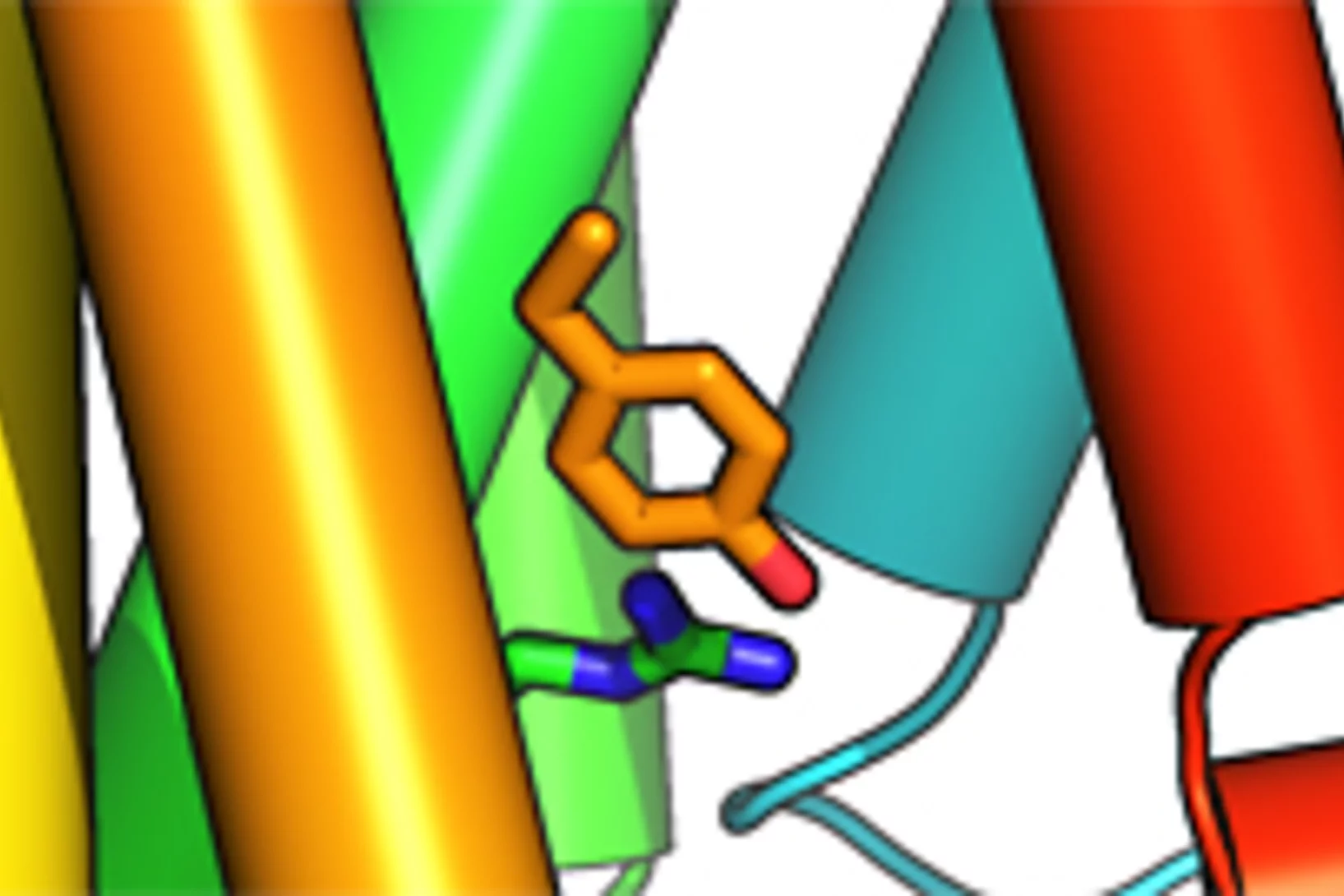
Lebende Zellen empfangen dauernd Informationen von aussen, die über Rezeptoren in das Zellinnere weitergeleitet werden. Genetisch bedingte Fehler in solchen Rezeptoren sind der Grund für zahlreiche Erbkrankheiten darunter verschiedene hormonelle Funktionsstörungen oder Nachtblindheit. Forschern des Paul Scherrer Instituts ist es nun erstmals gelungen, die exakte Struktur eines solchen fehlerhaften Rezeptors aufzuklären.This news release is only available in German.
Forscher der Universität Basel und des Paul Scherrer Instituts konnten im Nanomassstab zeigen, wie sich Karies auf die menschlichen Zähne auswirkt. Ihre Studie eröffnet neue Perspektiven für die Behandlung von Zahnschäden, bei denen heute nur der Griff zum Bohrer bleibt. Die Forschungsergebnisse wurden in der Fachzeitschrift «Nanomedicine» veröffentlicht.This news release is only available in German.
Mit einem Festakt hat das Paul Scherrer Institut (PSI) in Villigen (AG) heute an das zehnjährige Bestehen ihrer bedeutendsten Grossforschungsanlage erinnert. Seit der Inbetriebnahme im Sommer 2001 haben Tausende von Forschern aus Hochschule und Industrie an der Synchroton Lichtquelle Schweiz (SLS) qualitativ hochwertige Experimente durchgeführt. Ihre Forschung mündete in über 2000 wissenschaftlichen Publikationen und brachte darüber hinaus einen Nobelpreis sowie eine Vielzahl industrieller Anwendungen hervor.This news release is only available in German.
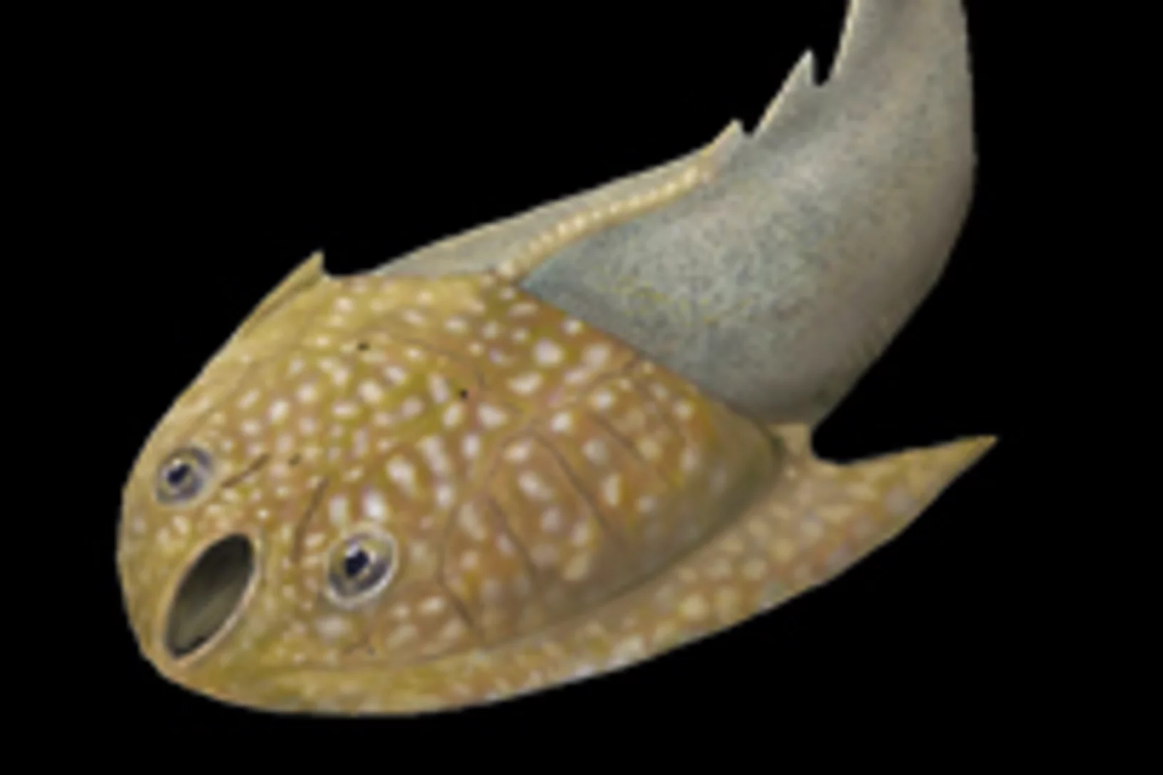
Reorganisation of the brain and sense organs could be the key to the evolutionary success of vertebrates, one of the great puzzles in evolutionary biology, according to a paper by an international team of researchers, published today in Nature. The study claims to have solved this scientific riddle by studying the brain of a 400 million year old fossilized jawless fish à an evolutionary intermediate between the living jawless and jawed vertebrates.
An international team of researchers has developed a new method for making detailed X-ray images of brain tissue, which has been used to make the myelin sheaths of nerve fibres visible. Damage to these protective sheaths can lead to various disorders, such as multiple sclerosis. The facility for creating these images of the protective sheaths of nerve cells is being operated at the Swiss Light Source (SLS), at the Paul Scherrer Institute.
At the beginning of the process of sight, light interacts with a protein molecule called Rhodopsin. This molecule contains the actual light sensor that is stimulated by the incoming light and changes its form, in order to trigger the rest of the process. Researchers have now managed to determine the exact structure of the Rhodopsin molecule in its short-lived, excited state. From this, they have obtained a precise picture of the first step of the process of sight.
In menschlichen Zellen finden sich stammesgeschichtlich sehr alte Funktionseinheiten, die als Centriolen bezeichnet werden. Ein Forscherteam vom PSI und der ETH Lausanne hat nun erstmals ein Modell für die Bildung der Centriolen vorgestellt. Das erstaunende Ergebnis ist, dass die Neuner-Symmetrie des Centriols durch die Fähigkeit eines einzelnen Proteins sich selbst zu organisieren zustande kommt.This news release is only available in French and German.
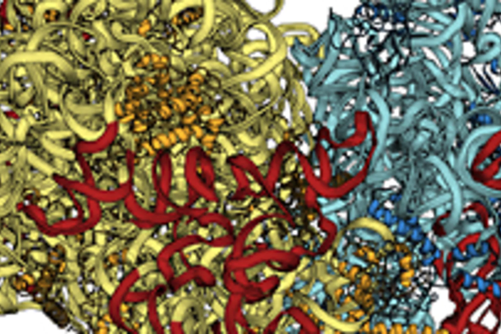
Ribosomes are the protein factories of the living cell and themselves very complex biomolecules. Now, a French research group has for the first time determined the structure of the ribosome in a eukaryotic cell à a complex cell containing a cell nucleus. An important part of the experiments was performed with synchrotron light at the Swiss Light Source SLS of the Paul Scherrer Institute.
For decades researchers have searched for magnetic monopoles à isolated magnetic charges that can move freely like electric charges. Now a team of researchers from the Paul Scherrer Institute and University College Dublin have been able to produce monopoles in the form of quasiparticles in an assembly of nanoscale magnets and have directly observed how they move.
