
Radionuclides are unstable atoms that emit radiation when they decay in an attempt to obtain stability. Doctors have utilized this trait for decades to diagnose patients. They inject the radionuclide into the bloodstream, so that its radiation can be given off directly from inside the body. This radiation makes its way out through the skin, where it can be captured by special-purpose cameras. Since the nuclide can be concentrated selectively at the sites of cancer cells or in organs with malfunctioning metabolism, the camera can locate these very accurately with the help of the emitted radiation. Important factors taken into consideration include the physical properties of the radionuclide in question, which decisively affects the quality and accuracy of the resulting images. Researchers at the Paul Scherrer Institute PSI have been working with different radionuclides to find those best suited for certain medical applications. They have a hot candidate.
More than 10,000 hospitals worldwide use radionuclides in medicine, and more than 90 percent of all such interventions are diagnostic: for early detection and more accurate localisation of cancer, or for functional examination of organs. The radionuclide most frequently used for these purposes is technetium-99m, which emits gamma rays when it decays. Doctors rely on it for roughly 35 million scans per year. There are modern diagnostic systems, however, that use other kinds of radiation instead of gamma rays and, thus, require other radiation sources. One of these is the so-called PET (positron emission tomography) scanner, a special-purpose camera that produces high-quality colour cross-sections of the body and even enables studies that follow the workings of the heart, kidneys, or brain in real-time. Yet for a PET scanner, radionuclides of a very special kind are needed. These must emit positrons when they decay – particles that have practically the same properties as electrons but carry a positive electric charge. Useful radionuclides that emit positrons are rare, and so far approximately ten different ones have been used for medical examination of patients. Not one of them is perfect. Therefore PSI researchers, with the help of the Institute's own proton accelerator facility, are looking for new radionuclides suitable for diagnostic tests with the PET scanner.
A difficult search
But how do you find such a radionuclide? Each has its own typical decay properties: the type of radiation, the half-life, and the radiation energy. To start with, you have to know which nuclides emit positrons in the first place – only these are even in the running,
explains PSI researcher Nick van der Meulen of the Laboratory for Radiochemistry as well as the Centre for Radiopharmaceutical Sciences. For that you need to assess which nuclear reaction is possible for a given atom and which precursor material is needed to achieve this exact nuclear reaction.
It is, then, crucial whether the nuclide generated out of the reaction gives off still more additional types of radiation. Gamma rays, for example, are not desired, as they are of no use for PET scanning, but contribute to the total radiation exposure. To permit later use with the patient, this should be kept as low as possible.
Van der Meulen must also keep the half-life of the radionuclide in mind. One of the most important characteristics of radionuclides, this indicates the time span after which half of the nuclide has decayed. If radionuclides used in medical tests have a long half-life of weeks or months, the patient receives too much radiation,
the researcher reports. On the other hand, with too short a half-life, in the range of minutes, the time from production to application is sometimes barely sufficient. The radionuclide decays before it can even be used. Thus materials with a half-life of a few hours are best suited for this application.
Aside from that, the radionuclides must also have the right energy range so that later, in the PET scanner, high resolution images will be produced.
A promising candidate
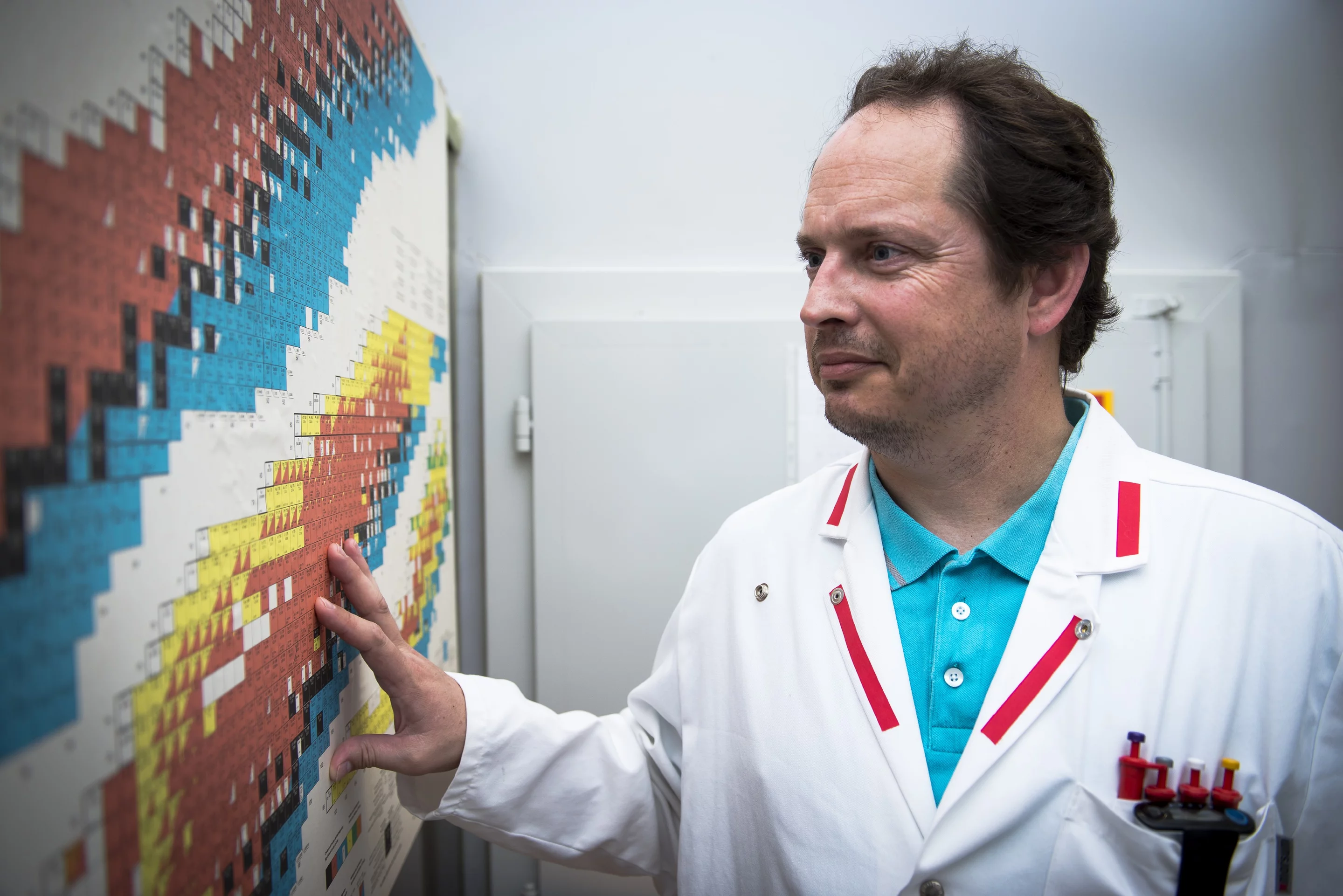
In recent years, van der Meulen alone has tested seven different radionuclides at the PSI. He considers one of them, scandium-44, especially promising for diagnostic PET imaging. It has a relatively short half-life of four hours and emits the desired positrons. In addition, it is easily linked to various molecules such as peptides, for example. This property is important in case it is used in the targeted search for tumours. A radionuclide, on its own, is not generally capable of finding cancer cells or organs in the body: the molecule a radionuclide is attached to can do that. It is either so specific that it recognizes the surface structure of a target cell and docks there, by means of a lock-and-key principle, or it is, like glucose for example, part of the body's natural metabolism. It then takes the same normal route as the body's own metabolic products, all the way through to excretion. During the process it sends out radiation, through its radioactive marking, that a special-purpose camera (such as the PET scanner) records.
The path to practice
In van der Meulen's opinion, scandium-44 has the potential to replace other radionuclides within a few years. Given its decay characteristics and particularly its half-life, images could still be taken with the PET scanner after many hours. In addition, the half-life would allow the radionuclide to be transported over a distance of several hundred kilometres – without having completely decayed when it reaches the hospital where it will be used. This means it can also be available to clinics that do not produce radionuclides on-site and must order them from facilities situated far away.
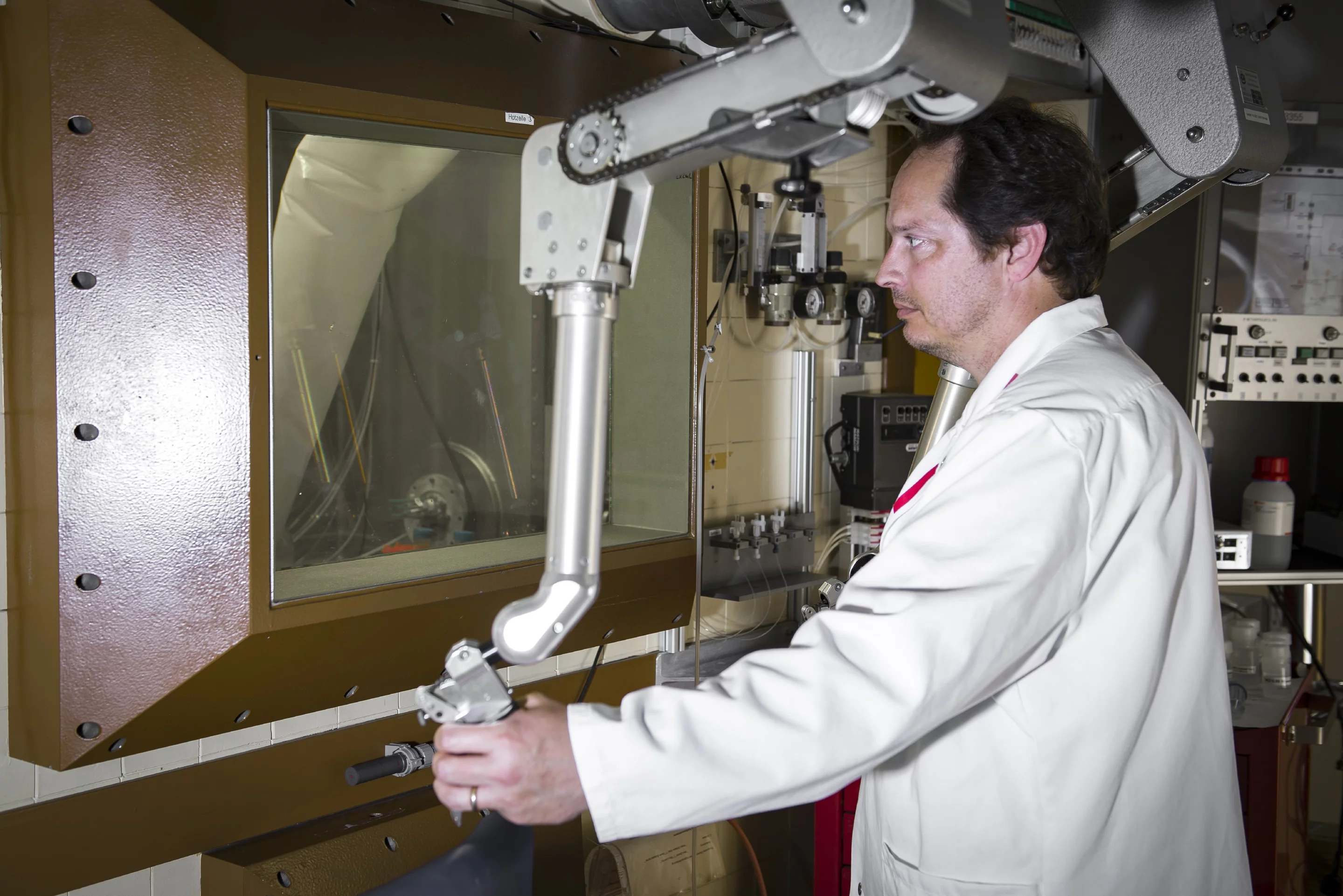
Van der Meulen and his work group have developed a cyclotron-based procedure for producing scandium-44 in a concentration and quantity as needed for medical purposes. Up to now, other research groups have only been able to produce this radionuclide in small activities by means of a cyclotron, or to obtain it from titanium-44 in a nuclide generator. A generator contains a mother nuclide, with a longer half-life, that decays into a daughter nuclide with a shorter half-life. For further use, the daughter nuclide is then separated out by chemical means. The titanium-44 itself is produced in a cyclotron. It takes weeks to produce the necessary titanium-44. What we do is, in comparison, simple,
van der Meulen says. We take our precursor material, calcium-44, put it in the beam, and fire protons at it for 90 minutes.
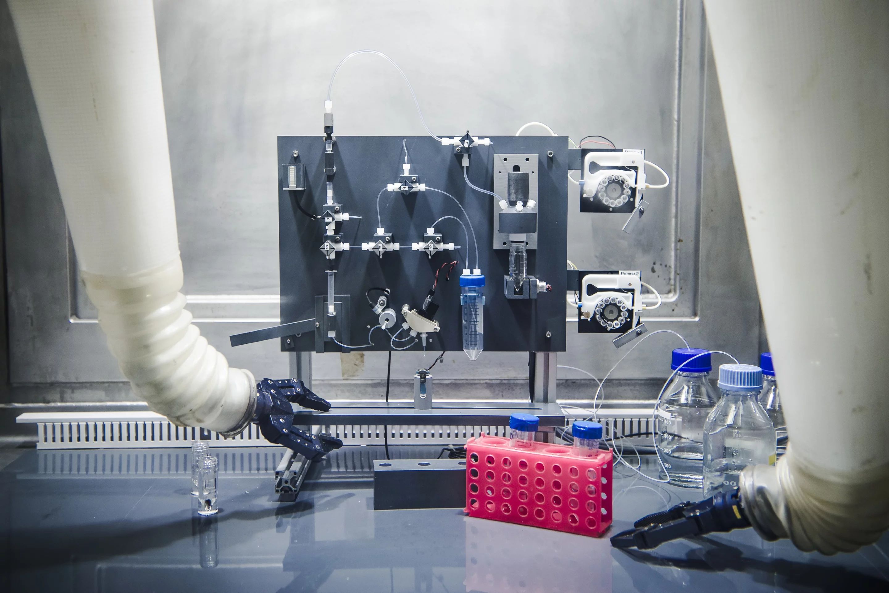
The chemist's stripped-down description represents a highly complex business. He has to precisely calculate, in advance, with what energy he wants to bombard the calcium so that, in the end, the scandium will be produced in the desired concentration and quantity and not too many reaction by-products will be created. These need to be removed in several purification steps, because scandium-44 must be as pure as possible. The remainder of the calcium is recycled.
In the whole process, one point is especially important for van der Meulen: The cyclotron we are using at the PSI is more powerful than other cyclotrons. If we want to develop something with our work that will later be useful to the patients, we need to have calculated all the parameters in such a way that they also work in a small, commercially available cyclotron at a hospital.
So far, he and his group are making good progress. In roughly five years he wants to have perfected the production of scandium-44 to the point where the method is technically reproducible and can be developed further for the market by interested companies.
Text: Sabine Goldhahn
Principle of positron emission tomography (PET)
Positron emission tomography (PET) makes structures and processes in the human body visible. For this method, radioactive atoms (radionuclides) that emit positrons when they decay are put into the body. Positrons are in a sense the antimatter equivalent of electrons. The two particles agree in practically all of their properties – e.g. they have the same mass. But the positrons have, for example, a positive electric charge, while the electrons carry a negative charge. If an electron and a positron come together, they wipe each other out, whereby two highly energetic light particles are created and fly off in more or less opposite directions. These two light particles can be observed with special detectors, and from their directions of flight it can be determined where the positron and the electron converged.Since there are very many electrons in the body, this process – which the experts refer to as annihilation – takes place very soon after the positron is generated. This makes it possible to reconstruct the position of the radionuclide in the body very well. But for positrons and electrons to annihilate each other, they can't be too fast. If the positron has a high kinetic energy and thus high speed when it is produced, it needs to be slowed down by the particles in its surroundings. In the process, however, it moves away from its point of origin, and this can compromise the spatial resolution. The kinetic energy of the positrons that are set free differs from one radionuclide to another.
Scandium-44 (44Sc) can be artificially produced in two ways:
1. From titanium-44 in a nuclide generator. The nucleus of titanium-44 can trap an electron from the atomic shell and thus transform itself into a nucleus of scandium-44. The titanium-44 can be produced from the stable scandium-45 with the help of a proton beam. In the process, scandium-44 captures a proton and immediately emits two neutrons. Radiochemists would describe this reaction using the formula: (45Sc(p,2n)44Ti->44Sc)
2. From calcium-44 in a cyclotron. In the process, the calcium-44 nucleus captures one proton and emits one neutron. Written as a formula: (44Ca(p,n)44Sc)
Additional information
- The article
Targeting cancer
provides an overview of the work of the Center for Radiopharmaceutical Sciences. - The articles
Developing a new drug against thyroid cancer
andMedicines made to order with pinpoint precision
describe the development and production of a drug at the Center for Radiopharmaceutical Sciences. - A new method that would channel radioactive substances right into the cell's nucleus is described in the article:
Hitting cancer from the inside
. - How researchers at PSI produce radionuclides:
In the focus of the protons
.
Contact
Dr. Nick van der Meulen, Group Leader for Radionuclide Development (Laboratory for Radiochemistry, PSI, and Centre for Radiopharmaceutical Sciences of the Paul Scherrer Institute PSI, ETH Zurich, and the University Hospital Zurich)Telephone: +41 56 310 50 87, e-mail: nick.vandermeulen@psi.ch