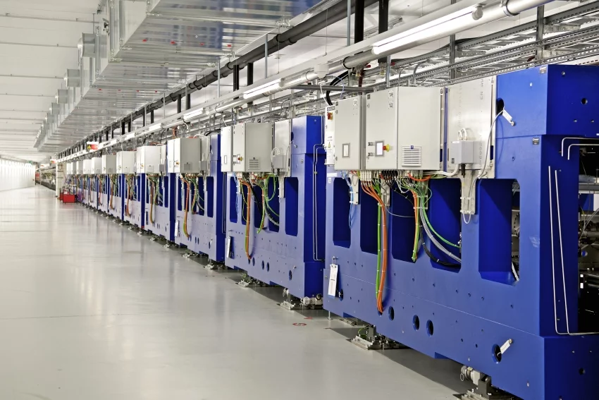
Die jüngste Grossforschungsanlage des PSI erzeugt sehr kurze Pulse von Röntgenlicht mit Lasereigenschaften. Damit können Forschende extrem schnelle Vorgänge wie die Entstehung neuer Moleküle bei chemischen Reaktionen verfolgen, die detaillierte Struktur lebenswichtiger Proteine bestimmen oder den genauen Aufbau von Materialien klären. Die Erkenntnisse erweitern unser Verständnis der Natur und führen zu praktischen Anwendungen wie etwa neuen Medikamenten, effizienteren Prozessen in der chemischen Industrie oder neuen Materialien in der Elektronik.
Mehr dazu unter Überblick SwissFEL
The role of long-lived reactive oxygen intermediates in the reaction of ozone with aerosol particles
The heterogeneous reactions of ozone with aerosol particles are of central importance to air quality. They are studied extensively, but the molecular mechanisms and kinetics remain unresolved. Based on new experimental data and calculations, we show that long-lived reactive oxygen intermediates (ROIs) are formed. The chemical lifetime of these intermediates exceeds 100 seconds, which is much longer than the surface residence time of molecular ozone (~ ns).
Recent increase in black carbon concentrations from a Mt. Everest ice core spanning 1860–2000 AD
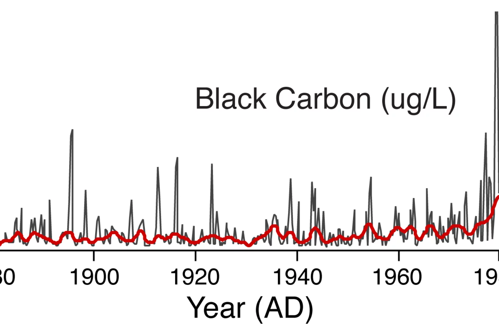
A Mt. Everest ice core spanning 1860à2000 AD and analyzed at high resolution for black carbon (BC) using a Single Particle Soot Photometer demonstrates strong seasonality, with peak concentrations during the winter‐spring, and low concentrations during the summer monsoon season. BC concentrations from 1975à2000 relative to 1860à1975 have increased approximately threefold, indicating that BC from anthropogenic sources is being transported to high elevation regions of the Himalaya.
Dem Rätsel der Centriolen-Bildung auf der Spur
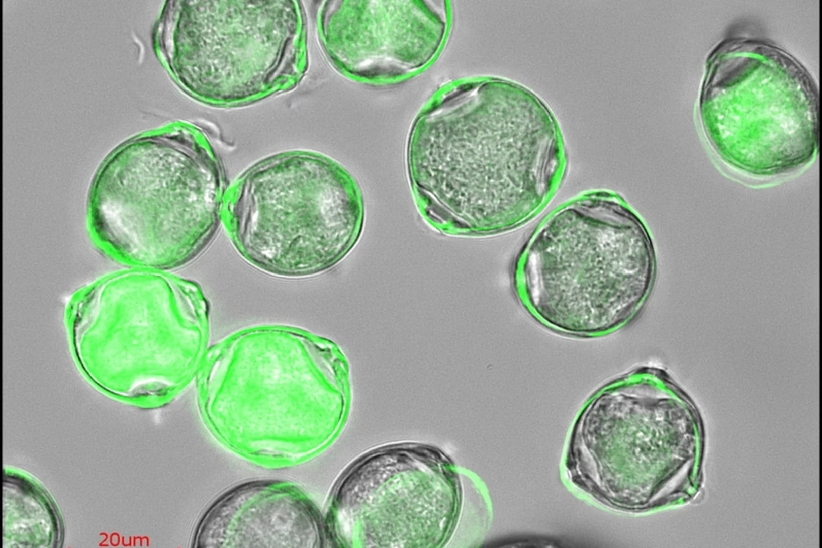
In menschlichen Zellen finden sich stammesgeschichtlich sehr alte Funktionseinheiten, die als Centriolen bezeichnet werden. Ein Forscherteam vom PSI und der ETH Lausanne hat nun erstmals ein Modell für die Bildung der Centriolen vorgestellt. Das erstaunende Ergebnis ist, dass die Neuner-Symmetrie des Centriols durch die Fähigkeit eines einzelnen Proteins sich selbst zu organisieren zustande kommt.
Wie stark ist die schwache Kraft?
Eine neue Messung der Lebensdauer des Myons à die genaueste Bestimmung einer Lebensdauer in der Welt der kleinen Teilchen à liefert einen hochgenauen Wert für einen Parameter, der für die Bestimmung der Stärke der schwachen Kernkraft entscheidend ist. Die Experimente wurden von einem internationalen Forschungsteam am Paul Scherrer Institut durchgeführt.
LaAlO3 - Buckling under pressure to hand over the charges
In this paper, we report on the change in the atomic structure of the conducting interface between the insulators LaAlO3 and SrTiO3 as a function of the LaAlO3 layer thickness. We discovered that the atoms at the interface buckle in an attempt to counteract the internal electric field produced when these two insulators touch one another.
Die Nanomaschinen des Lebens verstehen
Ribosomen sind die Proteinfabriken der lebenden Zellen à und selbst auch hochkomplexe Biomoleküle. Eine französische Forschungsgruppe hat nun erstmals die Struktur von Ribosomen in eukaryotischen Zellen bestimmt, also in komplexen Zellen, die über einen Zellkern verfügen. Ein wesentlicher Teil der Experimente wurde an der Synchrotron Lichtquelle Schweiz SLS des Paul Scherrer Instituts durchgeführt.
Observation of a ubiquitous three-dimensional superconducting gap function in optimally doped Ba0.6K0.4Fe2As2
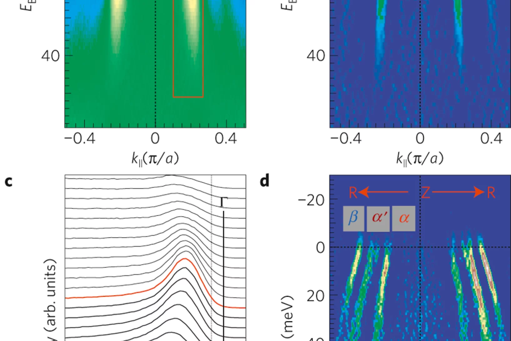
The iron-pnictide superconductors have a layered structureformed by stacks of FeAs planes from which the superconductivity originates. Given the multiband and quasi three-dimensional1 (3D) electronic structure of these high-temperature superconductors, knowledge of the quasi-3D superconducting (SC) gap is essential for understanding the superconducting mechanism.
Potenzial einer kostengünstigen Brennstoffzelle für Autos aufgezeigt
Das Paul Scherrer Institut PSI und die Belenos Clean Power AG haben ein Brennstoffzellensystem entwickelt, das das Potenzial aufweist, kostengünstig in einen Kleinwagen eingebaut zu werden, der dann über seine Lebenszeit ähnlich viel kosten würde wie ein herkömmliches Auto. Für diesen wichtigen Schritt in Richtung umweltfreundliche Mobilität erhalten PSI und Belenos den Watt d’Or 2011.
Benzin aus Wasser, CO2 und Sonnenlicht
Einem Forschungsteam um Aldo Steinfeld ist es gelungen, mit Solarenergie aus Wasser und Kohlendioxid Treibstoff zu erzeugen. Dazu haben die Wissenschaftler einen Solar-Reaktor entwickelt, in dem konzentrierte Sonnenstrahlung das dafür nötige thermochemische Verfahren antreibt.
Zukünftig auf demselben Chip: Daten speichern und verarbeiten
Forschern ist es gelungen, magnetisch polarisierte Elektronen mit elektrischen Feldern zu beeinflussen. Diese wichtige Entdeckung könnte es ermöglichen, die Eigenschaften von Elektronen in einem Computerchip gleichzeitig für Verarbeitung und Speicherung von Daten zu nutzen. In Zukunft könnte dies die Entwicklung erheblich sparsamerer und leichterer elektronischer Geräte aller Art ermöglichen.
Röntgenpreis for X-Ray research goes to Christian David
On 26th November 2010, Christian David, scientist at the Laboratory for Micro and Nanotechnology, received the Röntgenpreis for research in radiation science. David pioneered a method to enhance the quality of X-ray images. He received the award jointly with Franz Pfeiffer from Technische Universität München who worked closely together with him.
The award
Magnetisierte Bereiche in 3D sichtbar gemacht
Magnetisierbare Materialien sind nie völlig unmagnetisch, sondern enthalten immer magnetisierte Bereiche à die magnetischen Domänen. In einem Experiment am Helmholtz-Zentrum Berlin (HZB) konnten diese Domänen erstmals in ihrer dreidimensionalen Struktur abgebildet werden. Der Versuch beruhte auf einer Weiterentwicklung eines am Paul Scherrer Institut entstanden Verfahrens und nutzte neutronenoptische Komponenten, die am PSI hergestellt worden sind.
Effizienter Gentransfer nun auch in Säugerzellen möglich
Wissenschaftler am Paul Scherrer Institut entwickelten ein neues Verfahren, das auch zur Entwicklung von neuen Medikamenten genutzt werden kann.Die Gentechnik ist aus der modernen Biologie nicht mehr wegzudenken. Sie liefert Werkzeuge, mit denen Forscher Gene aus dem Erbgut von Zellen herausschneiden, verändern und einfügen können. Die stabile Einführung mehrerer Gene in Säugetierzellen gilt zwar als Schlüsseltechnologie, die verfügbaren Methoden waren bislang aber äusserst ineffizient. Wissenschaftler am PSI haben nun eine neuartige Technik entwickelt.
Was der Satz vom Igel über Flussschläuche in Supraleitern sagt
In einem starken Magnetfeld bilden Hochtemperatursupraleiter Flussschläuche à dünne Kanäle, in denen das Feld den Supraleiter durchdringen kann. Diese parallelen Schläuche ordnen sich meist in regelmässigen Mustern an. Nun haben zwei Physiker gezeigt, dass eine solche Anordnung von der Richtung des äusseren Magnetfelds abhängen muss. Grundlage dieser Ergebnisse ist eine mathematische Aussage, die als Satz vom Igel bekannt ist.
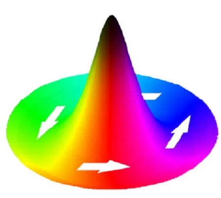
Direct Determination of Large Spin-Torque Nonadiabaticity in Vortex Core Dynamics
We use a pump-probe photoemission electron microscopy technique to image the displacement of
vortex cores in Permalloy discs due to the spin-torque effect during current pulse injection. Exploiting the
distinctly different symmetries of the spin torques and the Oersted-field torque with respect to the vortex
spin structure we determine the torques unambiguously, and we quantify the amplitude of the strongly
Magnetische Monopole auf Wanderschaft
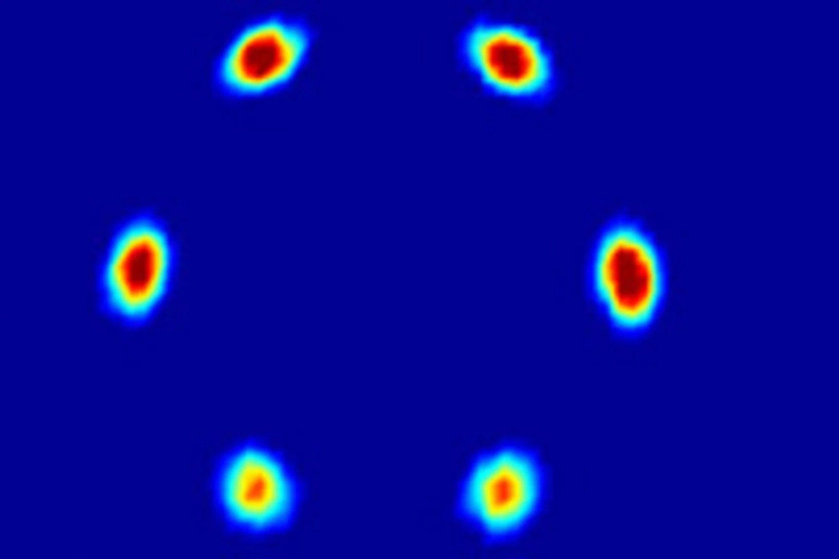
Seit Jahrzehnten suchen Forschende nach magnetischen Monopolen à einzelnen magnetischen Ladungen, die sich wie einzelne elektrische Ladungen alleine bewegen könnten. Nun ist es einem Team von Forschenden des Paul Scherrer Instituts und des University College Dublin gelungen, Monopole als Quasiteilchen in einer Anordnung von nanometergrossen Magneten zu erzeugen und ihre Bewegung unmittelbar zu beobachten.
Moving Monopoles Caught on Camera - researchers make visible the movement of monopoles in an assembly of nanomagnets
For decades, researchers have been searching for magnetic monopoles; isolated magnetic charges, which can move around freely in the same way as electrical charges – since magnetic poles normally only occur in pairs.
25 Jahre erfolgreiche Behandlung von Augentumoren am PSI
Heute haben die Physiker und Ärzte des PSI diesen Erfolg mit einem Festsymposium gefeiert. In Anwesenheit von geladenen Gästen aus Forschung, Medizin und Politik wurde dabei auch die brandneue Behandlungsanlage OPTIS 2 eingeweiht. Diese Bestrahlungseinrichtung befindet sich nicht nur technisch auf dem allerneusten Stand, sondern überzeugt auch durch ihre Patientenfreundlichkeit.
Fortschritt für die Knochen-Forschung
Hochauflösendes Verfahren zur Nano-Computertomographie entwickeltEin neuartiges Nano-Tomographie-Verfahren, das von einem Team der TU München, des Paul Scherrer Instituts und der ETH Zürich entwickelt wurde, erlaubt erstmals computertomographische Untersuchungen feinster Strukturen mit einer Auflösung im Nanometerbereich. Mit Hilfe der neuen Methode können etwa dreidimensionale Innenansichten fragiler Knochenstrukturen erstellt werden.
High-resolution method for computed nano-tomography developed
A novel nano-tomography method developed by a team of researchers from the Technische Universität München (TUM), the Paul Scherrer Institute (PSI) and the ETH Zurich opens the door to computed tomography examinations of minute structures at nanometer resolutions. The new method makes possible, for example, three-dimensional internal imaging of fragile bone structures. The first nano-CT images generated with this procedure was published in the renowned journal Nature on September 23, 2010.
Die Batterie der Zukunft hält länger
Der «swisselectric research award 2010» geht an den Chemiker Andreas Hintennach vom Paul Scherrer Institut. Dank seiner Forschung könnten Lithiumionen-Batterien in Zukunft deutlich langlebiger werden. Das Speichern von Strom wird somit umweltfreundlicher und kostengünstiger.
Paul Scherrer Institut erhält hohen Besuch aus Politik und Wirtschaft
Das Kernstück der neuen Grossforschungsanlage SwissFEL ging heute am Paul Scherrer Institut wie geplant in Betrieb. Ehrengast Bundesrat Didier Burkhalter drückte auf den roten Knopf, und die Anlage produzierte den ersten Elektronenstrahl.
Neue Karriere für lebenswichtiges Biomolekül möglich
Porphyrin, das als Teil des Hämoglobins den Sauerstofftransport im Blut möglich macht, könnte in leicht veränderter Form auch in technischen Geräten Verwendung finden. Forschende des Paul Scherrer Instituts PSI und der Universität Basel haben gezeigt, dass sich eine magnetische Eigenschaft des Moleküls chemisch ein- und ausschalten lässt, so dass dieses als winziger Schalter dienen könnte.
Gemeinsam forschen für bessere Batterien
Die Speicherung von elektrischer Energie ist eine der zentralen Fragen der Energiezukunft. Neue Batterietypen zu entwickeln, die mehr Energie speichern können als die heute verfügbaren, ist das Ziel eines Forschungsnetzwerks, das der weltweit grösste Chemiekonzern BASF gemeinsam mit dem Paul Scherrer Institut PSI und Forschungseinrichtungen aus Deutschland und Israel gegründet hat.
Universelles Gesetz für Veränderungen in Werkstoffen gefunden
In vielen wichtigen Werkstoffen findet man mehrere Phasen. Wird ein solcher Werkstoff erwärmt, können Atome von der einen Phase zur anderen wandern, so dass sich die Verteilung der Phasen ändert à und damit oft die Eigenschaften des Werkstoffs. Nun haben Forschende für einen wichtigen Fall einer solchen Veränderung gezeigt, dass es eine universelle Gesetzmässigkeit gibt, die den Vorgang beschreibt. Und zwar für alle Werkstoffklassen.
Halbleiter aus Kunststoff besser verstehen
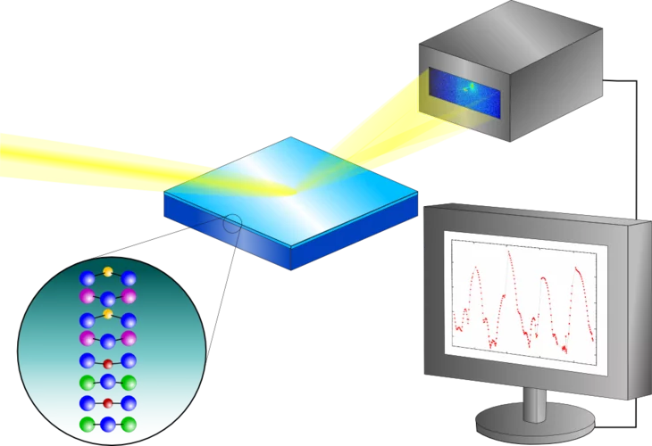
Halbleiter aus Polymermaterialien dürften in Zukunft immer mehr Bedeutung für die Elektronikindustrie bekommen à etwa als Grundlage von Transistoren, Solarzellen oder Leuchtdioden. Meist bestehen sie nicht aus einer einzelnen Substanz, weil sich ihre besonderen elektrischen Eigenschaften oft erst dann ergeben, wenn man mehrere verschiedene Polymere miteinander mischt. Forschende des Paul Scherrer Instituts und der Universität Cambridge ein Verfahren entwickelt, mit dem sie den detaillierten Aufbau des Materials sowohl im Inneren als auch an der Oberfläche bestimmen können.
Understanding plastic semiconductors better
New method allows important insights into polymer semiconductors
Semiconductors made from polymer materials are becoming increasingly important for the electronics industry – as a basis for transistors, solar cells or LEDs – showing important advantages when compared to conventional materials: they are lightweight, flexible and very cheap to produce.
Neues Röntgenverfahren unterscheidet, was bisher gleich aussah
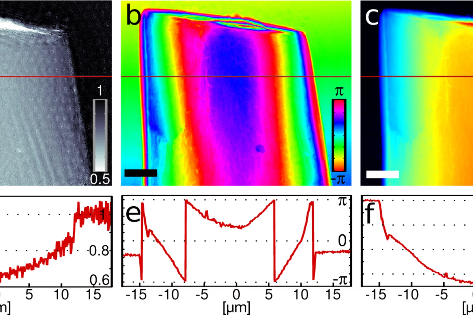
Auf Bildern, die mit Phasenkontrastverfahren erzeugt werden, kann man Gewebe unterscheiden, das auf gewöhnlichen Röntgenbildern fast gleich aussieht: etwa Muskeln, Knorpel, Sehnen oder Weichteiltumore. Forschende des Paul Scherrer Instituts und der Chinesischen Akademie der Wissenschaften haben das Verfahren so weiterentwickelt, dass es in Zukunft einfacher zu handhaben sein wird. Das könnte helfen, Tumore zu erkennen oder gefährliche Gegenstände im Gepäck sichtbar zu machen.
New X-ray technique distinguishes between that which previously looked the same
A new method forms the basis for the widespread use of an X-ray technique which distinguishing types of tissue that normally appear the same in conventional X-ray images
Proton kleiner als gedacht
Das Proton à einer der Grundbausteine der Materie à ist kleiner als bisher angenommen. Das haben Experimente eines internationalen Forschungsteams bewiesen, die am Paul Scherrer Institut PSI im schweizerischen Villigen durchgeführt worden sind.

